.webp&w=384&q=75)

Loading, please wait...
Loading, please wait...

Surgical correction of complex congenital defects often reveals rare, compound malformations. Specifically, performing a Cone procedure Ebstein anomaly repair in a patient with prior partial atrioventricular septal defect repair presents unique surgical hurdles. Partial atrioventricular septal defect typically involves an ostium primum atrial septal defect and cleft left atrioventricular valve. However, surgeons rarely encounter coexisting atypical Ebstein anomaly of the right-sided valve. In classical Ebstein anomaly, the septal and posterior tricuspid leaflets adhere downward into the inlet portion of the right ventricle. Consequently, this downward displacement atrializes a significant portion of the ventricular myocardium. In atypical presentations, the leaflet attachment patterns diverge markedly from classic morphology. Furthermore, prior patch closures and annular remodelling alter normal right ventricular landmarks. Because primary repair alters regional anatomy, scarring can severely distort the fibrous skeleton. Therefore, secondary severe right atrioventricular valve regurgitation develops over time. This severe regurgitation precipitates progressive right atrial enlargement, exercise intolerance, and refractory arrhythmias. Pediatric cardiac surgeons must carefully recognize these intertwined anatomical anomalies. Ultimately, accurate preoperative recognition prevents progressive right ventricular failure and guides precise surgical reconstructive strategy.
The Cone procedure represents the gold standard surgical approach for restoring anatomical valve competence. During this intricate operation, surgeons circumferentially mobilize the anomalous right atrioventricular valve tissue. First, the surgeon sharply detaches the displaced anterior, posterior, and septal leaflets from their anomalous ventricular attachments. Furthermore, the surgeon carefully preserves the primary cordal apparatus to maintain crucial leaflet geometry. Next, the surgeon longitudinally plicates the thinned, atrialized right ventricle. This plication effectively restores the functional ventricular chamber volume and reduces regional wall stress. Subsequently, the surgical team rotates and unifies the mobilized leaflet edges along their longitudinal axes. Consequently, this maneuver constructs a symmetric, cone-shaped valve structure that mirrors native leaflet coaptation. The surgical team then reattaches the circular base of this reconstructed cone to the true anatomic atrioventricular annulus. Additionally, surgeons often place an annuloplasty band or ring to reinforce annular stabilization. In patients with previous partial septal repair, the surgeon must diligently avoid compromising the nearby conduction system. Therefore, precise suture placement minimizes the occurrence of complete atrioventricular heart block. Ultimately, this comprehensive technical mobilization yields durable leaflet coaptation.
Operating in a reoperative congenital cardiac field introduces substantial operative complexity. In patients who previously underwent partial septal defect repair, extensive mediastinal and intracardiac adhesions typically obscure essential structures. Moreover, prior patch reconstruction of the atrial septum alters the anatomic course of the conduction axis. Specifically, the compact atrioventricular node and the bundle of His lie vulnerable near the post-repair junction. Therefore, cardiac surgeons must map the conduction axis meticulously during leaflet mobilization. In addition, the anomalous right valve tissue often exhibits dense fibrotic tethering to the ventricular septum. Consequently, freeing the leaflets without creating ventricular septal perforations demands extreme microsurgical delicacy. Furthermore, surgeons must simultaneously evaluate residual left atrioventricular valve regurgitation. If the left valve leaks, persistent regurgitation will elevate pulmonary pressures and jeopardize right heart recovery. Accordingly, preoperative multiplanar transesophageal echocardiography provides vital anatomical roadmaps. Advanced cardiac magnetic resonance imaging also delineates right ventricular stroke volumes and fibrosis. Thus, a multi-modality diagnostic protocol enables surgical teams to anticipate technical challenges before cardiopulmonary bypass begins. Careful reoperative planning ultimately ensures safe, repeatable reconstructive outcomes.
Successful cone reconstruction delivers profound hemodynamic and functional benefits. By restoring anatomical leaflet coaptation at the true annulus, the reconstruction eliminates significant right atrioventricular valve insufficiency. Consequently, right ventricular volume overload decreases rapidly following separation from cardiopulmonary bypass. Furthermore, longitudinal plication of the atrialized ventricle transforms non-contractile tissue into a stable fibrous anchor. This structural reduction significantly augments right ventricular fractional area change and global forward cardiac output. In addition, right atrial pressure decreases markedly, thereby mitigating hepatic congestion and peripheral systemic edema. Interventricular interactions also improve promptly as the ventricular septum regains a normal geometric configuration. Thus, left ventricular diastolic filling improves simultaneously, which enhances systemic perfusion. In the postoperative period, pediatric intensive care teams must maintain adequate preload while avoiding excessive inotropic stimulation. Moreover, vigilant monitoring helps identify transient rhythm disturbances or sluggish right ventricular recovery early. Most patients demonstrate substantial symptomatic improvement, experiencing enhanced functional exercise capacity and normal growth velocity. Therefore, effective anatomical repair successfully interrupts the adverse cycle of progressive right-sided cardiac failure.
Long-term valve repair durability remains a principal priority in congenital cardiac surgery. Historically, tricuspid valve replacement in children and young adults carried poor longevity. Artificial prosthetic valves incur high risks of rapid calcification, thromboembolism, and progressive patient-prosthesis mismatch. Conversely, the cone repair utilizes autologous native leaflet tissue, permitting dynamic growth in pediatric populations. Furthermore, preserving native living tissue eliminates the routine requirement for long-term anticoagulation. In developing nations like India, avoiding lifelong warfarin therapy delivers immense clinical and social advantages. Many pediatric patients residing in rural or semi-urban regions face barriers to frequent international normalized ratio monitoring. Therefore, durable native valve repair drastically reduces devastating hemorrhagic and embolic complications. Moreover, Indian tertiary pediatric cardiac centres increasingly encounter late presenters with complex, uncorrected or reoperated congenital defects. Establishing specialized cone reconstruction expertise in regional centers ensures accessible, top-tier surgical care for complex anomalies. Thus, continuous post-repair clinical and echocardiographic surveillance remains mandatory to assess valve stability. Ultimately, expanding cone reconstructive techniques elevates the standard of care for congenital heart disease nationwide.
The Cone procedure uses the patient's native valve leaflets rather than synthetic or bioprosthetic materials. Consequently, this autologous reconstruction preserves natural geometry, retains growth potential in growing children, and prevents early prosthetic degeneration. Furthermore, the repaired native valve eliminates the requirement for lifelong anticoagulation therapy. Therefore, patients experience substantially lower lifetime risks of thromboembolism, prosthetic endocarditis, and frequent reoperations, which drastically improves long-term cardiac survival and quality of life.
Prior atrioventricular septal defect repair creates dense myocardial adhesions and alters essential cardiac landmarks. Specifically, the atrial septal patch and previous suture lines distort the anatomical location of the cardiac conduction axis. Surgeons must therefore dissect leaflet attachments with extreme precision to prevent complete heart block. Additionally, coexistence of atypical Ebstein anatomy causes abnormal leaflet tethering. Consequently, surgeons must achieve complete 360-degree leaflet delamination while carefully preserving delicate subvalvular structures to avoid tearing.
In India, pediatric cardiac teams prioritize structured follow-up protocols combining serial clinical evaluations and transthoracic echocardiography. Postoperative monitoring specifically tracks right ventricular dimension reduction, residual regurgitation, and functional status. Moreover, clinicians must remain vigilant for latent rhythm disturbances, such as supraventricular tachycardia or heart block. Because patient accessibility to tertiary cardiac centers varies widely across regions, teams educate families regarding red-flag symptoms and facilitate communication with local pediatricians to maintain seamless continuity of care.
Disclaimer: This content is for informational and educational purposes only... Refer to the latest local and national guidelines for clinical practice.
References

Read summarized clinical updates, watch expert medical content, and earn CME certifications right from your smartphone.
Open your smartphone camera to scan and install.





A review of the Cone procedure for severe right atrioventricular valve insufficiency in atypical Ebstein anomaly following partial AVSD repair, emphasizing operative technique and ventricular recovery.
Today

Preclinical research reveals that the hydrophilic extract of the inner bran fraction of rice attenuates skeletal muscle proteolysis in protein-deficient rats, restoring glutathione and polyamine metabolites while downregulating Atrogin-1 and FoxO1 expression.
Today
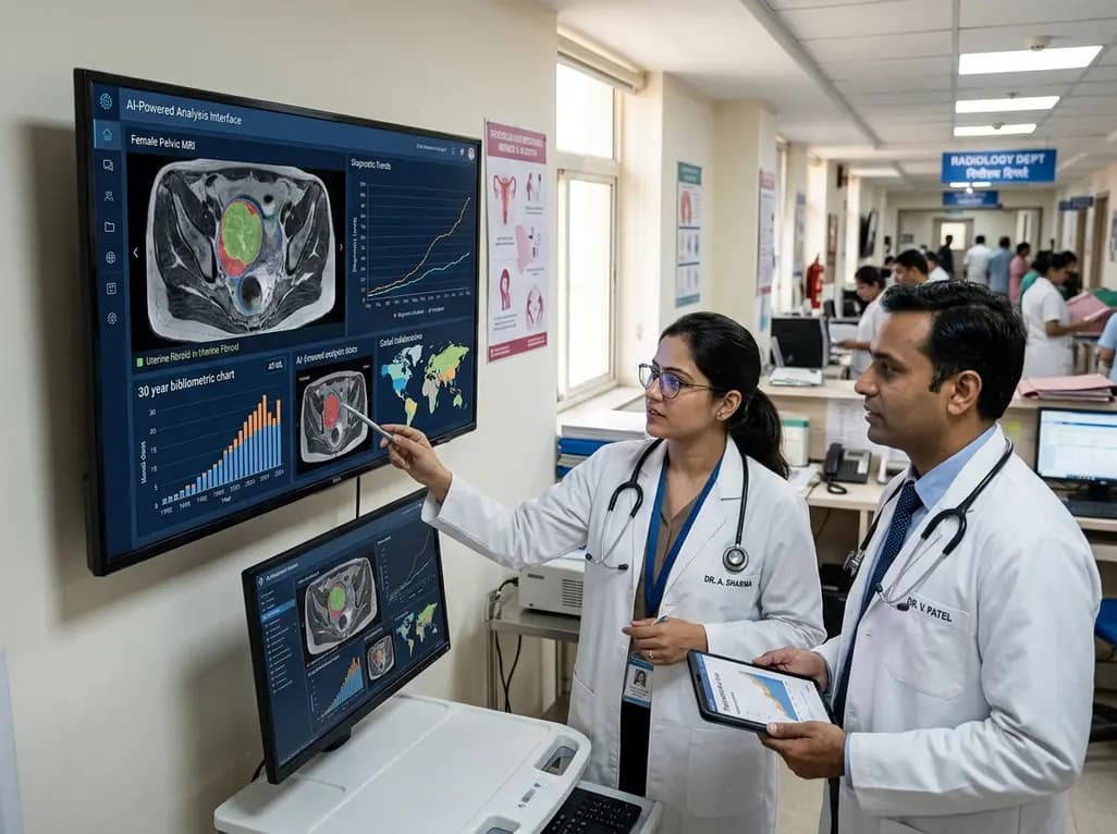
A 30-year bibliometric analysis highlights key research trends in AI in women's health from 1992 to 2024. Discover how evolving computational models, global institutional collaborations, and clinical decision support tools are shaping maternal, oncological, and reproductive care worldwide.
Today

A Level I trauma center study shows a brief bedside mental health intervention boosts 30-day follow-up screening adherence by 84% in nonviolent injury patients. However, outcomes for violence survivors reveal critical care gaps, underscoring the urgent need for specialized community violence intervention programs.
Today

Recent data from Karnataka reveals a threefold surge in genitourinary and renal deaths over ten years. Driven by uncontrolled diabetes, hypertension, and emerging lifestyle hazards, this epidemiological shift highlights an urgent need for proactive screening and multidisciplinary nephrology management in primary care.
Today
