

Loading, please wait...
Loading, please wait...

Detecting an arachnoid cyst in preterm infants presents significant diagnostic challenges for neonatologists and pediatric neurologists. Arachnoid cysts represent benign, congenital intra-arachnoid fluid collections situated between the layers of the arachnoid membrane. Although these lesions frequently remain asymptomatic, clinicians occasionally discover them during neuroimaging for unrelated conditions. For example, a preterm infant born at 30 weeks and 6 days gestation presented with respiratory distress requiring immediate resuscitation. However, prenatal ultrasonography had previously revealed lateral ventricular dilation alongside an expansive posterior fossa cystic lesion. Consequently, the neonatal team initiated targeted diagnostic protocols immediately following stabilization. In premature neonates, physiologic vulnerability often complicates the clinical picture. Therefore, clinicians must distinguish acute perinatal pulmonary compromise from neurological impairment induced by mass effect. Fortunately, detailed physical examinations in this infant demonstrated normal primitive reflexes and no focal neurological deficits. Moreover, continuous interdisciplinary observation ensures immediate recognition of subtle neurological changes. Thus, establishing an accurate baseline neurological profile remains critical for guiding subsequent management decisions in preterm neonates.
Advanced neuroimaging modalities play an indispensable role in delineating neonatal intracranial architecture. Specifically, prenatal ultrasonography provides the earliest window into fetal brain development by identifying abnormal intracranial fluid collections. When sonographers observe posterior fossa cystic lesions, they must assess ventricular symmetry and brainstem morphology. In this preterm case, postnatal serial cranial ultrasonography provided rapid, non-invasive bedside evaluation within the neonatal intensive care unit. Furthermore, cranial sonography precisely delineated the hypoechoic nature of the lesion without patient transport risks. Subsequently, clinicians performed brain magnetic resonance imaging to obtain superior tissue contrast and multiplanar anatomical clarity. High-resolution magnetic resonance imaging confirmed a large retrocerebellar cyst with cerebrospinal fluid signal intensity across all sequences. In addition, the imaging demonstrated preserved cerebellar architecture without vermian agenesis. Consequently, multi-sequence imaging excluded complex posterior fossa malformations and verified the extra-axial arachnoid boundary. Therefore, combining prenatal ultrasonography, bedside cranial sonography, and magnetic resonance imaging establishes a robust diagnostic foundation. Consequently, clinicians achieve diagnostic certainty without exposing unstable neonates to unnecessary transport hazards.
Accurate differentiation of posterior fossa cystic lesions remains essential for predicting prognosis and directing therapy. For instance, clinicians must distinguish retrocerebellar arachnoid cysts from Dandy-Walker malformations, Blake pouch cysts, and mega cisterna magna. In Dandy-Walker malformations, magnetic resonance imaging typically demonstrates vermian hypoplasia alongside cystic enlargement of the fourth ventricle. In contrast, an arachnoid cyst characteristically displaces an intact cerebellum anteriorly without intrinsic parenchymal malformations. Furthermore, Blake pouch cysts exhibit persistent non-perforation of the embryonic area, producing classic fourth ventricular communication. Mega cisterna magna, on the other hand, represents a benign anatomical variant with normal cerebellar vermis morphology. In addition, rare conditions like epidermoid cysts can mimic arachnoid cysts on preliminary sonography. However, advanced diffusion-weighted magnetic resonance imaging readily distinguishes these entities by demonstrating restricted diffusion characteristics. Therefore, clinicians must meticulously examine anatomical relationships on sagittal and axial imaging planes before finalizing management strategies. Consequently, precise diagnostic characterization avoids misdiagnosis and prevents inappropriate interventions.
Historically, neurosurgeons often favored prompt surgical decompression for large intracranial cysts to eliminate potential mass effect. However, modern pediatric neurosurgery increasingly advocates conservative observational management for stable, asymptomatic neonates. In this preterm infant, the clinical team adopted a watchful waiting protocol despite the substantial size of the cyst. Because serial clinical evaluations demonstrated normal fontanelle tension, urgent surgical fenestration remained unnecessary. Moreover, serial cranial ultrasound scans confirmed stable ventricular measurements without progressive hydrocephalus. Consequently, avoiding invasive neurosurgery protected the fragile neonate from intraoperative blood loss, infection, and anesthetic neurotoxicity. In contrast, emerging signs of intracranial hypertension, such as macrocephaly or persistent vomiting, necessitate surgical intervention. Under such circumstances, pediatric neurosurgeons typically perform endoscopic cyst fenestration into the basal cisterns. In addition, longitudinal head circumference tracking provides reliable objective data regarding stable intracranial dynamics. Therefore, conservative management represents a safe initial pathway when supported by rigorous multidisciplinary surveillance. Ultimately, this approach prioritizes neurodevelopment while reserving operative intervention strictly for progressive symptoms.
Managing intracranial cystic malformations in resource-diverse healthcare settings requires pragmatic, evidence-based clinical protocols. In Indian neonatal intensive care units, high-frequency bedside cranial ultrasonography serves as the primary diagnostic tool. Because point-of-care ultrasound is accessible and safe, clinicians can perform serial evaluations without transporting fragile neonates. Furthermore, tertiary centers equipped with pediatric neuro-imaging facilities should confirm sonographic findings through magnetic resonance imaging. Clinicians must also prioritize transparent communication with parents regarding the benign natural history of asymptomatic arachnoid cysts. In many instances, prenatal diagnosis generates extreme parental anxiety, triggering requests for premature surgical intervention. Therefore, neonatologists and pediatricians must offer reassuring counseling, emphasizing that clinical stability justifies conservative monitoring. In addition, routine immunization and developmental stimulation should proceed normally unless specific neurosurgical contraindications emerge. Consequently, establishing standardized referral networks ensures that pediatric neurosurgeons evaluate enlarging lesions promptly. By combining prudent resource utilization with disciplined clinical vigilance, clinicians optimize outcomes across diverse practice environments.
Neurosurgical intervention becomes necessary when an arachnoid cyst causes progressive intracranial hypertension, obstructive hydrocephalus, or focal neurological deficits. Specifically, clinical indicators include a bulging anterior fontanelle, rapidly accelerating head circumference, persistent vomiting, bradycardia, or cranial nerve palsies. In these urgent scenarios, pediatric neurosurgeons typically perform endoscopic cyst fenestration or cystoperitoneal shunting. However, asymptomatic and stable cysts do not warrant surgical intervention, as operative risks often outweigh theoretical benefits.
Cranial ultrasound serves as an ideal bedside screening and monitoring modality for neonates because it avoids radiation, sedation, and patient transport. However, bone acoustic shadowing can restrict acoustic windows in the posterior fossa. In contrast, magnetic resonance imaging provides superior multiplanar anatomical resolution and excellent soft-tissue characterization. Therefore, clinicians utilize magnetic resonance imaging for definitive anatomical diagnosis and rely on serial cranial sonography for routine bedside tracking of ventricular size in stable neonates.
The overall prognosis for infants with isolated, asymptomatic arachnoid cysts remains largely favorable when clinicians implement structured observational monitoring. Most children attain normal developmental milestones, cognitive skills, and motor coordination. However, comprehensive neurodevelopmental surveillance is essential because posterior fossa lesions can occasionally induce subtle cerebellar coordination deficits or speech delays. Consequently, early developmental screening and physical therapy referrals ensure prompt intervention if functional impairments or learning challenges emerge during childhood growth.
Disclaimer: This content is for informational and educational purposes only... Refer to the latest local and national guidelines for clinical practice.
References

Read summarized clinical updates, watch expert medical content, and earn CME certifications right from your smartphone.
Open your smartphone camera to scan and install.





A case report of a preterm infant with a large posterior fossa arachnoid cyst highlights the efficacy of prenatal and postnatal neuroimaging alongside conservative management for asymptomatic cases, minimizing unnecessary surgical morbidity.
Today

Failing to account for competing events like mortality in longitudinal geriatric studies produces biased risk estimates and misleading inferences. This review highlights key methodological principles, insights from the Precipitating Events Project, and practical approaches for clinical research appraisal.
Today

A study in JAMIA Open shows lightweight open-source reasoning LLMs achieve up to 95% sensitivity and accelerate evidence synthesis screening by nearly twentyfold, presenting a cost-effective, scalable AI solution for biomedical research and systematic reviews.
Today

A landmark study details how an enterprise pediatric remote patient management model expanded from a single cardiology pilot to 16 diverse specialty programs by combining centralized digital operations with specialty clinical leadership.
Today
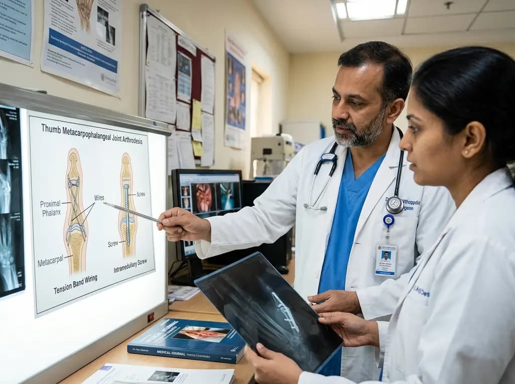
A comparative review of tension band wiring and intramedullary screw fixation in thumb metacarpophalangeal arthrodesis, evaluating union rates, hardware complications, and clinical outcomes.
Yesterday

A breakthrough ultrathin multimodal sensor decouples physical hemodynamic monitoring from disposable electrochemical calibration, achieving long-term wearable glucose prediction with 81.36% accuracy and a 9.12% MARD. This offers a robust, drift-free solution for outpatient metabolic surveillance.
Today
