.webp&w=384&q=75)

Loading, please wait...
Loading, please wait...
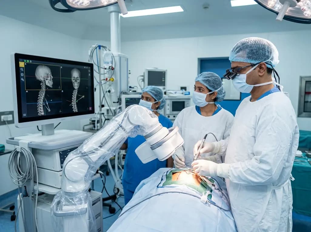
Surgical intervention within the central nervous system demands uncompromising accuracy, high-resolution anatomical visualization, and real-time intraoperative guidance. In a landmark collaboration, Amrita Hospital Faridabad has signed a Memorandum of Understanding with German medtech pioneer Brainlab to establish North India's first Centre of Excellence for Brain and Spine. This strategic partnership introduces advanced robotic intraoperative imaging directly into tertiary neurosurgical suites. Consequently, surgical teams can access instantaneous high-definition data while performing intricate cranial and spinal procedures. The centre also functions as a specialized knowledge forum for surgical training, technology demonstrations, and multi-institutional academic collaboration across India.
Neurosurgical procedures historically depended on static preoperative imaging studies, such as diagnostic CT and MRI scans. However, intraoperative anatomical distortion, commonly termed brain shift, rapidly degrades the surgical accuracy of rigid preoperative coordinates. Furthermore, dynamic spinal decompression and patient repositioning inevitably alter baseline spinal alignment. Robotic intraoperative imaging directly overcomes these challenges by generating updated anatomical models during ongoing procedures. Consequently, surgeons do not have to rely on guesswork or outdated navigation registration.
Brainlab's Loop-X mobile robotic imaging platform anchors this advanced Centre of Excellence in Faridabad. The system features independent, motorized robotic positioning that glides seamlessly into the surgical perimeter. Moreover, its wireless control tablet allows surgeons to direct scan trajectories without breaking the sterile barrier. Operating room staff execute high-resolution 2D fluoroscopy and 3D cone-beam computed tomography scans in seconds. Therefore, the surgical team visualizes neural anatomy, delicate vascular margins, and dense bony structures without halting surgical progress. As a result, the technology minimizes surgical trauma and substantially reduces intraoperative revision rates.
Traditional intraoperative cross-sectional imaging frequently requires transferring anesthetized patients out of the operating room to stationary CT suites. This complex transport introduces significant safety risks, airway vulnerability, and disruptive delays. In contrast, mobile cone-beam computed tomography brings advanced cross-sectional diagnostics directly to the operating table. The Loop-X device incorporates high-frequency X-ray sources and flat-panel digital detectors that rotate around the patient. Importantly, the automated non-isocentric imaging capabilities adjust scans to target anatomies of varied dimensions.
Furthermore, the mobile robot communicates synchronously with contemporary image-guided navigation suites. Surgeons quickly register intraoperative scans with preoperative diagnostic plans, establishing dynamic instrument tracking in real time. Consequently, clinicians navigate complex skull-base corridors and dysplastic pedicles with sub-millimeter precision. In addition, the device drastically curtails unnecessary radiation exposure. Its robotic collimation feature focuses radiation strictly on the targeted anatomical volume, shielding adjacent healthy tissue. Therefore, both patient safety and staff occupational health receive robust protection throughout lengthy cranial or deformity correction cases.
Spinal instrumentation in compromised anatomy, such as severe idiopathic scoliosis or osteoporotic fractures, presents severe technical challenges. A misplaced pedicle screw risks catastrophic neurologic deficit, vascular injury, or construct instability. Fortunately, mobile robotic imaging allows immediate intraoperative verification of hardware placement before wound closure. Surgeons can confirm screw purchase, cage positioning, and cortical integrity before waking the patient. Consequently, hospital readmissions for hardware revision decline substantially.
Moreover, the Centre of Excellence significantly elevates surgical neuro-oncology standards. Resecting high-grade gliomas and skull-base meningiomas requires maximal safe tumor clearance while preserving eloquent functional zones. By refreshing the navigation field following initial tumor debulking, surgeons accurately delineate residual tumor margins. In addition, the platform supports minimally invasive spine surgery, reducing muscular dissection and intraoperative blood loss. Patients subsequently experience diminished postoperative pain, lower surgical site infection rates, and faster functional rehabilitation. Thus, the integration of advanced imaging platforms establishes a new benchmark for tertiary surgical care across North India.
Beyond clinical operative excellence, the newly established centre serves as a critical academic and educational powerhouse. India requires expanded specialized training pathways for junior neurosurgeons and spine fellows in computer-assisted surgery. Therefore, Amrita Hospital and Brainlab designed this facility as a hands-on training hub for emerging surgical technologies. Trainees, fellows, and practicing surgeons will master modern navigation ergonomics, automated robotics, and digital imaging physics under experienced faculty supervision.
Furthermore, the centre encourages structured peer-to-peer knowledge sharing and clinical protocol standardization across the National Capital Region and beyond. Multi-angle visualization and high-speed data transmission enable live-streamed surgical demonstrations and interactive procedural masterclasses. In addition, clinical researchers will utilize the high-fidelity platform to track surgical outcomes, accuracy metrics, and radiation dose optimization protocols. Consequently, this initiative bridges the divide between cutting-edge medtech engineering and routine surgical practice, inspiring reproducible clinical excellence across domestic healthcare institutions.
The establishment of this specialized Centre of Excellence reflects the rapid technological modernization underway within Indian tertiary healthcare. Historically, advanced intraoperative robotic imaging remained limited to isolated international research institutes. However, bringing these integrated systems to Faridabad ensures that patients across North India access world-class neurosurgical precision locally. Minimizing procedural morbidity and revision rates directly lowers total treatment costs and alleviates the emotional burden on families.
Additionally, the initiative establishes a comprehensive blueprint for public and private healthcare integration. When healthcare providers invest in collaborative infrastructure, they elevate entire clinical departments simultaneously. The combined deployment of robotics, navigation, and digital ergonomics reduces surgeon fatigue during complex multi-hour procedures. Consequently, patient safety metrics and long-term functional survival improve dramatically across neurotrauma, spine deformity, and complex oncology cohorts. Ultimately, this milestone heralds a transformative phase in precise, patient-centric neurosurgical care.
Q1: What is the main advantage of the Loop-X mobile robotic imaging robot in surgery?
The Loop-X platform brings automated 2D and 3D cone-beam CT imaging directly into the operating suite. Consequently, surgical teams capture real-time anatomical updates and verify implant placement without transporting the patient to a radiology suite. The device pairs with surgical navigation systems, substantially reducing procedural delays, cutting overall radiation exposure through smart collimation, and improving instrument tracking accuracy during complex spine and brain surgeries.
Q2: How does intraoperative 3D imaging improve safety during complex spine surgery?
Complex spine operations require sub-millimeter precision when placing pedicle screws and interbody cages near the spinal cord and major blood vessels. Intraoperative 3D cone-beam CT enables surgeons to verify exact hardware trajectories and cortical bone integrity before completing the procedure. Therefore, surgeons detect and rectify any hardware malposition immediately, significantly decreasing neurological complications and preventing the need for secondary revision surgeries.
Q3: How does this Centre of Excellence benefit medical training in India?
The Centre of Excellence serves as a dedicated academic platform where neurosurgeons, orthopedic spine specialists, and operating room staff receive structured hands-on training. Clinicians gain direct exposure to robotic imaging systems, computer-assisted navigation, and digital surgical workflows. Furthermore, through technology demonstrations, standardized workshops, and collaborative clinical research, the facility accelerates the clinical adoption of advanced surgical techniques across regional healthcare centers.
Disclaimer: This content is for informational and educational purposes only. It does not constitute medical advice or replace professional judgment. Refer to the latest local and national guidelines for clinical practice.
References

Read summarized clinical updates, watch expert medical content, and earn CME certifications right from your smartphone.
Open your smartphone camera to scan and install.





North India's first Centre of Excellence for Brain and Spine at Amrita Hospital Faridabad integrates Brainlab's Loop-X mobile robot. This landmark facility combines cone-beam computed tomography with surgical navigation to elevate precision, optimize operative workflows, and enhance specialized clinician training.
Today

A breakthrough study demonstrates that BAP31 protects against acute lung injury by inhibiting ferroptosis in alveolar epithelial cells. Through activating the AKT/GSK3β/Nrf2 pathway and upregulating GPX4 and HMOX1, BAP31 preserves the alveolar-capillary barrier, offering novel avenues for critical care management.
Today
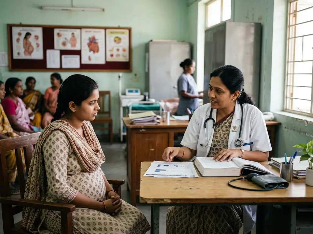
A recent longitudinal study evaluated the link between prenatal loss of control eating and cardiovascular health using Life's Essential 8 frameworks. While direct associations across pregnancy were non-significant, the high prevalence of dysregulated eating highlights critical implications for obstetric practice.
Today

Explore modern perioperative management protocols for clinical xenotransplantation. Learn how costimulation blockade, anti-inflammatory perfusion, and coagulopathy rescue overcome interspecies hurdles.
Today

At Homa CME 2026 at Hyderabad's T-Hub, healthcare leaders emphasized moving beyond routine metrics through digital calculators and precision stratification. Discover why characterizing individual insulin secretion, resistance patterns, and vascular risks transforms chronic metabolic care in India.
Today
