.webp&w=384&q=75)

Loading, please wait...
Loading, please wait...
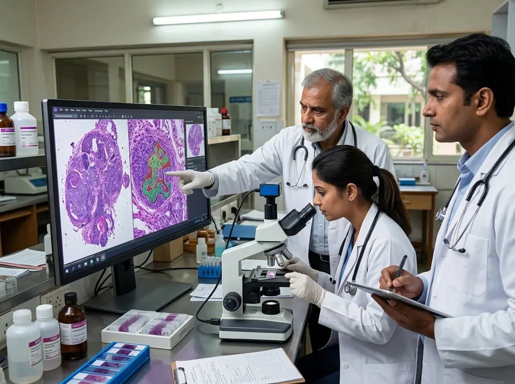
Oral cavity squamous cell carcinoma (OSCC) represents a major global health challenge with substantial disease burden. Accurate pathological staging dictates treatment pathways, prognosis, and patient survival. Elective neck dissection remains the standard surgical approach for clinically node-negative disease to address occult lymph node metastases. Consequently, pathologists must examine dozens of resected cervical lymph nodes for subtle metastatic deposits. Routine hematoxylin and eosin (H&E) staining serves as the primary diagnostic modality in global laboratory workflows. However, identifying tiny micrometastases or isolated tumor cells on standard H&E slides remains remarkably challenging and labor-intensive. Pathologists frequently order cytokeratin (CK) immunohistochemistry (IHC) to confirm ambiguous findings or detect occult tumor clusters. Although cytokeratin staining improves sensitivity, it substantially increases diagnostic costs, turnaround times, and laboratory workloads. Therefore, clinical teams need reliable, automated computational solutions to screen standard histological sections. Deep learning models provide unprecedented computational capabilities to analyze complex cellular morphology directly from routine digital slides. Recent advances demonstrate that artificial intelligence can segment subtle malignant cells with high precision. By augmenting human diagnostic performance, these algorithmic frameworks help pathologists identify early nodal spread while reducing the necessity for expensive confirmatory staining protocols.
To address the diagnostic bottleneck, researchers developed an innovative two-stage deep learning framework for precise tumor segmentation. The investigation utilized a custom expert-annotated dataset comprising primary OSCC resections and cervical lymph node specimens. Initially, the computational model underwent extensive pre-training on primary tumor morphology to master diverse architectural patterns. Subsequently, the investigators fine-tuned the network on lymph node metastases to refine its sensitivity for subtle metastatic deposits. A senior pathologist generated comprehensive pixel-level segmentation masks for all training and validation datasets. Furthermore, the pathologist incorporated cytokeratin immunohistochemistry confirmation when clinically indicated to guide the ground-truth annotations accurately. The development team implemented a standardized pre-processing pipeline to preserve histological detail across whole-slide images. This pipeline ensured uniform color normalization, optimal resolution scaling, and artifact reduction across diverse tissue samples. Consequently, the network learned to recognize distinct cytological alterations, dysplastic nuclear features, and architectural irregularities within complex lymphoid tissue. By integrating robust primary tumor features before nodal fine-tuning, the model achieved exceptional generalizability. Thus, the system demonstrated a reliable capacity to differentiate malignant squamous cells from dense surrounding germinal centers, sinus histiocytosis, and reactive lymphoid architecture without requiring explicit chemical stains.
The performance evaluation on a held-out test cohort revealed remarkable quantitative metrics for the deep learning model. During the initial pre-training stage on primary tumors, the model achieved a precision of 0.8484 and a recall of 0.8682. Following specific fine-tuning on lymph node metastases, the model demonstrated improved metrics, reaching a precision of 0.8778 and a recall of 0.8607. Overall diagnostic accuracy reached 0.9193, while diagnostic specificity attained 0.9458 across the evaluation dataset. These robust numbers highlight the model's powerful ability to discriminate between malignant deposits and benign lymphoid parenchyma. Additionally, the investigators performed a granular case-level analysis to evaluate model behavior across varying degrees of diagnostic difficulty. While macroscopic metastatic foci achieved excellent overlap, complex micrometastatic cases yielded lower Dice similarity coefficients. This finding highlights the inherent difficulty of segmenting tiny, scattered tumor clusters embedded within dense inflammatory backgrounds. Nevertheless, the algorithm maintained high sensitivity across challenging specimens, flagging suspicious regions that human observers might overlook during rapid screening. Moreover, the low false-positive rate prevents unnecessary clinical alarm. Therefore, the computational architecture provides reliable objective assistance, establishing a solid foundation for clinical implementation in high-volume pathology departments.
A pivotal component of the validation study involved qualitative and spatial comparisons between AI predictions and cytokeratin staining. In cases where immunohistochemistry was available, the AI-predicted tumor boundaries exhibited remarkable spatial concordance with cytokeratin-positive regions. The deep learning system successfully delineated subtle tumor nests on routine H&E sections that closely mirrored the brown chromogenic staining seen on matched cytokeratin slides. Consequently, the algorithm demonstrated that standard H&E morphology contains sufficient optical and structural information for reliable metastasis identification. By approximating the diagnostic accuracy of immunohistochemistry, this computational approach provides significant clinical and operational value. Routine implementation of such AI tools could dramatically decrease the volume of confirmatory cytokeratin stains ordered by laboratories. Furthermore, reducing immunohistochemical testing conserves tissue blocks for downstream molecular testing, minimizes expensive antibody consumption, and shortens turnaround times. Pathologists can utilize AI heatmaps to verify clear-cut negative or positive lymph nodes immediately. As a result, laboratories can reserve immunohistochemistry exclusively for genuinely ambiguous or borderline cases. Ultimately, this workflow optimization enhances laboratory efficiency while preserving rigorous diagnostic precision across oncology centers.
Integrating AI segmentation into surgical pathology workflows offers profound advantages for head and neck oncology. Head and neck surgeons frequently perform selective or comprehensive neck dissections containing dozens of lymph nodes per patient. Pathologists must scrutinize hundreds of histological fields under high power, creating substantial diagnostic fatigue and time pressure. Implementing an automated computational assistant helps triaging slides effectively, prioritizing positive specimens, and highlighting micro-metastatic regions. In resource-limited settings where digital pathology is emerging, this technology can bridge critical expertise gaps and standardize diagnostic quality. Moreover, accurate nodal staging directly impacts subsequent clinical management, such as the decision to administer adjuvant radiotherapy or chemoradiotherapy. Missing occult metastases leads to undertreatment and disease recurrence, whereas overestimating nodal spread causes unnecessary treatment toxicities. By providing consistent, objective, and reproducible nodal evaluations, AI algorithms support multidisciplinary tumor boards in selecting personalized treatment strategies. Furthermore, the combination of computational precision and pathologist expertise establishes a safer diagnostic safety net. As digital pathology infrastructure continues to expand globally, AI-assisted lymph node screening will play a crucial role in modern cancer care.
Deep learning models analyze high-resolution digital whole-slide images to identify subtle cellular and structural alterations characteristic of malignancy. The algorithm evaluates dysplastic nuclear features, nuclear-to-cytoplasmic ratios, cellular crowding, and abnormal architectural arrangements within lymphoid tissue. By training on thousands of expert-annotated primary tumors and lymph node specimens, the neural network learns to differentiate subtle metastatic clusters from surrounding reactive lymphocytes, histiocytes, and endothelial cells directly on standard hematoxylin and eosin slides.
Artificial intelligence models cannot completely eliminate the need for cytokeratin immunohistochemistry in complex pathology practice. Although deep learning achieves high concordance with cytokeratin staining, highly ambiguous lesions, severe tissue artifacts, and isolated single tumor cells may still require immunohistochemical confirmation. Instead, AI serves as an effective screening and decision-support tool. It substantially reduces the routine volume of immunohistochemistry orders while allowing pathologists to reserve specialized staining for genuinely uncertain or borderline cases.
AI-assisted lymph node evaluation significantly enhances diagnostic sensitivity, reduces pathologist screening fatigue, and speeds up turnaround times for surgical pathology reports. Furthermore, the technology helps standardize diagnostic accuracy across different healthcare centers by highlighting occult micrometastases that human observers might miss. Consequently, patients benefit from more accurate pathological staging, which ensures appropriate selection for adjuvant radiotherapy or chemoradiotherapy while avoiding unnecessary treatment delays and excess laboratory costs associated with immunohistochemistry.
Disclaimer: This content is for informational and educational purposes only and is not intended to serve as medical advice, diagnosis, or treatment. Healthcare professionals must exercise independent clinical judgment. Refer to the latest local and national guidelines for clinical practice.
References

Read summarized clinical updates, watch expert medical content, and earn CME certifications right from your smartphone.
Open your smartphone camera to scan and install.





A two-stage deep learning framework segments occult cervical lymph node metastases in oral cancer from routine H&E slides with 91.9% accuracy, offering strong concordance with cytokeratin staining and potential workflow savings.
last month
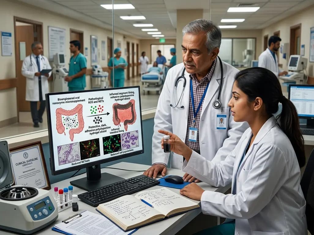
A bioengineered oral nanozyme microsupplement converts pathological ROS into a protective polydopamine hydrogel shield. By curbing neutrophil extracellular traps and balancing T cells, this platform outperforms standard 5-ASA in preclinical colitis models, opening new paths for sustained remission.
Today
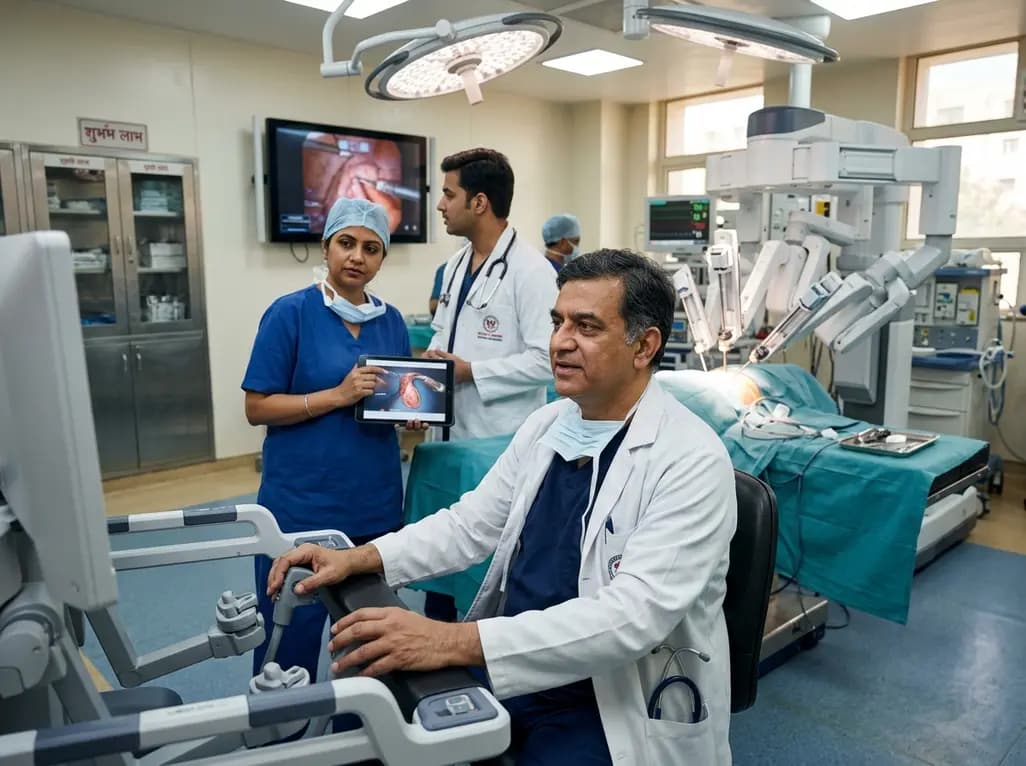
India experiences an exponential rise in robotic-assisted procedures, with Delhi NCR leading system installations and clinical volume. Discover how multidisciplinary surgical adoption, remote telesurgery breakthroughs, and standardized international consensus guidelines are shaping surgical care nationwide.
Today

Radiofrequency catheter ablation of the cavotricuspid isthmus successfully terminates typical atrial flutter. This article reviews the mechanisms driving coronary sinus electrogram changes, wavefront activation pathways, and clinical criteria to verify bidirectional block during electrophysiology studies.
Today

A study comparing four knee flexion strength tests after ACL reconstruction using hamstring tendon autografts reveals marked variability in limb symmetry indices. Clinicians must recognize that different strength testing modalities yield divergent return-to-sport clearance rates.
Today

A breakthrough study leverages natural language processing to accurately identify incident medication-related osteonecrosis of the jaw and track antiresorptive drug holidays from clinical narratives in electronic health records, advancing osteoporosis pharmacovigilance and dental safety.
Yesterday
