.webp&w=384&q=75)

Loading, please wait...
Loading, please wait...

Modern clinical ophthalmology increasingly relies on cellular-level imaging to characterize progressive macular and retinal degenerations. Specifically, adaptive optics flood illumination ophthalmoscopy provides remarkable visualization of individual photoreceptors in living human retinas. However, clinicians face a major bottleneck during routine workflow. The availability of specialized software tools for adaptive optics image analysis remains remarkably scarce, particularly when quantifying degenerated photoreceptor mosaics in pathological eyes. Commercial programs frequently fail because engineers train them predominantly on healthy retinal architecture. Furthermore, writing custom mathematical scripts in statistical environments such as R requires extensive programming expertise that many clinicians lack. Generative artificial intelligence, particularly large language models like GPT-4, offers a revolutionary solution to this dilemma. Researchers can now prompt advanced language models to write, troubleshoot, and refine complex scientific code without manual programming from scratch. Consequently, this artificial intelligence paradigm bridges the gap between laboratory computational tools and bedside clinical research. By translating conversational prompts into functional image processing code, generative platforms open unprecedented avenues for retinal diagnostics. Thus, generative scripting holds immense transformative potential for high-resolution ocular diagnostics and personalized medicine.
In a recent exploratory investigation, visual scientists evaluated whether GPT-4 could independently construct a viable R script for ophthalmic research. The team specifically tasked the generative model with processing raw adaptive optics flood illumination ophthalmoscopy data. Rather than relying on a single automated output, the investigators employed an iterative prompt-engineering workflow. In total, the development required fifty-four distinct iterations of conversational instruction, diagnostic debugging, and systematic algorithmic tuning. Through this trial-and-error cycle, the language model progressively refined code functions for image filtering, background contrast equalization, and brightness thresholding. Additionally, the developers introduced specialized routines to detect individual cone photoreceptors as discrete luminous structures, known computationally as blobs. Subsequently, an independent computational researcher, who remained naive to the initial coding phases, verified the R code against novel image datasets. This verification phase ensured that the script avoided idiosyncratic programming errors and ran dependably on separate operational machines. Therefore, the iterative protocol established a reproducible blueprint for non-programmers seeking customized analytical scripts in specialized retinal imaging domains.
To evaluate real-world utility, researchers tested the generative script on images gathered from both healthy volunteers and individuals with Stargardt disease. Inherited juvenile macular dystrophies like Stargardt disease present profound disruption of photoreceptor mosaics, characterized by dark cones, cellular loss, and distorted reflective signals. Existing commercial algorithms struggle tremendously under these pathological conditions because irregular reflective patterns confuse standard edge-detection mechanisms. Therefore, robust verification requires comparing script outputs directly against human expert observations. In this exploratory trial, investigators compared automated cone counts from five retinal image snippets against manual annotations from two independent expert graders. Furthermore, the team contrasted these values with results produced by pre-existing commercial adaptive optics analysis software. Notably, the GPT-4-generated R script demonstrated a measurable capacity to identify and quantify photoreceptor blobs across varying disease severities. Although discrepancies existed between automated algorithmic totals and human manual counts, the custom script processed disrupted retinal regions that typically halt legacy software. Consequently, these initial benchmarks prove that generative AI scripting can generate functional computational frameworks for challenging clinical cohorts.
The operational core of the generated script depends on mathematical blob detection to delineate packed cone photoreceptors. While healthy foveal and parafoveal photoreceptors display uniform hexagonal tiling, inherited macular dystrophies fundamentally disrupt this spatial geometry. In degenerate retinae, fluctuating optical reflectivity and dense cellular debris frequently generate imaging artifacts. Consequently, simple intensity-based blob detection scripts can misinterpret cellular debris or illumination hotspots as true photoreceptor inner segments. The preliminary GPT-4 script successfully identified basic punctate structures, yet it lacked sophisticated contextual spatial modeling. Furthermore, the exploratory script did not undergo rigorous mathematical cross-validation against multi-center histological or multimodal imaging ground truths. Human graders still outperformed the automated code when deciphering borderline cellular margins and low-contrast regions. Additionally, generative language models occasionally insert obsolete software packages or subtle algorithmic syntax errors during code generation. Clinicians must recognize that a functional script does not equal a clinically validated diagnostic tool. Therefore, extensive prospective optimization remains vital before implementing such algorithmic tools in regulated medical decision-making.
Establishing sensitive, objective cellular biomarkers represents a crucial objective for modern ophthalmic therapeutics and clinical trials. In inherited conditions such as Stargardt disease, structural photoreceptor loss often precedes measurable functional decay on standard visual field testing. Consequently, regulatory bodies increasingly demand surrogate endpoints that capture microscopic therapeutic preservation before macroscopic vision loss occurs. High-resolution adaptive optics imaging directly measures cone density, rod spacing, and mosaic regularity in vivo. Nevertheless, multi-center clinical trials require rapid, scalable, and highly standardized image processing to evaluate thousands of retinal frames efficiently. Custom artificial intelligence scripting can eliminate tedious manual counting that currently consumes hundreds of research hours. Moreover, accessible generative models democratize software development for academic clinicians worldwide, including specialized tertiary eye hospitals across India. By enabling rapid prototyping of bespoke analytical pipelines, artificial intelligence reduces the operational costs of rare disease research. Ultimately, validated open-source scripts will accelerate the discovery of novel therapeutic agents, gene therapies, and neuroprotective interventions.
Adaptive optics flood illumination ophthalmoscopy significantly improves retinal evaluation by correcting ocular wavefront aberrations in real time. Standard fundus cameras cannot overcome optical imperfections in the human cornea and crystalline lens. In contrast, adaptive optics technology provides cellular-level resolution, allowing clinicians to visualize individual cone photoreceptors directly. This microscopic visualization facilitates the early detection of localized photoreceptor loss in inherited retinal dystrophies long before conventional imaging detects macroscopic structural damage.
Researchers required fifty-four iterations because large language models cannot construct flawless image analysis software in a single prompt. Initial code drafts frequently lacked proper syntax, omitted vital image filtering functions, or misidentified non-cellular artifacts as photoreceptors. Through iterative debugging, the investigators progressively supplied error messages, refined algorithmic parameters, and adjusted mathematical thresholding. This persistent trial-and-error cycle ensured the final script successfully performed contrast equalization, background subtraction, and consistent cellular blob detection.
Clinicians cannot use generative artificial intelligence scripts immediately in clinical practice without extensive formal validation. Although GPT-4 accelerates coding workflows, unvalidated scripts often yield inaccurate cell counts in pathological tissues with heavy scarring or atrophy. Medical software must undergo rigorous testing across diverse patient cohorts to guarantee diagnostic safety. Furthermore, regulatory agencies mandate strict adherence to medical device software guidelines before any automated computational tool directly guides patient management.
Disclaimer: This content is for informational and educational purposes only... Refer to the latest local and national guidelines for clinical practice.
References
Pas JA et al. Adaptive Optics Image Analysis Using Generative AI (GPT-4) Scripting: Exploratory Study. JMIR Form Res. 2026 Sep 14. doi: 10.2196/94906. PMID: 42735400.
Waisberg E, Ong J, Masalkhi M, et al. GPT-4 and medical image analysis: strengths, weaknesses and future directions. Ann Eye Sci. 2023;8:32.
Song H, Pugliese AG, Rossi EA, et al. Adaptive optics scanning laser ophthalmoscopy in Stargardt disease reveals decreased cone and rod densities. Invest Ophthalmol Vis Sci. 2013;54(15):1743.
Gill J, Moosajee M, Dubis AM. Cellular imaging of inherited retinal diseases using adaptive optics. Eye. 2019;33(11):1683-1698.

Read summarized clinical updates, watch expert medical content, and earn CME certifications right from your smartphone.
Open your smartphone camera to scan and install.





A proof-of-principle study demonstrates how GPT-4 generated an R script to automate adaptive optics image analysis, quantifying retinal cone photoreceptors in Stargardt disease and highlighting both the promise and validation requirements of generative AI in ophthalmic imaging.
Today
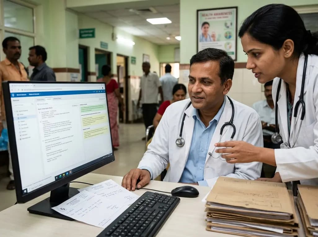
A crossover study demonstrates that locally deployed large language models with exact-match knowledge augmentation significantly elevate outpatient prescription review accuracy to 97.2%, curbing hallucinations and securing patient data without requiring complex cloud infrastructure.
Today
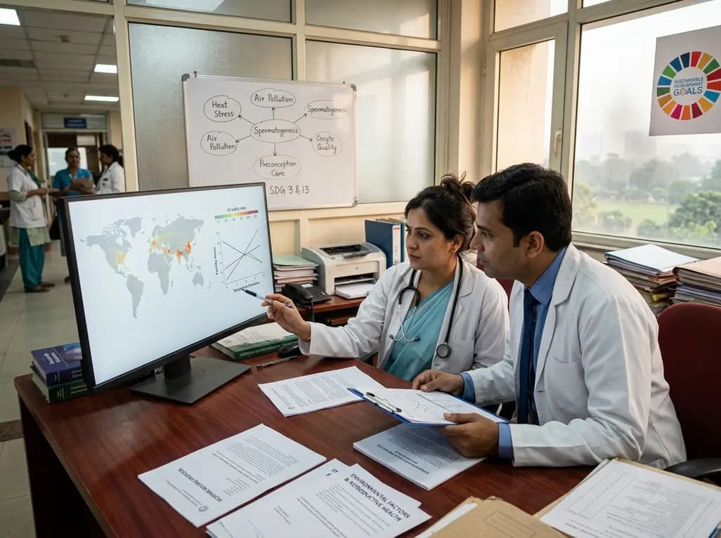
A new scoping review protocol systematically maps global evidence on how climatic shifts, ambient heat, and pollution impact human fertility. Discover key physiological mechanisms, clinical preconception strategies, and critical public health alignments under Sustainable Development Goals 3 and 13.
Today
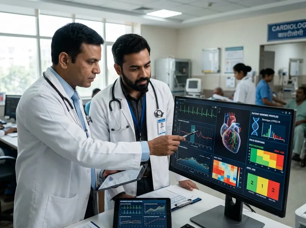
Pharmacogenomics and artificial intelligence are revolutionizing cardiology by tailoring therapies to individual genetic profiles and clinical data, reducing adverse drug events, and improving cardiovascular patient outcomes.
Today
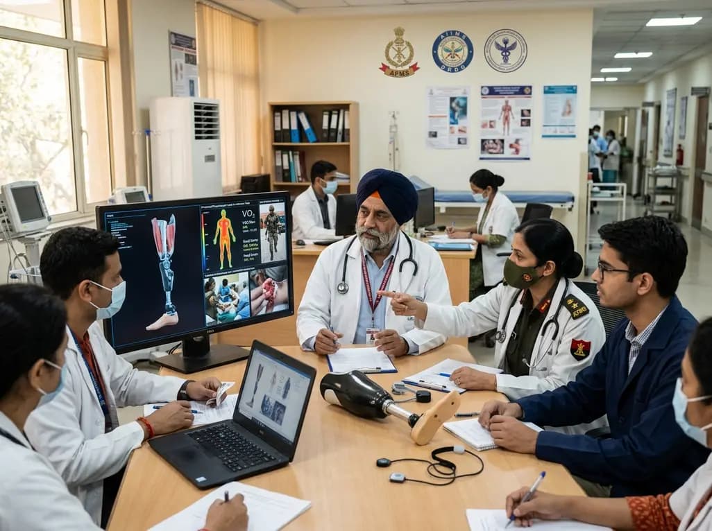
India's premier scientific bodies have joined the Armed Forces Medical Services to solve unique physiological and operational challenges faced by soldiers. The national collaboration covers combat casualty care, artificial intelligence diagnostics, bionic prosthetics, and extreme-environment physiological resilience.
Today
A retrospective cohort study evaluates the short- and long-term impacts of SARS-CoV-2 infection on patients with Graves' disease receiving antithyroid drug therapy, highlighting thyroid status destabilization, clinical symptom exacerbations, and post-viral sequelae.
Today
