.webp&w=384&q=75)

Loading, please wait...
Loading, please wait...

Preoperative assessment of skeletal fragility remains vital for successful spinal instrumentation. Historically, surgeons have relied on dual-energy X-ray absorptiometry to detect osteopenia and osteoporosis. However, degenerative changes, osteophytes, and vascular calcifications often create falsely elevated mineral density readings on standard absorptiometry. Consequently, spine specialists require reliable, non-invasive imaging tools to evaluate local skeletal health. The vertebral bone quality score has recently emerged as an innovative diagnostic parameter derived directly from routine lumbar T1-weighted magnetic resonance imaging. Furthermore, this score quantifies marrow changes by evaluating the ratio between vertebral body signal intensity and cerebrospinal fluid. Because magnetic resonance imaging captures subtle alterations in marrow fat infiltration, it provides valuable structural data. Clinicians frequently order these scans during regular assessments, avoiding additional radiation exposure or extra cost. Consequently, integrating this scoring mechanism into preoperative workflows allows clinicians to detect compromised bone integrity early. Ultimately, this objective measurement offers invaluable diagnostic guidance.
Bone strength depends on both static mineral density and dynamic metabolic remodeling. Nevertheless, conventional imaging modalities fail to illustrate ongoing cellular activity within the skeletal matrix. Bone turnover markers provide critical physiological insights by reflecting realtime enzymatic activity of osteoblasts and osteoclasts. Specifically, markers such as osteocalcin reflect active bone formation, whereas beta-CrossLaps indicate active osteoclastic resorption. In addition, serum levels of 25-hydroxyvitamin D3 and parathyroid hormone orchestrate systematic calcium homeostasis. When metabolic turnover shifts toward excessive resorption, patients experience compromised microarchitecture and delayed skeletal healing. Unfortunately, surgeons frequently overlook these serum markers during standard surgical evaluations due to logistical delays. However, dynamic metabolic changes directly dictate the mechanical stability of pedicle screws and interbody cages. Therefore, establishing a reliable connection between routine imaging parameters and turnover markers fulfills a crucial diagnostic need. By aligning metabolic shifts with imaging metrics, clinicians can better appreciate patient-specific cellular environments prior to lumbar surgery.
A recent retrospective cohort study evaluated 234 consecutive patients who underwent lumbar spine surgery. Researchers measured preoperative vertebral bone quality scores and categorized participants into high, middle, and low cohorts based on established thresholds. Additionally, investigators analyzed serum bone turnover markers alongside computed tomography Hounsfield units and absorptiometry T-scores. Comparative evaluations revealed striking variations in metabolic markers across different score categories. Specifically, patients with higher imaging scores demonstrated significantly lower levels of osteocalcin and 25-hydroxyvitamin D3, confirming suppressed osteoblast function. Meanwhile, serum beta-CrossLaps increased markedly in high-scoring individuals, indicating accelerated bone resorption. Furthermore, multivariate regression confirmed that age, sex, body mass index, and distinct turnover markers independently predicted score variations. Most importantly, the imaging score demonstrated stronger statistical correlations with dynamic metabolic markers than traditional T-scores. In fact, patients with osteopenia who exhibited elevated scores displayed profound bone resorption. Thus, these data confirm that the scoring tool reliably mirrors dynamic cellular remodeling.
These physiological correlations carry immediate clinical significance for orthopedic and neurosurgical practices. In spinal fusion surgery, rigid mechanical fixation relies entirely on robust bone-implant interfaces. If an unrecognized high turnover state persists, osteoclasts rapidly resorb bone adjacent to pedicle screws. Consequently, patients face heightened risks of screw loosening, cage subsidence, and pseudarthrosis. Furthermore, when surgeons identify an elevated vertebral bone quality score, they can anticipate underlying metabolic dysfunction even if traditional absorptiometry suggests normal density. This realization enables surgical teams to modify operative strategies proactively. For example, surgeons may select cement-augmented fenestrated screws or deploy wider interbody cages to distribute axial loads effectively. In addition, multidisciplinary teams can initiate targeted metabolic therapies before elective operations. Prescribing anabolic agents like teriparatide stimulates osteoblast recruitment, whereas antiresorptive medications suppress aggressive osteoclast activity. Therefore, identifying metabolic deficits early helps prevent construct failures and enhances long-term spinal fusion success.
Dual-energy X-ray absorptiometry has long served as the reference standard for diagnosing osteoporosis. However, lumbar absorptiometry exhibits severe limitations in patients with advanced degenerative disease. Specifically, aortic calcification, facet arthropathy, and endplate sclerosis falsely elevate areal bone mineral density measurements. Although computed tomography Hounsfield units provide regional quantitative data without superimposition artifacts, computed tomography exposes patients to ionizing radiation. In contrast, the magnetic resonance imaging-based vertebral bone quality score avoids ionizing radiation entirely while evaluating deep trabecular fat conversion. As bone quality deteriorates, yellow marrow gradually replaces red hematopoietic marrow within vertebral spongiosa. Because T1-weighted sequences highlight lipid deposition, this scoring method captures early pathophysiological marrow remodeling before cortical bone loss occurs. Furthermore, standard surgical workups already incorporate lumbar magnetic resonance imaging for decompression planning. Thus, clinicians can opportunistically extract vital bone quality metrics without subjecting patients to supplemental appointments, costs, or radiation. Consequently, this imaging modality represents an exceptionally practical and holistic screening method.
In India, the burden of skeletal fragility and hypovitaminosis D remains exceptionally widespread among aging individuals. Unfortunately, routine preoperative bone mineral density screening through dual-energy absorptiometry is underutilized across semi-urban and public hospitals due to limited machine accessibility. In contrast, magnetic resonance imaging facilities are widely accessible and routinely utilized for diagnosing lumbar spinal pathology. Therefore, Indian spine surgeons and radiologists can readily adopt this imaging calculation to screen surgical candidates for silent metabolic bone fragility. Moreover, identifying elevated scores early enables timely medical optimization in resource-constrained settings. Clinicians can promptly correct pervasive vitamin D deficiencies and optimize dietary calcium intake before surgery. Additionally, endocrinologists and orthopedic surgeons can collaborate to prescribe cost-effective antiresorptive or osteoanabolic regimens. This integrated approach mitigates expensive revision surgeries caused by hardware failure, reducing the overall financial burden on families. Consequently, implementing opportunistic magnetic resonance screening advances value-based orthopedic care across the country.
Clinicians calculate the score by measuring median signal intensities within L1 to L4 vertebral bodies on non-contrast T1-weighted sagittal images. Then, they divide this value by the signal intensity of cerebrospinal fluid at L3. This standardized ratio objectively quantifies fatty marrow infiltration independent of scanner variations.
Degenerative spinal changes and vascular calcifications frequently inflate DXA T-scores artificially, masking metabolic bone deficits. In contrast, magnetic resonance imaging directly reflects adipocyte expansion and marrow transformation. These cellular shifts mirror ongoing alterations in osteoblast and osteoclast activity, making the score an accurate reflection of dynamic bone metabolism.
Surgeons identifying elevated scores can modify surgical tactics by employing cement augmentation, larger implants, or bicortical screw purchases to prevent construct loosening. Furthermore, clinicians can order targeted bone turnover marker panels and initiate preoperative osteoanabolic or antiresorptive therapy to stimulate bone formation and accelerate fusion healing.
Disclaimer: This content is for informational and educational purposes only... Refer to the latest local and national guidelines for clinical practice.
References

Read summarized clinical updates, watch expert medical content, and earn CME certifications right from your smartphone.
Open your smartphone camera to scan and install.





A recent study highlights the utility of the MRI-derived vertebral bone quality (VBQ) score in evaluating dynamic bone turnover markers. VBQ scores correlate closely with bone formation and resorption markers, enabling spine surgeons to detect impaired bone metabolism before instrumentation.
Today

A randomized controlled trial demonstrates that theory-driven personalized mobile messaging reduces the risk of consecutive walking goal failures by 33 percent, highlighting the vital role of digital health tools in sustaining long-term exercise habits and chronic disease prevention.
Today

Prelacteal feeding persists in South Asian communities despite known clinical risks. This review examines determinants from Chitral, pathophysiological hazards, and pediatric strategies to eliminate harmful feeds and support exclusive breastfeeding.
Today
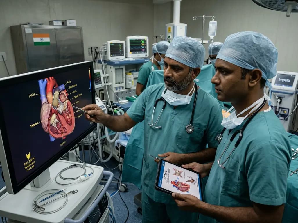
Transcatheter mitral valve replacement in patients with degenerated bioprostheses carries a substantial risk of LVOT obstruction. Combining leaflet modification via the BATMAN technique with mechanical circulatory support offers a viable, life-saving solution for anatomically challenging surgical candidates.
Today
A comparative cohort study demonstrates that patients undergoing total hip arthroplasty after prior hip arthroscopy achieve comparable functional recovery, low complication rates, and equivalent implant survivorship when conversion occurs after six months.
Today

A cross-sectional study of 320 adults evaluated the link between habitual fasting duration and dietary quality using Nutrition Quotient scores, highlighting the clinical necessity of balancing time-restricted eating with nutrient density and meal quality in routine metabolic practice.
Today
