

Loading, please wait...
Loading, please wait...
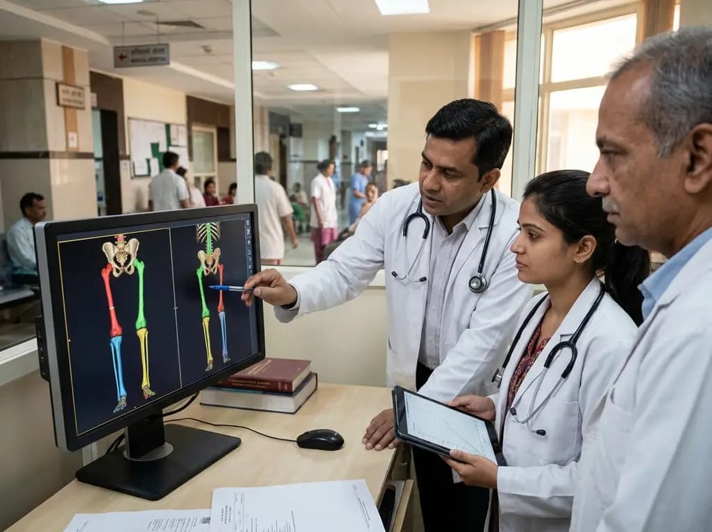
Pediatric orthopedic surgeons frequently manage leg length discrepancy in growing children to restore biomechanical balance. For decades, clinicians relied on historical growth charts to determine when growth plates stop elongating. However, recent scientific advances indicate that standing height often conceals true lower extremity behavior. When physicians base surgical timing on overall height velocity, they risk serious clinical miscalculations. Consequently, contemporary evidence urges clinicians to separate appendicular skeletal maturation from axial skeletal progression.
Surgical epiphysiodesis provides an effective and minimally invasive technique to equalize limbs in adolescent patients. Nevertheless, accurate surgical timing remains the most critical determinant of therapeutic success. Historically, pediatric surgeons utilized the classical Anderson-Green growth-remaining charts to guide their operative plans. These foundational models established an enduring clinical dogma suggesting that male lower extremity growth continues until seventeen years of age. Therefore, practitioners often calculated residual distal femoral and proximal tibial elongation up to this late adolescent threshold. Unfortunately, this assumption frequently leads to overestimating remaining physeal growth. Consequently, surgical intervention is often delayed or miscalculated, creating inadvertent overcorrection or persistent deformity. In clinical practice across India, where patients frequently present late in adolescence, precise timing becomes even more critical. When surgeons assume that legs continue growing simply because standing height is increasing, they misjudge physeal closure. Thus, modern pediatric orthopedics requires updating these antiquated paradigms to ensure optimal anatomical outcomes and prevent avoidable revision surgeries.
A landmark 2026 study examined 3D automated anthropometric scans of 1110 healthy children to assess segmental growth dynamics. By mapping standardized skeletal landmarks, researchers isolated lower limb trajectories from standing height progression. The findings demonstrated a striking physiological discrepancy, termed segmental growth dissociation. Specifically, lower extremity growth plateaus far earlier than total stature. In adolescent males, lower limb elongation essentially ceases between 14.5 and 15.5 years of age. Standing height, however, continues to expand steadily until 17.5 to 18.5 years. Therefore, boys experience a three-year temporal gap where only the spinal column elongates. Females exhibit a comparable two-year dissociation. In girls, lower extremity growth arrests between 12.5 and 13.5 years, whereas spinal growth continues until 14.5 to 15.5 years. Because overall height continues to rise, clinicians often mistake spinal lengthening for persistent leg growth. This critical diagnostic error creates the false illusion that long bones possess remaining potential. Consequently, orthopedists must recognize that the axial and appendicular skeletons operate on distinct biological timelines.
For over seven decades, the medical community accepted the 17-year rule as an immutable biological standard. Yet, recent biomechanical discoveries expose this rule as a clinical illusion driven entirely by late adolescent spinal elongation. The original Anderson-Green datasets evaluated standing stature and distal physes using limited radiographic tools. As a result, those earlier investigators conflated trunk elongation with lower extremity expansion. When contemporary clinicians apply these classical curves, they consistently overestimate residual lower extremity growth. This statistical distortion poses genuine risks for pediatric patients scheduled for temporary or permanent epiphysiodesis. If a surgeon operates under the belief that two years of growth remain, overcorrection inevitably occurs when the physis closes sooner. Furthermore, skeletal maturity varies significantly between different ethnic populations. Applying Western historical datasets to contemporary Asian cohorts introduces substantial predictive bias. Therefore, orthopedic specialists must replace outdated assumptions with precise, segment-specific measurements. Recognizing the earlier cessation of long bone elongation protects young patients from unintended iatrogenic deformities.
To resolve the limitations of historical curves, researchers developed an adaptive growth prediction framework using modern 3D anthropometry. By applying monotonic smoothing to high-resolution national anthropometric scans, investigators mapped isolated physeal trajectories across adolescence. The authors validated a simplified linear growth hypothesis for individual long bone physes through rigorous simulation analyses. Although human maturation involves complex hormonal cascades, individual growth plates demonstrate remarkably steady linear elongation before abrupt physiological closure. Consequently, modelling bone growth as a simple linear trajectory provides an intuitive, highly practical alternative for busy surgical teams. This simplified approach maintains exceptional predictive accuracy while dramatically reducing user calculation errors. Moreover, developers integrated these modernized population-specific algorithms into an interactive, customizable web application. Indian orthopedic clinicians can leverage such digital platforms to perform rapid, individualized growth simulations at the bedside. Rather than relying on static paper charts, surgeons can adjust parameters according to chronological age, skeletal maturity, and verified segment measurements.
Applying these anthropometric insights directly benefits orthopedic decision-making across Indian healthcare settings. In India, pediatricians and orthopedic surgeons regularly encounter adolescents with untreated congenital limb deficiencies, post-infectious physeal arrests, and post-traumatic deformities. Frequently, these young patients present relatively late during pubertal growth spurts. If clinicians assume that male limbs grow until seventeen, they may delay epiphysiodesis inappropriately. As a result, the opportunity to equalize limb discrepancies through simple guided growth vanishes, forcing families toward complex surgical limb lengthening. Furthermore, nutritional factors and population-specific growth variations in Indian children demand caution when extrapolating classical Western datasets. Clinicians must routinely decouple lower extremity assessments from total stature velocity. In daily practice, doctors should measure sitting height alongside standing height to evaluate trunk-to-limb proportions accurately. Combining radiographic bone age assessment with modern segmental growth models enables clinicians to determine operative timing with superior precision. Ultimately, transitioning to segment-specific algorithms safeguards surgical success and improves long-term functional mobility for growing adolescents.
Segmental growth dissociation describes the temporal desynchronization between axial spinal growth and appendicular lower extremity elongation during adolescence. While standing height continues to rise during late puberty, leg growth plates close significantly earlier. Specifically, male lower limb growth ceases around 15 years while the spine elongates until 18 years. Recognizing this dissociation prevents clinicians from assuming that ongoing height gains represent continued long bone development.
The classical Anderson-Green model assumes that male lower limb growth persists until seventeen years of age. Because the model conflates spinal elongation with ongoing leg growth, it overestimates remaining physeal potential. Consequently, if an orthopedic surgeon performs epiphysiodesis expecting several centimeters of future growth, the operated limb may fall short or overcorrect. Modern data prove that long bone plates fuse years earlier than previously assumed.
Clinicians should determine epiphysiodesis timing by evaluating segment-specific limb parameters and skeletal age rather than total standing height alone. Surgeons must account for earlier physeal closure, intervening before age 14 in boys and age 12 in girls when indicated. Utilizing modern digital growth calculators and linear physeal models prevents dangerous delays, ensuring equal leg lengths without requiring invasive osteotomies or complex distraction lengthening.
Disclaimer: This content is for informational and educational purposes only and does not constitute medical advice, diagnosis, or treatment. Healthcare professionals should exercise their independent clinical judgment when evaluating patient care options. Refer to the latest local and national guidelines for clinical practice.
References

Read summarized clinical updates, watch expert medical content, and earn CME certifications right from your smartphone.
Open your smartphone camera to scan and install.





Modern 3D anthropometry reveals that pediatric leg growth ceases years before spinal elongation, exposing the clinical illusion of the classical Anderson-Green model. Learn how segmental growth dissociation improves surgical timing for epiphysiodesis in adolescent leg length discrepancy.
Today
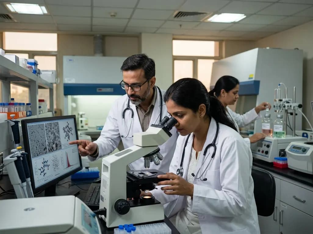
New research reveals that parenteral nutrition-based infusion solutions significantly improve biologics formulation stability. By reducing hydrophobic aggregation and maintaining positive zeta potential, these solutions protect recombinant antibodies like trastuzumab and viruses under severe physical stress.
Today

A KGMU study reveals over half of women with breast abnormalities fail to undergo diagnostic confirmation, leading to advanced-stage detection. Discover the socio-cultural barriers, clinical risks of incomplete resection, and community-level solutions needed to bridge the critical screening-to-diagnosis gap in India.
Yesterday

Recent findings from the VESUVE study demonstrate that brain MRI detects intracranial lesions in over 76% of patients with infective endocarditis. Acute ischemic stroke and subarachnoid hemorrhage independently elevate six-month mortality, underscoring the critical prognostic utility of neuroimaging.
Today

A meta-analysis of 18 studies reveals that bariatric surgery does not significantly alter circulating homocysteine levels, regardless of follow-up duration. Clinicians must maintain vigilance over post-surgical micronutrient supplementation to prevent hyperhomocysteinemia.
Today
