.webp&w=384&q=75)

Loading, please wait...
Loading, please wait...

Vogt-Koyanagi-Harada disease represents an aggressive autoimmune disorder that targets melanocytes throughout the uveal tract. Clinicians often encounter severe visual deterioration during the acute uveitic phase due to multifocal exudative retinal detachments and diffuse choroiditis. Traditionally, ophthalmologists relied heavily on invasive fluorescein and indocyanine green angiography to evaluate posterior segment involvement. However, non-invasive imaging has advanced rapidly in recent years. Recent clinical investigations have revealed remarkable topographic concordance when detecting UWF-OCTA choroidal granulomas compared to conventional dye angiography. Consequently, this non-invasive modality offers ophthalmologists an unprecedented opportunity to visualize stromal inflammation across both central and peripheral retinochoroidal zones with minimal patient discomfort.
Vogt-Koyanagi-Harada disease predominantly affects pigmented populations, making it especially relevant to clinical practice across India and Southeast Asia. The underlying pathology involves a T-lymphocyte-mediated autoimmune reaction directed against melanocyte-associated antigens. In the acute stage, this inflammation manifests as diffuse bilateral stromal choroiditis. Furthermore, non-necrotizing granulomas proliferate rapidly within the choroidal stroma, causing structural thickening and secondary exudative neurosensory detachments. Patients typically present with sudden bilateral blurring of vision, ocular pain, photophobia, and profound headache. In addition, prodromal auditory and meningeal manifestations often accompany these ocular complaints. If clinicians miss the therapeutic window, progressive stromal scarring and chronic recurrent inflammation can lead to permanent visual loss. Therefore, clinicians must identify these choroidal inflammatory lesions as early as possible to institute aggressive systemic corticosteroid and immunomodulatory therapy.
For several decades, indocyanine green angiography served as the gold standard for visualizing the choroidal vasculature and stromal lesions. In acute Vogt-Koyanagi-Harada disease, indocyanine green angiography characteristically reveals hypofluorescent dark dots that reflect non-perfused or granulomatous choroidal areas. Moreover, fuzzy stromal vessels and disc hyperfluorescence frequently highlight the severity of active inflammation. Nevertheless, this dye-based modality carries meaningful clinical limitations. First, intravenous administration of indocyanine green dye requires vein cannulation and prolongs clinic chair time. Second, clinicians must manage risks of adverse reactions, including nausea, vasovagal collapse, and severe anaphylaxis in iodine-sensitive patients. Third, frequent longitudinal angiograms create substantial cumulative dye exposure and financial burden for patients. Consequently, retina specialists have actively sought reliable, rapid, and non-invasive alternatives that can deliver equivalent spatial information without pharmacological risks.
Ultra-widefield optical coherence tomography angiography has transformed how clinicians evaluate the peripheral chorioretinal architecture. By capturing backscattered light from moving red blood cells, this modern technology reconstructs depth-resolved vascular maps across wide retinal fields without intravenous contrast. In acute Vogt-Koyanagi-Harada disease, the technology reliably highlights UWF-OCTA choroidal granulomas as distinct areas of choriocapillaris flow deficit and focal flow voids. Moreover, these flow voids correspond topographically to the classic hypofluorescent lesions observed on intermediate-phase indocyanine green angiography. Because the field of view captures far peripheral quadrants beyond the vortex veins, specialists can detect active peripheral foci that standard narrow-field scans consistently overlook. Thus, the correlation between structural flow signal loss and dye hypocyanescence validates widefield perfusion mapping as an accurate surrogate for active choroidal inflammation.
Understanding the physical basis of these imaging findings is essential for accurate clinical interpretation. When inflammatory granulomas infiltrate the choroidal stroma, they induce mechanical compression and severe hypoperfusion of adjacent choriocapillaris lobules. In addition, the dense inflammatory cellular infiltrates cause optical signal attenuation, casting shadow artifacts on underlying vascular slabs. Therefore, both true microvascular non-perfusion and signal shadowing contribute to the prominent flow voids visible on en-face angiograms. Interestingly, comparative studies show that the spatial margins of these flow voids match the hypofluorescent dots on indocyanine green angiography with remarkable geometric precision. Furthermore, serial widefield scans confirm that as systemic immunosuppression quells the granulomatous infiltrate, choriocapillaris flow signals rapidly return. This dynamic behavior proves that these non-invasive vascular deficits reflect reversible inflammatory ischemia rather than irreversible capillary atrophy.
Managing acute Vogt-Koyanagi-Harada disease requires prolonged systemic therapy, typically beginning with pulsed intravenous methylprednisolone followed by slowly tapered oral corticosteroids. However, clinical exam findings and standard optical coherence tomography can be misleadingly reassuring. Exudative retinal detachments frequently resolve within several days of treatment initiation, yet subclinical choroidal inflammation often smolders underneath. If physicians taper immunosuppressive medications prematurely, recurrent panuveitis and depigmentation of the fundus inevitably occur. Fortunately, widefield optical coherence tomography angiography provides an objective, repeatable biomarker to guide medication titration. By tracking the disappearance of peripheral and posterior flow voids over sequential appointments, clinicians can confirm complete histological resolution of choroidal granulomas. Consequently, this non-invasive approach protects patients from premature treatment withdrawal while simultaneously avoiding the repeated dye injections demanded by conventional angiographic protocols.
Integrating advanced widefield imaging modalities holds profound clinical implications for ophthalmic centres throughout India. Because Vogt-Koyanagi-Harada disease represents an important cause of bilateral uveitis in Indian tertiary eye care centres, clinicians require efficient diagnostic pathways. Many peripheral and semi-urban patients face substantial challenges in accessing frequent dye-based angiographic evaluations. Implementing rapid, non-invasive imaging protocols minimizes systemic complications, reduces hospital stay duration, and lowers overall procedural costs. Moreover, widefield platforms allow clinicians to screen pediatric patients, pregnant women, and individuals with severe kidney disease who cannot tolerate contrast agents. Nevertheless, practitioners must remember that optical artifacts, media opacities, and patient movement can sometimes degrade scan quality. Therefore, clinicians should use widefield angiography alongside clinical biomicroscopy and enhanced-depth imaging to formulate comprehensive treatment decisions for every affected patient.
UWF-OCTA delivers high-resolution, non-invasive vascular mapping that correlates closely with indocyanine green angiography. While indocyanine green angiography detects choroidal granulomas through hypofluorescent dark dots caused by dye exclusion, widefield angiography reveals them as focal flow voids within the choriocapillaris. In addition, widefield angiography avoids intravenous dye risks, eliminates allergic complications, and saves considerable chair time. However, indocyanine green angiography still penetrates thicker exudative fluid collections more effectively during severe acute presentations.
Although widefield optical coherence tomography angiography provides exceptional non-invasive monitoring, it does not yet completely replace dye-based imaging in every scenario. Baseline indocyanine green angiography remains invaluable for assessing deep choroidal vessel hyperpermeability, diffuse stromal leakage, and severe cases with dense subretinal detachments. Nevertheless, widefield angiography serves as a powerful adjunct that dramatically reduces the necessity for repeated dye injections during longitudinal therapeutic follow-up, ensuring safe and objective disease tracking.
Choroidal flow voids on widefield angiography result primarily from two combined pathological mechanisms during active acute inflammation. First, non-necrotizing granulomas physically compress adjacent choriocapillaris vessels, causing focal microvascular ischemia and perfusion loss. Second, dense inflammatory infiltrates and overlying subretinal fluid scatter incoming laser light, producing optical shadowing artifacts on deeper choroidal slabs. As corticosteroid therapy resolves these granulomas, flow signals gradually reappear, proving that these vascular deficits represent reversible inflammatory changes.
Disclaimer: This content is for informational and educational purposes only and should not be considered as medical advice or used as a substitute for consulting a qualified healthcare professional. While the information presented is based on evidence and clinical guidelines available up to 2026, healthcare providers should exercise their clinical judgment and refer to the latest local and national guidelines for clinical practice.
References

Read summarized clinical updates, watch expert medical content, and earn CME certifications right from your smartphone.
Open your smartphone camera to scan and install.





UWF-OCTA provides non-invasive widefield perfusion mapping of choroidal granulomas in acute Vogt-Koyanagi-Harada disease, matching ICGA findings and improving monitoring of uveal inflammation.
Today

Preclinical research reveals that the hydrophilic extract of the inner bran fraction of rice attenuates skeletal muscle proteolysis in protein-deficient rats, restoring glutathione and polyamine metabolites while downregulating Atrogin-1 and FoxO1 expression.
Today
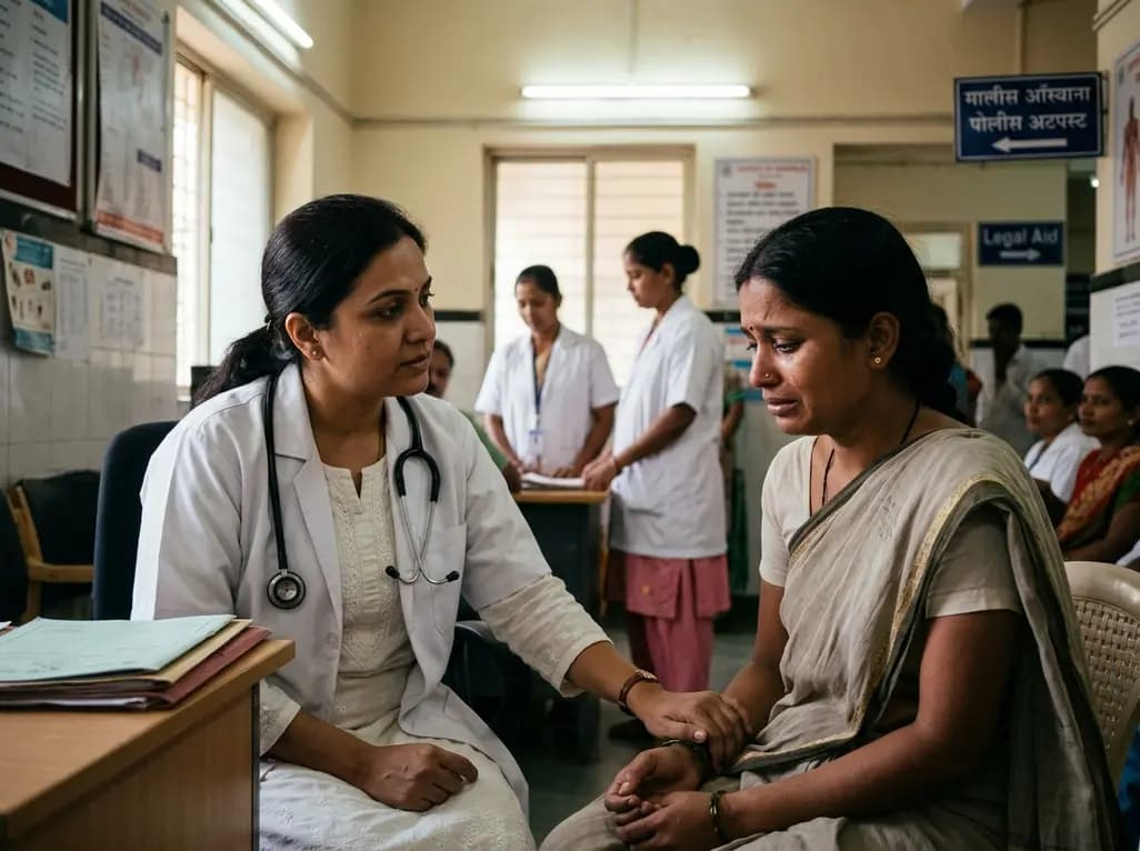
A recent qualitative study evaluates institutional gaps across health, police, and justice sectors in supporting survivors of intimate partner violence. Healthcare providers must bridge procedural fragmentation with empathetic, trauma-informed care and cross-sector coordination to safeguard vulnerable patients.
Today
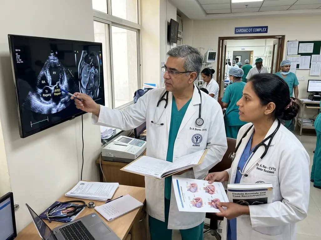
A review of the Cone procedure for severe right atrioventricular valve insufficiency in atypical Ebstein anomaly following partial AVSD repair, emphasizing operative technique and ventricular recovery.
Today
A cross-sectional study reveals a strong positive correlation between problematic internet use and kinesiophobia in adults aged 18-45 with non-specific low back pain, highlighting how digital overuse reinforces movement fear and complicates musculoskeletal recovery.
Today
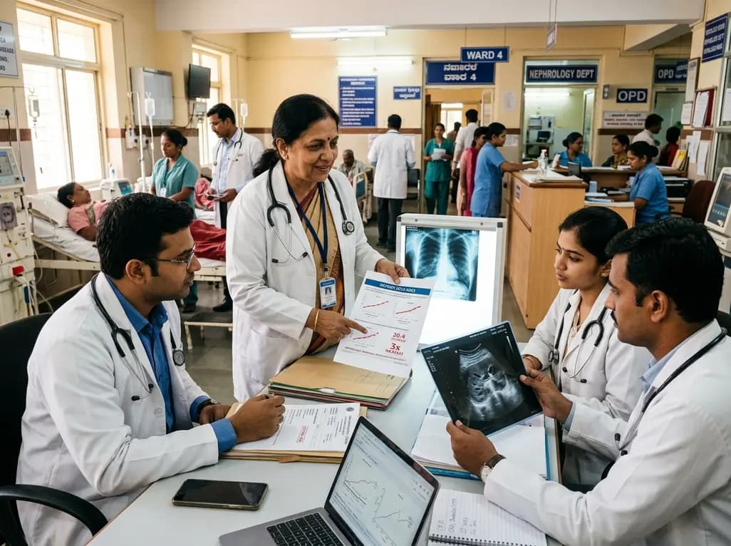
Recent data from Karnataka reveals a threefold surge in genitourinary and renal deaths over ten years. Driven by uncontrolled diabetes, hypertension, and emerging lifestyle hazards, this epidemiological shift highlights an urgent need for proactive screening and multidisciplinary nephrology management in primary care.
Today
