.webp&w=384&q=75)

Loading, please wait...
Loading, please wait...
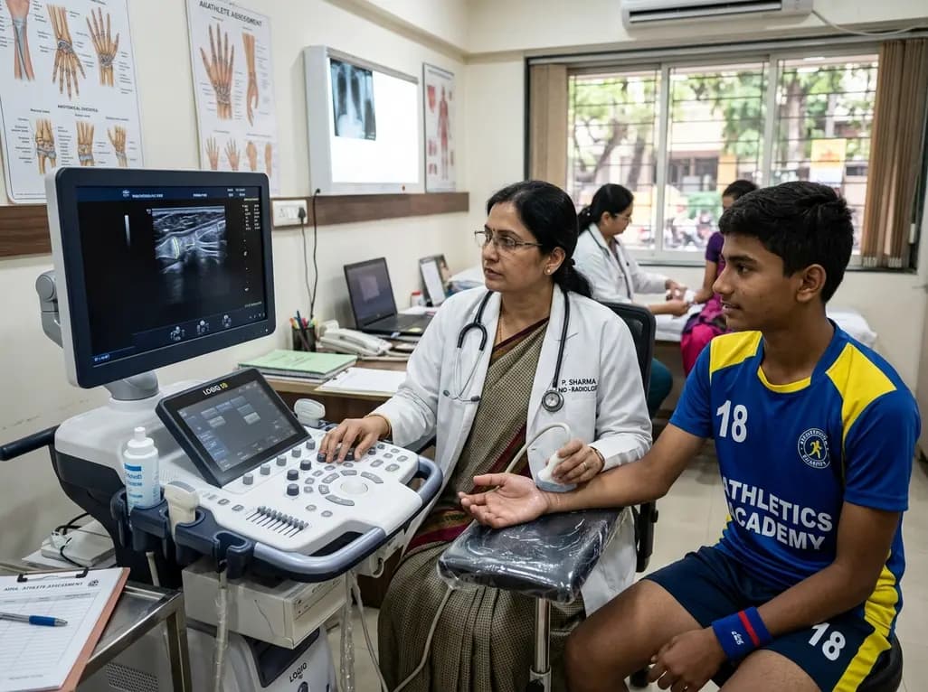
In youth sports development, chronological age often fails to reflect biological maturation, leading to selection biases that favor early-maturing adolescents. Evaluating biological age through non-invasive techniques is becoming increasingly vital in pediatric sports medicine. A recent exploratory study examined ultrasound skeletal maturity estimates among 716 South African and German adolescents participating in competitive football, athletics, and general school programs. Using ultrasound technology, researchers analyzed skeletal age across geographic and discipline boundaries. Their findings highlight significant variation in biological development, emphasizing the necessity of integrating objective biological maturity tracking into youth athletic pathways worldwide.
Biological maturation rate varies greatly among adolescents of the same chronological age. Consequently, traditional youth sports systems often unintentionally favor early maturers who possess temporary physical advantages in strength, height, and speed. Ultrasound skeletal maturity assessment offers a safe, radiation-free method to evaluate skeletal age by scanning growth plates in the hand and wrist.
Historically, clinicians relied on hand-wrist radiographs to determine skeletal age. However, ionizing radiation exposure limits repetitive screening in healthy adolescent athletes. Quantitative ultrasound systems solve this challenge by using acoustic velocity and attenuation to gauge epiphyseal ossification. Thus, sports physicians and coaches can monitor growth spurts without radiologic risk.
Furthermore, accurate maturation assessment helps differentiate between physical talent and temporary developmental advancement. When talent identification relies solely on physical performance, late-maturing youth are frequently overlooked or dropped from developmental academies. Implementing routine ultrasound evaluations provides objective biological context. As a result, sporting organizations can implement bio-banding, a process that groups young athletes by biological maturity rather than birth date. This approach ensures fairer competition, fosters technical skill development, and reduces dropout rates among talented but late-maturing participants across diverse sporting disciplines globally.
The exploratory study evaluated 233 adolescent girls and 483 adolescent boys in South Africa and Germany. In the South African cohort, male track and field athletes displayed advanced skeletal maturity compared to football players. Moreover, football players were significantly more mature than age-matched school learners from the same region. South African female track athletes similarly demonstrated advanced skeletal age compared to school controls.
These patterns indicate a strong maturity selection bias within South African youth sports pathways. Specifically, coaches and talent scouts systematically recruit adolescents who are biologically older than their chronological age. Because athletics demands raw power and sprinting capability, early-maturing boys gain a distinct competitive edge during selection trials.
However, relying heavily on biological advancement can obscure underlying technical potential. Additionally, environmental and socioeconomic factors influence growth trajectories in low- and middle-income countries. Therefore, comparing athletic cohorts directly with regional school controls is essential for accurate baseline measurements. Without localized control groups, talent development programs risk mistaking pubertal timing for long-term athletic capacity. Consequently, South African sports federations must refine selection frameworks to support youth across all developmental stages, ensuring that late-maturing individuals receive equal training opportunities and professional guidance.
In contrast to South African findings, the German cohort exhibited distinct developmental patterns across sports. German male athletes showed no significant maturity differences between football players, track athletes, and school controls. Interestingly, German female football players were less biologically mature than their counterparts in athletics or school groups.
Furthermore, cross-country comparisons revealed that South African adolescents were overall delayed in skeletal maturity compared to German peers. These geographical variations reflect complex interactions between genetic backgrounds, nutritional status, and environmental conditions. Consequently, standardized growth curves derived from high-income European populations may not directly apply to African youth.
Understanding these international disparities is critical for sports medicine practitioners and international governing bodies. For instance, global sports academies frequently apply uniform talent identification metrics across diverse geographic regions. However, if baseline biological maturity varies by population, uniform benchmarks create systematic disadvantages for specific demographic groups. Therefore, clinicians must interpret skeletal age assessments within population-specific contexts. By establishing localized normative data, sports scientists can accurately track adolescent growth spurts. Ultimately, this tailored clinical approach prevents premature athlete dropouts, supports optimal physical conditioning, and promotes equitable international competition standards across youth developmental pathways.
Incorporating ultrasound technology into routine pediatric assessments provides substantial clinical and practical benefits. Because ultrasound devices emit no ionizing radiation, clinicians can safely conduct serial measurements throughout adolescence. Consequently, sports physicians can closely track peak height velocity, the phase of rapid growth during pubertal development.
Tracking growth velocity is clinically vital because rapid bone elongation temporarily alters musculoskeletal biomechanics. Specifically, tendon tightness, muscle inflexibility, and temporary coordination deficits frequently occur during adolescent growth spurts. As a result, young athletes face an elevated risk for overuse injuries, such as Osgood-Schlatter disease and apophysitis. By identifying growth spurts early through ultrasound screening, clinical teams can adjust training loads proactively.
Moreover, non-invasive portable ultrasound devices allow on-site testing at academies, schools, and training camps. This accessibility enables large-scale population screening, which was previously impractical due to radiological limitations and financial costs. Additionally, digital ultrasound algorithms generate immediate objective skeletal age scores, reducing inter-observer variability inherent in manual x-ray scoring systems. Therefore, ultrasound technology bridges the gap between clinical endocrinology and field-based sports performance, providing medical staff with accurate data to safeguard growing athletes during critical physical growth phases.
The findings from this landmark study carry important implications for pediatricians, endocrinologists, and sports medicine specialists. Clinicians must advocate for holistic evaluation models that combine biological maturity tracking with skill assessment. Furthermore, medical professionals working with youth sports teams should educate coaches and administrative bodies about biological age variation.
When managing adolescent athletes, physicians should routinely evaluate biological age alongside chronological milestones. For example, if a young player presents with recurrent joint pain or performance stagnation, understanding their skeletal age clarifies whether growth acceleration is contributing to symptoms. Consequently, treatment plans can incorporate load management, targeted physical therapy, and flexibility training tailored to the patient's maturation phase.
In addition, sports medicine practitioners in low- and middle-income nations must lead research efforts to establish population-specific reference values. Because environmental factors influence pubertal timing, relying on external international standards can lead to misclassification. By establishing robust local control cohorts, researchers can better differentiate normal population variation from selection bias in elite academies. Ultimately, integrating non-invasive maturity assessment into clinical practice protects young athletes' physical health, prevents overuse injuries, and ensures sustainable long-term athletic development.
Ultrasound technology evaluates skeletal maturity by transmitting sound waves through specific bone sites, such as the distal radius, ulna, and hand physis. The device measures acoustic transmission speed and attenuation, which correlate directly with epiphyseal ossification and cortical density. Advanced algorithms then calculate a skeletal age without exposing young athletes to ionizing radiation. This allows safe, repeated assessments during key adolescent growth phases.
Biological maturity tracking is essential because chronological age does not reflect individual growth rates during adolescence. Early-maturing youth often gain physical advantages in power, height, and speed, leading to biased selection in competitive academies. Tracking biological age allows sports organizations to implement bio-banding, ensuring fair competition, preventing talent loss among late-maturing individuals, and allowing coaches to tailor training loads accurately during growth spurts.
During adolescent growth spurts, rapid bone elongation outpaces muscle and tendon lengthening, resulting in temporary flexibility loss and altered biomechanics. Consequently, young athletes experience increased susceptibility to overuse injuries, traction apophysitis such as Osgood-Schlatter disease, and coordination deficits. Identifying biological growth acceleration enables clinicians and coaches to adjust training volume proactively, prescribe targeted stretching, and implement load management strategies to prevent severe injury.
Disclaimer: This content is for informational and educational purposes only. It does not constitute medical advice, diagnosis, or treatment. Always consult a qualified healthcare provider for personalized medical recommendations. Refer to the latest local and national guidelines for clinical practice.
References
Wik EH et al. Ultrasound-derived skeletal maturity estimates of boys and girls selected to football and athletics development programmes: an exploratory study of 716 South African and German adolescents. Ann Hum Biol. 2026 Dec undefined. doi: 10.1080/03014460.2026.2700420. PMID: 42560736.
Cumming SP et al. Biological maturation in youth sport: Application of bio-banding and talent identification principles. Sports Med. 2017;47(11):2115-2127.
Lloyd RS et al. Long-term athletic development: Part 1: A pathway for optimizing athletic development in youth. J Strength Cond Res. 2012;26(8):2291-2300.

Read summarized clinical updates, watch expert medical content, and earn CME certifications right from your smartphone.
Open your smartphone camera to scan and install.





An exploratory study of 716 South African and German adolescents revealed significant maturity differences across sports pathways and populations, highlighting the clinical value of ultrasound skeletal maturity tracking for youth athletic development and injury prevention.
last month
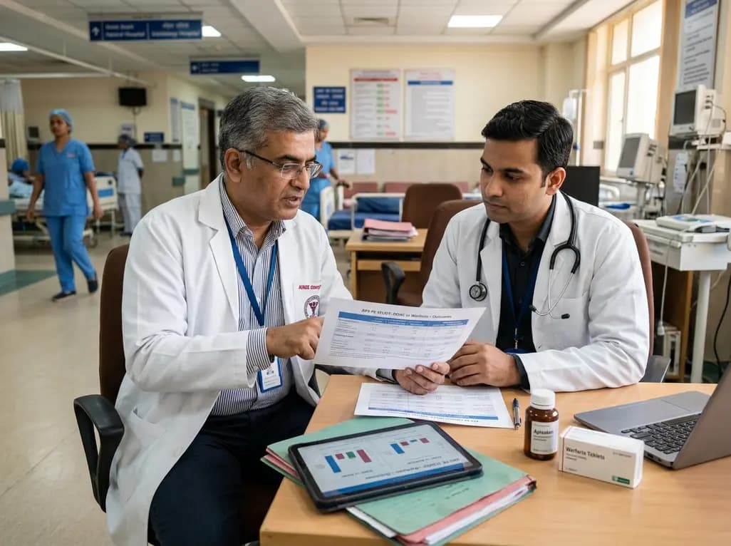
A retrospective study compares DOACs and warfarin for pulmonary thromboembolism in non-triple-positive antiphospholipid syndrome, demonstrating comparable recurrence rates and bleeding risks across both therapeutic cohorts.
Today
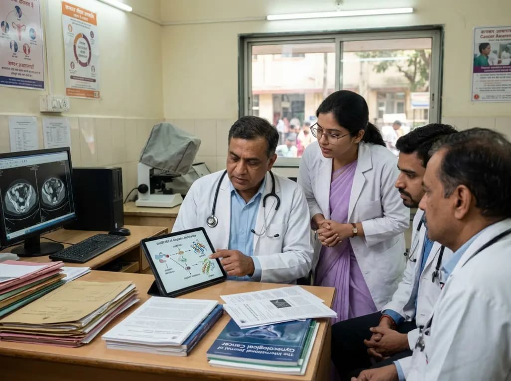
A landmark study reveals that circMCM3 promotes cisplatin resistance in cervical squamous cell carcinoma by recruiting USP49 to stabilize PTBP1 and CCND1. Targeting this newly identified molecular axis presents a promising therapeutic avenue to restore chemotherapy sensitivity and improve gynaecological outcomes.
Today
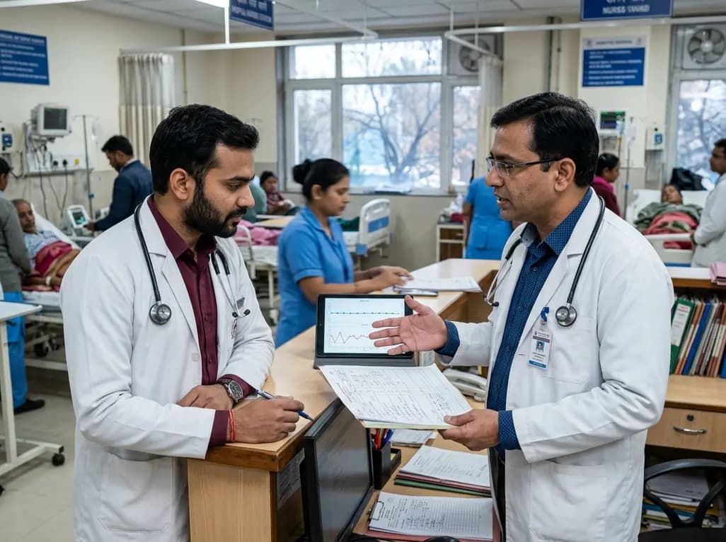
A 24-week trial shows that patients starting amlodipine/benazepril maintain stable blood pressure across all seasons, whereas those on benazepril/hydrochlorothiazide require significantly more up-titration when initiated in winter (18.9% vs. 5.0%), highlighting amlodipine's superior seasonal hemodynamic stability.
Today

A rare case of a pediatric pathological femoral fracture caused by occult telangiectatic osteosarcoma demonstrates the risks of misdiagnosis after trauma, where intramedullary nailing prompted iatrogenic tumor dissemination and required modular total endoprosthetic reconstruction.
Today
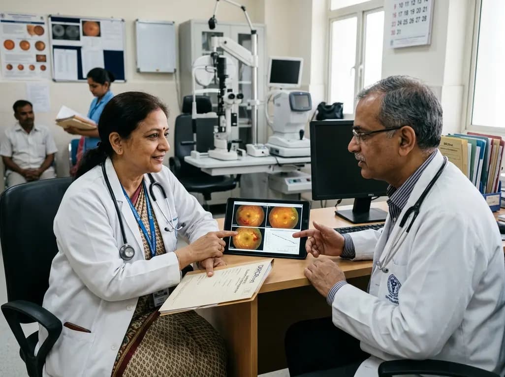
A comprehensive analysis of DRCRnet studies reveals that 15.1% of participants discontinue diabetic retinopathy clinical trials. Identifying baseline demographic and clinical predictors helps ophthalmologists and endocrinologists mitigate real-world treatment dropouts and protect long-term visual outcomes.
Today
