

Loading, please wait...
Loading, please wait...

Breast cancer remains the most prevalent malignancy among women globally, demanding rapid and precise identification of suspected lesions. Ultrasound imaging serves as a frontline, noninvasive diagnostic modality because it provides real-time tissue assessment without ionizing radiation. However, conventional sonographic interpretation depends heavily on operator experience, which frequently introduces subjective variability and diagnostic ambiguity. To address these clinical dilemmas, investigators increasingly leverage ultrasound deep radiomics to convert standard acoustic patterns into objective, high-dimensional markers. Consequently, automated algorithms can extract sub-visual textural features that escape the unaided human eye. Recent research introduces the WinDWS-SwinNet framework, an attention-driven architecture designed specifically for comprehensive lesion characterization. This novel model processes brightness-mode sonograms to differentiate benign masses from invasive malignancies with remarkable consistency. Furthermore, the system establishes a new benchmark for computational scalability by reducing heavy processing overhead. Because dense breast tissue frequently obscures lesions on mammography, sonography represents an indispensable pathway for early cancer detection. Therefore, integrating advanced computational tools into frontline diagnostic workflows promises to minimize unnecessary biopsies while improving clinical decision-making.
Standard convolutional networks often struggle with the complex acoustic artifacts and speckle noise inherent in ultrasound scans. In contrast, the newly designed WinDWS-SwinNet architecture utilizes windowed depthwise separable convolutions paired with shifted window self-attention mechanisms. This hybrid design captures both localized micro-architectural boundaries and broad global context within suspicious breast tissue. Moreover, the lightweight architecture ensures exceptional computational efficiency, enabling practical deployment on standard clinical workstations without requiring supercomputing hardware. Before classification occurs, an optimized image preprocessing algorithm significantly enhances subtle ultrasound radiomic features. This vital preprocessing step sharpens lesion margins, standardizes pixel intensity distributions, and effectively suppresses extraneous acoustic noise. As a result, the model reliably preserves critical morphological markers, including irregular angular margins, internal vascular patterns, and posterior acoustic alterations. Clinicians know that subtle acoustic attenuation frequently signals underlying tissue malignancy. Furthermore, the attention layers dynamically weigh relevant regions within the lesion while ignoring surrounding normal fibroglandular parenchyma. Therefore, the network prevents spurious background artifacts from distorting diagnostic calculations, bridging the gap between computational research and daily clinical workflows.
Evaluating artificial intelligence on multi-institutional datasets remains crucial for establishing true real-world generalizability. To achieve this standard, researchers trained and tested WinDWS-SwinNet across a multicentric retrospective repository encompassing 3,256 brightness-mode ultrasound images from five independent clinical centers. The cohort included 2,068 biopsy-proven benign lesions alongside 1,188 confirmed malignant neoplasms, establishing a balanced spectrum of clinical pathology. Consequently, the model demonstrated exceptional diagnostic efficacy during rigorous testing. It achieved an outstanding overall test accuracy of 95.79% and an area under the receiver operating characteristic curve of 0.96. For benign lesions, the network exhibited 95.11% precision paired with 98.51% sensitivity, effectively preventing erroneous cancer misdiagnoses. Similarly, the system attained 97.13% precision and 90.86% sensitivity when identifying malignant masses. Crucially, the researchers validated the architecture against an independent, unseen external dataset, achieving a persistent accuracy of 95.36%. This remarkable consistency proves that the preprocessing and attention algorithms resist overfitting across disparate scanner hardware and varied imaging protocols. In addition, these performance metrics surpass many conventional convolutional models, offering reliable noninvasive diagnostic verification for clinicians.
In routine oncologic practice, clinicians require far more granular insight than simplistic binary categorizations can provide. Distinguishing between simple fibroadenomas, complex cysts, inflammatory conditions, and invasive ductal carcinomas dictates completely different patient management pathways. Fortunately, the WinDWS-SwinNet platform excels at intricate multipathology classification, directly addressing this critical clinical necessity. During comprehensive multi-class evaluations, the model achieved an overall accuracy of 93%, with specific class-wise accuracies ranging remarkably between 94% and 99.5%. By differentiating distinct pathological subtypes noninvasively, the network accurately mirrors actual diagnostic workflows in tertiary healthcare settings. Furthermore, identifying indolent benign entities with high certainty prevents unnecessary core needle biopsies and minimizes emotional distress for patients. Conversely, immediate identification of aggressive malignant phenotypes accelerates direct referral to multidisciplinary tumor boards and surgical oncologists. In addition, the system provides nuanced textural differentiation for atypical lesions that often mimic cancer during standard sonography. Such capabilities substantially refine the traditional Breast Imaging Reporting and Data System categorization process, especially for ambiguous BI-RADS 3 and 4 lesions. Therefore, deep radiomic phenotyping helps radiologists optimize follow-up intervals while efficiently conserving critical hospital resources.
Traditional deep learning architectures frequently operate as opaque black boxes, preventing clinicians from understanding why an algorithm generates a specific prediction. This lack of diagnostic transparency severely hampers the widespread adoption of artificial intelligence in daily healthcare. WinDWS-SwinNet directly resolves this fundamental limitation by embedding explainable attention mechanisms into its core feature extraction pipeline. Specifically, the model generates intuitive visual saliency maps that clearly highlight the exact morphological regions driving its classification output. Radiologists can instantly verify whether the network bases its assessment on true pathological landmarks, such as microlobulated margins, hypoechoic cores, or acoustic shadowing, rather than peripheral imaging artifacts. Consequently, this interpretability fosters clinical trust and facilitates seamless collaborative decision-making between computational software and medical specialists. Additionally, deploying lightweight, scalable models onto portable sonography platforms can democratize expert-level diagnostic precision in secondary community clinics, particularly across resource-constrained regions in India. Because ultrasound units are portable and radiation-free, automated screening helps frontline workers triage suspicious masses swiftly. Ultimately, combining high predictive accuracy with transparent spatial reasoning transforms artificial intelligence into a dependable diagnostic partner for global healthcare teams.
Conventional computer-aided detection relies on hand-crafted mathematical formulas to analyze predefined geometric features like mass borders and aspect ratios. In contrast, ultrasound deep radiomics combines self-learning deep convolutional neural networks with high-throughput phenotypic data extraction. This advanced methodology captures complex, sub-visual tissue heterogeneity and multi-scale texture variations that standard computer algorithms miss entirely. Consequently, deep radiomic systems achieve superior diagnostic accuracy and deliver robust multipathology classification across diverse patient cohorts.
No computational system can entirely replace histopathological verification, which remains the definitive gold standard for cancer diagnosis. Instead, WinDWS-SwinNet functions as an advanced decision-support tool designed to reduce unnecessary biopsies for indolent, benign lesions. By providing high-confidence subtyping with 98.51% benign sensitivity, the architecture helps radiologists safely downgrade ambiguous BI-RADS 3 and 4 lesions to regular observation. Consequently, physicians can reserve invasive biopsy procedures for truly suspicious, high-risk malignancies.
Explainability remains essential because uninterpretable black-box models prevent clinicians from verifying the underlying biological rationale behind diagnostic predictions. Without transparent reasoning, physicians cannot distinguish between genuine pathological features and confounding acoustic artifacts. By generating visual attention heatmaps that clearly delineate lesion borders and internal acoustic textures, explainable models build clinical confidence. Moreover, this transparency satisfies stringent regulatory standards and ensures safer, highly collaborative decision-making between radiologists and artificial intelligence systems.
Disclaimer: This content is for informational and educational purposes only and does not constitute medical advice, diagnosis, or treatment. Refer to the latest local and national guidelines for clinical practice.
References

Read summarized clinical updates, watch expert medical content, and earn CME certifications right from your smartphone.
Open your smartphone camera to scan and install.





A multicentric study introduces WinDWS-SwinNet, an explainable ultrasound deep radiomics framework achieving 95.79% accuracy for breast lesion classification and 93% accuracy for multipathology subtyping, reducing diagnostic uncertainty and unnecessary biopsies.
Today
This methodological viewpoint establishes ten rules to constrain noise and enhance precision across image scoring, database handling, and data analysis in rheumatology research.
Today

A study in JAMIA Open shows lightweight open-source reasoning LLMs achieve up to 95% sensitivity and accelerate evidence synthesis screening by nearly twentyfold, presenting a cost-effective, scalable AI solution for biomedical research and systematic reviews.
Yesterday
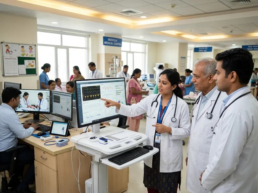
A landmark study details how an enterprise pediatric remote patient management model expanded from a single cardiology pilot to 16 diverse specialty programs by combining centralized digital operations with specialty clinical leadership.
Yesterday
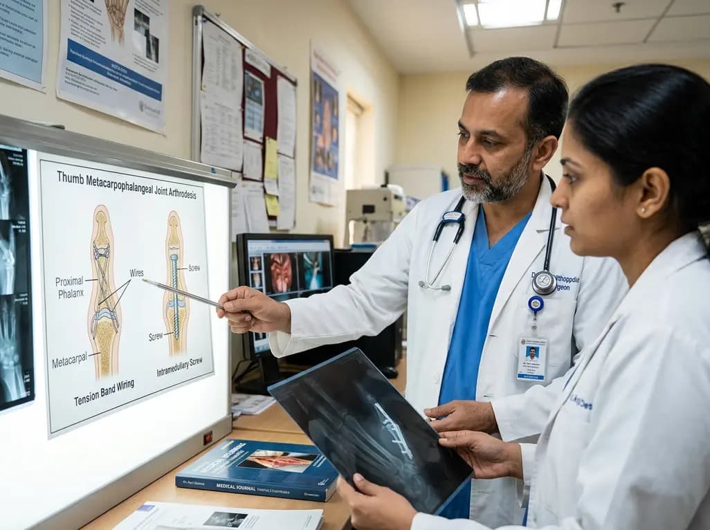
A comparative review of tension band wiring and intramedullary screw fixation in thumb metacarpophalangeal arthrodesis, evaluating union rates, hardware complications, and clinical outcomes.
Yesterday
A nationwide observational study from Norway investigates whether SGLT2 inhibitor therapy improves 5-year metabolic parameters in kidney transplant recipients with post-transplantation diabetes mellitus (PTDM), finding comparable glycemic, renal, and blood pressure profiles between user and non-user cohorts.
Today
