

Loading, please wait...
Loading, please wait...

Advanced imaging techniques provide critical objective endpoints in modern musculoskeletal medicine. Consequently, rigorous imaging studies in rheumatology have transformed clinical decision-making and disease monitoring. Researchers have dedicated substantial resources to enhancing the diagnostic sensitivity of ultrasound, computed tomography, and magnetic resonance imaging. However, investigators have historically paid inadequate attention to constraining unwarranted experimental noise.
Historically, investigators have prioritized diagnostic sensitivity over experimental consistency. They frequently developed granular scoring systems to detect microscopic structural alterations in inflamed joints. Consequently, researchers often neglected the cumulative effect of measurement variations. Regulatory agencies enforce stringent standards to minimize analytical noise during novel drug development. Therefore, academic researchers must adopt similar rigor when evaluating clinical therapeutics. Complex statistical adjustments cannot rescue trials marred by poor image acquisition or inconsistent interpretation. Furthermore, eye-catching P values cannot compensate for baseline inaccuracies. True scientific credibility relies on maximizing the signal-to-noise ratio rather than generating complex mathematical models. By suppressing unwanted variation, clinicians can identify true therapeutic responses with smaller sample cohorts. As a result, trials become substantially more economical and scientifically reliable. Minimizing noise also protects patients from misleading conclusions regarding drug efficacy. Methodologists now recognize that precision represents an indispensable prerequisite for clinical translation. Ultimately, robust data collection protocols lay the foundation for therapeutic discoveries that truly improve long-term patient outcomes in daily rheumatologic practice.
Unwarranted noise enters clinical research through multiple independent pathways. First, technical acquisition errors occur when radiographers alter patient positioning or scanner settings between sequential visits. These discrepancies introduce artificial differences that mimic real disease progression. Second, reader variability introduces substantial measurement error during visual interpretation. Different radiologists often apply scoring definitions inconsistently, particularly in borderline cases. In addition, temporal fatigue compromises diagnostic consistency during prolonged reading sessions. To counter these systemic pitfalls, research teams must implement standardized image acquisition protocols across all investigational sites. Technicians should position anatomical structures identically at baseline and follow-up examinations. Moreover, centers must harmonize scanner parameters and post-processing algorithms across multi-center initiatives. When centers standardize imaging acquisitions, they eliminate baseline confounding factors before reading begins. Methodologists also advocate for automated technical quality checks prior to final evaluation. By systematically eliminating acquisition flaws, investigators ensure that observed radiographic alterations reflect genuine biological phenomena rather than machine artifacts. Consequently, downstream statistical evaluations yield higher fidelity and greater clinical utility.
Visual evaluation remains a major source of analytical noise across clinical trials. Therefore, expert consensus highlights reader calibration as a fundamental methodological safeguard. Central reading facilities should train independent readers simultaneously using curated training libraries. Furthermore, adjudicators must assess inter-reader agreement prior to formal trial commencement. Investigators should also re-score existing baseline images when discrepancies emerge. Blinding readers to chronological sequence and treatment allocation prevents cognitive bias. However, when measuring progressive structural damage, reading paired radiographs in known temporal order can reduce noise. Methodologists frequently debate this strategy, yet empirical studies support its efficiency in detecting minor progression. Additionally, research leaders should implement regular reader retraining intervals during long clinical trials. Reader drift occurs naturally as radiologists adjust their subjective scoring thresholds over time. Structured re-calibration sessions effectively prevent this gradual divergence in scoring accuracy. By combining reader calibration with rigorous re-scoring strategies, study teams preserve longitudinal consistency. As a result, the precision of imaging endpoints increases significantly without requiring larger participant cohorts.
Measurement precision depends heavily on disciplined database management and transparent analytical workflows. Flawed data entry and inappropriate missing-data imputations introduce substantial artificial variance into clean datasets. Consequently, data managers must institute automated logic checks that flag impossible score trajectories immediately. For example, joint space width cannot spontaneously improve without surgical or biologic intervention. Furthermore, investigators should rigorously audit archival databases to identify historical data entry anomalies. Correcting past analytical errors restores integrity to valuable longitudinal observational cohorts. In addition, statisticians must select analytical procedures that appropriately reflect the underlying distribution of imaging scores. Researchers often apply parametric tests to highly skewed radiographic scores, introducing avoidable bias. Instead, biostatisticians should utilize specialized longitudinal models that account for bounded scoring scales and zero-inflation. By refining analytical models, researchers avoid generating inflated significance claims. Thus, clean database architecture directly supports robust, reproducible therapeutic evaluations. Ultimately, precise computational analysis transforms raw radiological measurements into dependable clinical evidence that clinicians can trust.
Implementing these ten methodological rules carries direct implications for clinical research and patient care. While imaging data appears inherently objective, human interpretation and data handling heavily influence trial outcomes. Clinicians frequently rely on published clinical trial results to guide difficult management choices in refractory disease. Therefore, biased or noisy imaging metrics can lead practitioners toward suboptimal therapeutic strategies. By reducing noise at the scoring and analysis stages, researchers provide clearer evidence regarding structural preservation. Moreover, these methodological enhancements lower trial costs by maximizing statistical efficiency. Smaller patient cohorts can reliably demonstrate structural benefits when researchers eliminate background measurement error. However, experts emphasize that these ten principles currently represent methodological hypotheses requiring formal prospective validation. Methodologists must conduct comparative research to quantify the specific noise reduction achieved by each distinct rule. In India and across global centers, incorporating standardized protocols will strengthen investigator-initiated studies. Ultimately, embracing systematic noise reduction ensures that radiographic endpoints deliver objective, reproducible guidance for improving joint health worldwide.
Regulatory agencies require robust statistical precision when evaluating novel therapeutics for rheumatoid arthritis and related diseases. Unwarranted noise from image acquisition or reader variation obscures genuine treatment differences, leading to erroneous conclusions. Furthermore, excessive variance artificially deflates study power, compelling sponsors to recruit needlessly large patient cohorts. By systematically reducing measurement noise, investigators maximize the signal-to-noise ratio, ensuring that structural preservation endpoints meet stringent regulatory standards with greater diagnostic accuracy and statistical certainty.
Reader calibration aligns scoring thresholds across radiologists through targeted pre-trial training and iterative benchmark evaluations. Without calibration, individual readers interpret subjective scoring boundaries differently, creating significant inter-observer variability. Calibrated scoring sessions harmonize criteria for assessing joint space narrowing, erosions, and synovitis. Moreover, regular retraining throughout lengthy trials prevents temporal reader drift. Consequently, calibrated readers generate reproducible measurements, reducing analytical noise and ensuring that observed changes reflect true disease activity rather than subjective human error.
Yes, re-scoring archival datasets significantly improves statistical power by correcting historical interpretation errors and standardizing scoring procedures across longitudinal time points. Initial reading rounds often suffer from reader variability or inconsistent protocols. By applying modern consensus rules and reading paired images with updated blinding strategies, researchers markedly reduce measurement variance. This precision gain uncovers subtle therapeutic differences previously obscured by background noise, enabling investigators to derive robust secondary discoveries without recruiting additional patients.
Disclaimer: This content is for informational and educational purposes only... Refer to the latest local and national guidelines for clinical practice.
References

Read summarized clinical updates, watch expert medical content, and earn CME certifications right from your smartphone.
Open your smartphone camera to scan and install.





This methodological viewpoint establishes ten rules to constrain noise and enhance precision across image scoring, database handling, and data analysis in rheumatology research.
Today

A study in JAMIA Open shows lightweight open-source reasoning LLMs achieve up to 95% sensitivity and accelerate evidence synthesis screening by nearly twentyfold, presenting a cost-effective, scalable AI solution for biomedical research and systematic reviews.
Yesterday
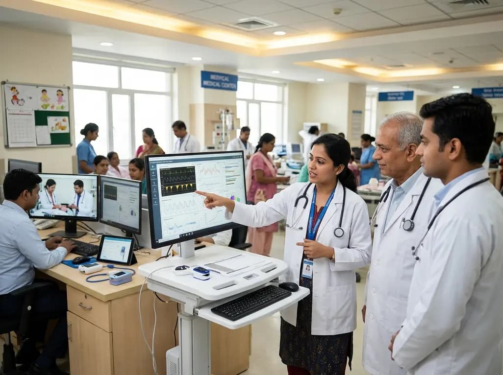
A landmark study details how an enterprise pediatric remote patient management model expanded from a single cardiology pilot to 16 diverse specialty programs by combining centralized digital operations with specialty clinical leadership.
Yesterday

A comparative review of tension band wiring and intramedullary screw fixation in thumb metacarpophalangeal arthrodesis, evaluating union rates, hardware complications, and clinical outcomes.
Yesterday
A nationwide observational study from Norway investigates whether SGLT2 inhibitor therapy improves 5-year metabolic parameters in kidney transplant recipients with post-transplantation diabetes mellitus (PTDM), finding comparable glycemic, renal, and blood pressure profiles between user and non-user cohorts.
Today
