

Loading, please wait...
Loading, please wait...
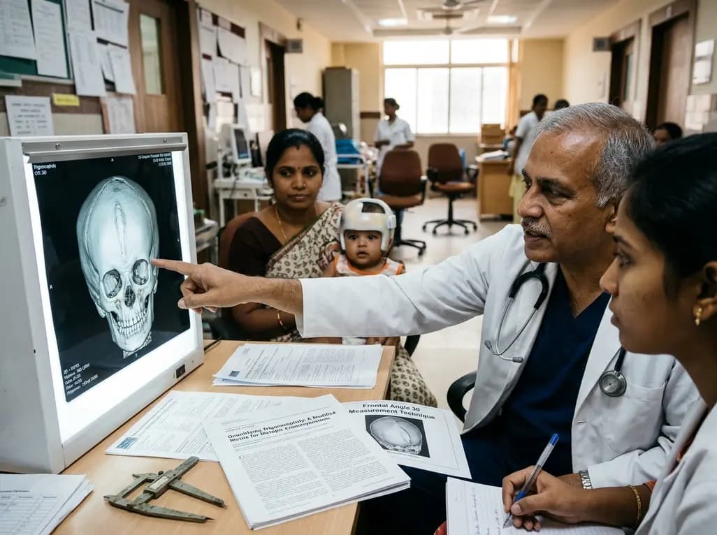
Premature closure of the metopic suture creates trigonocephaly, a distinct craniofacial deformity characterized by forehead narrowing and hypotelorism. To correct this, surgeons perform minimally invasive strip craniectomy paired with postoperative helmet orthosis. However, objective outcome assessment has long posed significant logistical challenges. Conventional surface scanning requires expensive proprietary platforms. Consequently, researchers have validated an accessible method for trigonocephaly quantification using widely available software.
Metopic craniosynostosis accounts for nearly ten to fifteen percent of all craniosynostosis cases. Historically, pediatric surgeons corrected trigonocephaly with open fronto-orbital advancement and cranial vault remodeling. Although open reconstruction produces immediate calvarial reshaping, it carries notable risks. Specifically, extensive procedures involve prolonged anesthesia, substantial blood loss, and prolonged pediatric intensive care stays. Consequently, minimally invasive endoscopic suturectomy has emerged as a favored alternative over the last two decades. During this operation, surgeons resect the fused metopic ridge through tiny scalp incisions before four months of age. Afterwards, the infant wears a custom cranial remolding helmet to guide expanding cerebral growth.
Nevertheless, evaluating cranial morphology after endoscopic release remains challenging. Many centers historically relied on serial computed tomography scans to monitor cranial vault expansion. However, ionizing radiation carries demonstrable oncological risks in infants. Therefore, clinicians actively avoid repeated postoperative radiography. While stereophotogrammetry provides non-invasive digital tracking, commercial optical scanners demand expensive capital investment. Craniofacial teams urgently need practical, radiation-free assessment protocols.
To overcome hardware barriers, investigators evaluated an adapted angle measurement technique within existing clinical workflows. Specifically, the craniofacial team established Frontal Angle 30 (FA30) as an accessible metric for trigonocephaly quantification. The investigators retrospectively evaluated ten non-syndromic infants who underwent endoscopic suturectomy and helmet orthosis. Clinicians captured surface scans across three standardized milestones: preoperatively, at six months post-surgery, and at treatment completion. Additionally, the researchers evaluated twelve healthy pediatric controls to determine diagnostic discrimination.
Furthermore, raters compared preoperative surface scans against baseline interfrontal angles measured on diagnostic computed tomography. Multiple independent craniofacial clinicians performed repeated measurements to determine interobserver and intraobserver reliability. Statistical analyses utilized Intraclass Correlation alongside Spearman rank testing to evaluate correlation with radiographic benchmarks. Crucially, the clinicians measured all angles using free, ubiquitous software already installed in remolding clinics. This pragmatic workflow completely eliminates the need for expensive secondary workstations. As a result, outpatient clinics can evaluate morphologic outcomes objectively without burdening hospital budgets.
The success of this minimally invasive protocol depends on combining precise surgical release with dynamic orthotic shaping. First, the neurosurgeon performs an endoscopic suturectomy, removing a narrow strip of bone along the closed midline seam. Because infants undergo surgery under four months of age, their calvarial bones remain exceptionally pliable. Moreover, minimal tissue dissection drastically reduces blood loss and avoids blood transfusion. Most pediatric patients return home safely within twenty-four hours of surgery.
However, bone release alone cannot fully eliminate established frontal triangularity. Therefore, custom cranial remolding orthosis serves as an essential companion therapy. Orthotists obtain digital surface scans within days after surgery to fabricate an individualized helmet. This cranial orthosis applies gentle, targeted contact over the prominent frontal keel while leaving spacious expansion zones over constricted temporal areas. Consequently, natural brain growth exerts outward hydraulic force directly into these designated relief channels. Families maintain orthotic compliance for twenty-three hours daily, attending regular adjustments throughout the infant's first year.
The quantitative data revealed remarkable, statistically significant improvements in forehead symmetry. Specifically, the average Frontal Angle 30 increased from 129.85 degrees preoperatively to 134.08 degrees at six months post-surgery. This substantial correction proved highly significant, reflecting rapid cranial expansion during early infancy. Interestingly, measurements between six months and treatment completion showed virtually no statistical difference, plateauing at 134.13 degrees. This outcome demonstrates that the primary calvarial expansion occurs during the first six months of orthotic therapy.
Additionally, statistical comparisons showed a significant distinction between surgical patients and unaffected controls, who averaged 141.99 degrees. Thus, while strip craniectomy and helmeting normalize forehead shape, calvarial contours retain minor anatomical differences compared to healthy peers. Notably, the study demonstrated high Intraclass Correlation coefficients for both interrater and intraobserver assessments. Furthermore, preoperative FA30 measurements strongly correlated with interfrontal angles measured on diagnostic computed tomography. Consequently, surface scanning reliably mirrors underlying osseous architecture without exposing infants to harmful ionizing radiation.
These clinical findings offer clear advantages for pediatric craniofacial units, particularly in resource-conscious health settings. In India, where access to dedicated stereophotogrammetry equipment remains restricted, cost-effective alternatives are invaluable. Because this measurement technique uses free, accessible software, surgical centers can track outcomes without additional overhead costs. Furthermore, parents appreciate objective numerical evidence detailing their infant's progressive calvarial improvement over time.
Nevertheless, clinicians must interpret these measurement trajectories with comprehensive clinical awareness. Because angular expansion largely concludes by six months, surgical teams can reassure families regarding procedural efficacy early. However, orthotists must maintain helmet therapy until twelve months to prevent secondary deformational relapse. Moreover, while FA30 quantifies regional forehead flattening accurately, clinicians must still monitor supraorbital retrusion and ocular hypotelorism. In addition, larger multi-center cohorts should validate this modified workflow across diverse patient demographics. Ultimately, adopting reproducible, accessible measurement protocols elevates pediatric surgical care, promotes transparent auditing, and ensures equitable craniofacial follow-up.
Frontal Angle 30 evaluates calvarial curvature using non-invasive surface scans instead of ionizing computed tomography. Standard interfrontal angle assessments rely heavily on bony landmarks visible only on axial CT slices. In contrast, Frontal Angle 30 measures external soft-tissue and skeletal contours at an accessible thirty-degree reference plane. Consequently, this technique allows clinicians to monitor ongoing cranial remodeling safely in outpatient settings without repeated radiation exposure or sedation.
Infants experience their most rapid brain growth during the initial post-operative months. Therefore, endoscopic strip craniectomy combined with helmet orthosis capitalizes primarily on this early period of calvarial expansion. By six months post-surgery, cranial remolding achieves the vast majority of frontal widening. Although orthotic therapy continues until twelve months of age to consolidate morphology, subsequent skeletal changes become gradual. Consequently, the Frontal Angle 30 stabilizes, showing minimal numerical variation between six months and treatment completion.
Yes, accessible tools integrated into remolding workflows provide excellent reliability for routine monitoring. Traditional stereophotogrammetry setups demand substantial capital investment and proprietary software platforms. In contrast, standardized surface measurement protocols utilize existing scanner infrastructure already present in orthotic clinics. Furthermore, this study demonstrated strong interrater reliability and significant correlation with computed tomography benchmarks. As a result, community craniofacial centers can successfully perform objective longitudinal tracking without acquiring costly specialized stereophotogrammetric devices.
Disclaimer: This content is for informational and educational purposes only and does not constitute medical advice, diagnosis, or treatment regimens. Healthcare professionals must exercise independent clinical judgment and correlate findings with patient presentations. Refer to the latest local and national guidelines for clinical practice.
References

Read summarized clinical updates, watch expert medical content, and earn CME certifications right from your smartphone.
Open your smartphone camera to scan and install.





A modified Frontal Angle 30 measurement provides an accessible, radiation-free method for trigonocephaly quantification. This novel workflow reliably assesses cranial vault remodeling following minimally invasive strip craniectomy and helmet orthosis therapy in pediatric metopic craniosynostosis.
Today
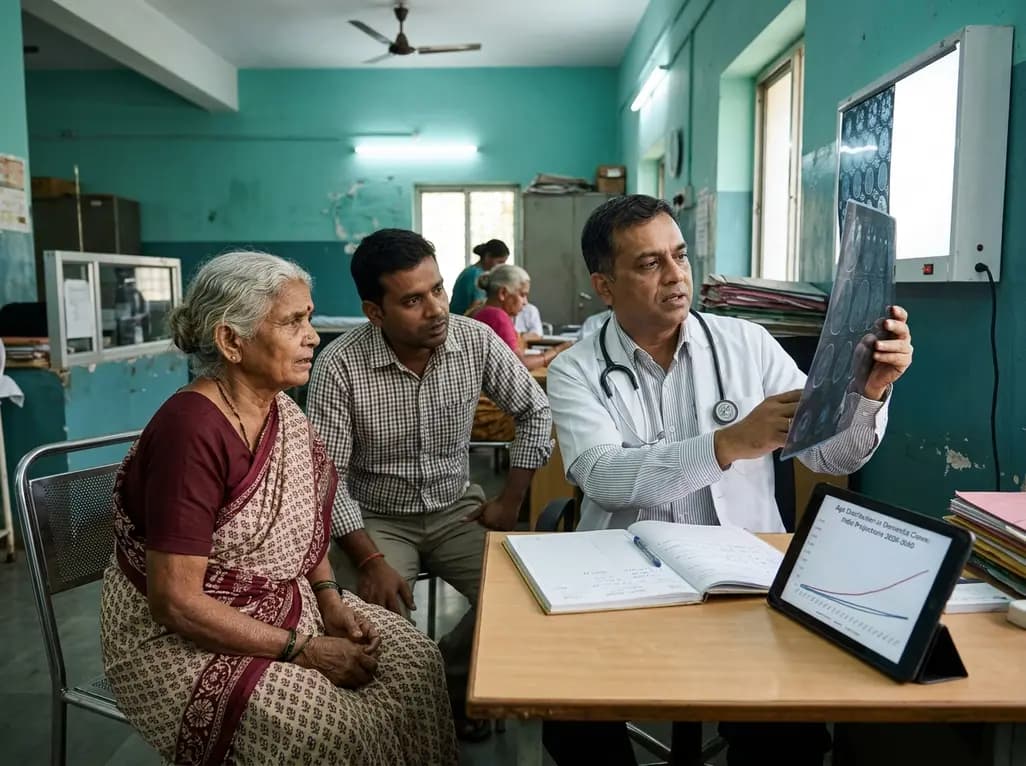
New Global Burden of Disease findings reveal that while age-standardised rates remain stable, absolute dementia cases are surging worldwide. Population ageing and metabolic risks like elevated fasting glucose demand aggressive midlife screening and systemic primary care interventions.
Today

A recent community-partnered study highlights critical gaps in newborn screening follow-up in high-infant-mortality areas. Discover actionable strategies for clinicians to improve antenatal education, train community healthcare workers, and optimize timely interventions for metabolic and congenital disorders.
Today
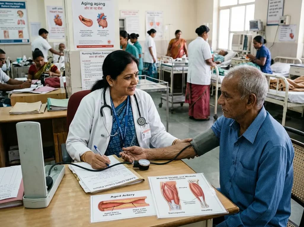
A multicenter longitudinal study of the CHARLS and HRS cohorts demonstrates that elevated cumulative pulse pressure and high-stable trajectories significantly increase incident sarcopenia risk in older adults, particularly in those with heart disease, linking arterial stiffness directly to progressive muscle loss.
Today
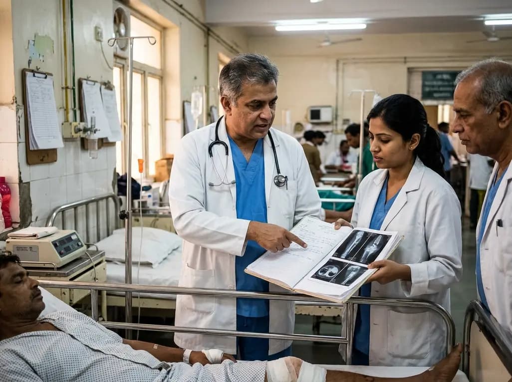
A 491-patient cohort study in Sudan reveals critical insights into war trauma reconstructive surgery, analyzing injury patterns, soft-tissue procedures, and a 25.5% complication rate in resource-limited conflict zones.
Today
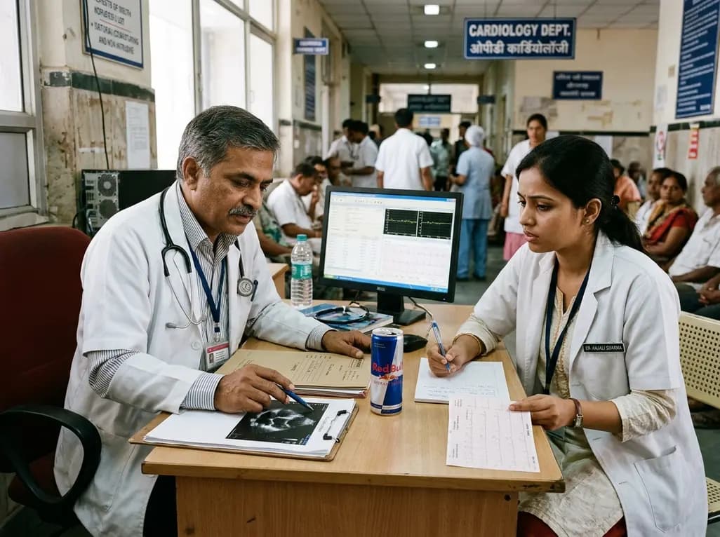
The Food Safety and Standards Authority of India has challenged Red Bull over its marketing claims, demanding scientific evidence for the term energy drink. With products packing massive amounts of caffeine and added sugar, clinicians must recognize the critical cardiovascular and metabolic implications for young patients.
Today
