

Loading, please wait...
Loading, please wait...
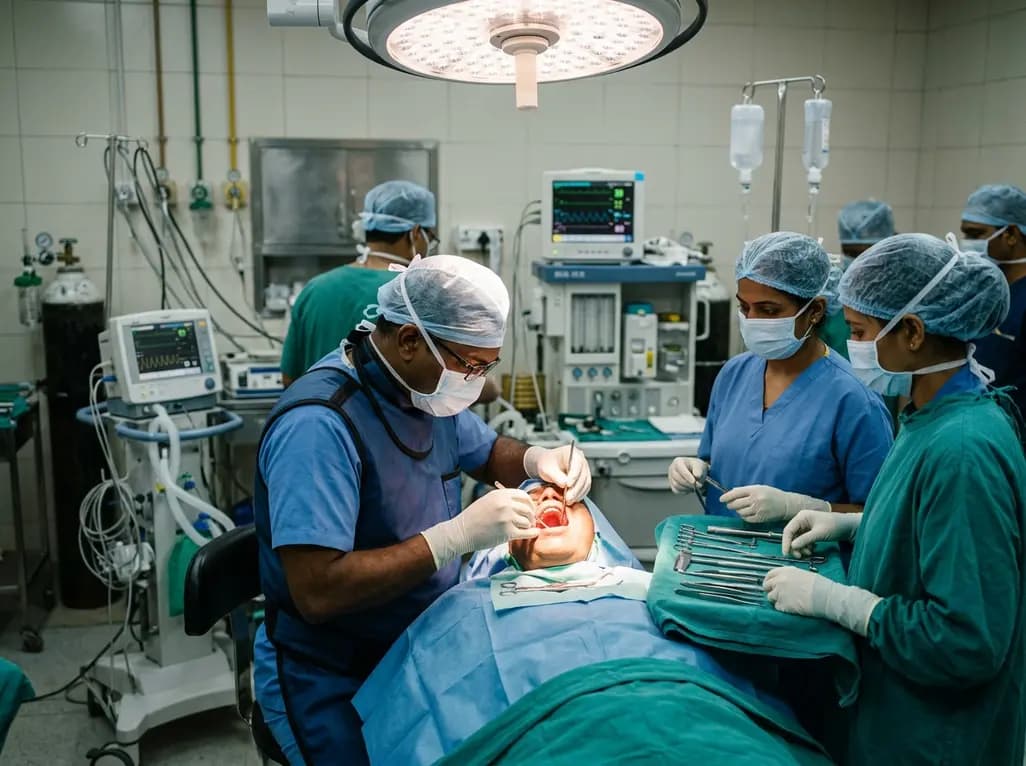
Surgeons traditionally approach upper jaw pathologies through extensive external facial incisions. For decades, the standard approach required a Weber-Ferguson transfacial incision, which leaves a permanent facial scar and creates substantial psychological morbidity. However, innovative surgical advances now enable a completely scarless transoral hemimaxillectomy for severe midfacial pathologies. This sophisticated surgical approach allows complete midfacial bone resection without any external cut on the patient's skin. Furthermore, clinicians can perform immediate microvascular autologous reconstruction in the very same setting. Consequently, patients achieve radical tumor clearance alongside outstanding cosmetic and functional recovery.
Fibrous dysplasia represents a progressive, non-neoplastic bone disorder where abnormal fibro-osseous tissue gradually replaces normal trabecular bone. Although benign, the expanding lesions frequently invade vital anatomical architecture of the craniomaxillofacial region. In particular, extensive involvement of the maxillary sinus, palate, and orbital margins can compress adjacent cranial nerves and displace orbital structures. When conservative surgical interventions such as recontouring or bone shaving fail, recurrent disease often exhibits aggressive local expansion. For example, progressive orbital narrowing threatens visual acuity and causes severe globe displacement. In many complex clinical scenarios, patients undergo multiple conservative recontourings before definitive surgery becomes inevitable. Therefore, surgical teams require definitive ablation rather than repeated debulking when vital functional structures face compression. However, performing radical resections in younger adults traditionally demanded disfiguring facial skin incisions. Consequently, surgeons sought less invasive methods that achieve en bloc extirpation without sacrificing external appearance. Pure transoral access provides an ideal answer to these challenges.
The surgical team accomplishes a transoral hemimaxillectomy without any external facial skin cuts or endoscopic assistance. First, the surgeon makes a deep gingivolabial vestibular incision extending from the anterior midline to the ipsilateral maxillary tuberosity. Next, the surgeon incises the hard palatal mucosa along the midline and the posterior palatal boundary to expose underlying bone. Subperiosteal dissection exposes the anterior and lateral walls of the maxilla up to the inferior orbital rim. Furthermore, the operator carefully preserves the infraorbital neurovascular bundle whenever oncologically feasible. The surgical team then performs precision osteotomies through the frontomaxillary junction, zygomaticomaxillary buttress, and pterygomaxillary junction. Subsequently, the surgeon applies Walsham forceps to down-fracture the hemimaxilla. This controlled down-fracture safely separates the bony attachments and delivers the entire surgical specimen directly into the oral cavity. As a result, the entire hemimaxilla exits cleanly without lacerating facial soft tissues. Thus, the technique completely preserves facial mimetic muscles, facial skin integrity, and native cutaneous innervation.
A complete hemimaxillectomy creates a massive three-dimensional space that communicates directly with the sinonasal cavities and orbit. Without vascularized reconstruction, the defect causes midfacial collapse, palatal fistulas, severe hypernasal speech, and orbital floor descent. Therefore, surgeons must obliterate this extensive dead space during the primary operative session. A free rectus abdominis muscle flap offers outstanding bulk, pliable contour, and a long vascular pedicle. Clinicians harvest the rectus muscle through a lower abdominal incision, which yields ample vascularized tissue without facial donor-site morbidity. Subsequently, the microvascular team tunnels the deep inferior epigastric vessels into the neck for microvascular anastomosis. The surgical team safely secures the muscle flap within the maxillectomy cavity to support the orbital floor and restore the palate. Moreover, the robust blood supply promotes rapid mucosalization across the oral surface. As a result, the transposed flap seals the oral cavity from the nasal passage effectively. Consequently, patients avoid chronic crusting, persistent speech compromise, and severe midfacial concavity.
Functional recovery forms an essential pillar of comprehensive midfacial reconstructive success. Following vascularized soft tissue transfer, the oral mucosal layer re-epithelializes over the transposed muscle within several weeks. Furthermore, the supportive tissue matrix provides a stable base for prosthetic dental appliances. By seven months postoperatively, prosthodontists can fabricate a customized removable partial denture. This denture restores dental occlusion, stabilizes masticatory load, and replenishes maxillary arch architecture. Consequently, patients resume an unrestricted diet without food nasal regurgitation or chewing pain. In addition, the sealed palatal contour restores normal intraoral air pressure dynamics during phonation. Therefore, the patient achieves clear, intelligible speech and normal swallow function. Long-term longitudinal follow-up confirms that patients maintain stable prosthetic retention without mucosal breakdown. Ultimately, combining precise mucosal resection with vascularized flap transfer bridges the historic gap between radical ablative surgery and holistic oral rehabilitation.
Traditional approaches using a Weber-Ferguson incision frequently result in prominent facial scars, lower eyelid ectropion, and facial edema. In contrast, the purely transoral approach avoids any external cutaneous scarring, preserving natural facial symmetry and youthful tissue tone. Over long-term surveillance exceeding a decade, patients demonstrate no evidence of recurrent fibrous dysplasia. Serial computed tomography scans verify stable orbital volume and clear surgical margins without bony regrowth. Moreover, the muscular free flap maintains appropriate soft tissue volume, preventing progressive enophthalmos or facial depression. Surgeons also observe high levels of psychological satisfaction because the procedure spares patients from stigmatizing surgical scars. Thus, this scarless technique achieves true curative intent while protecting subjective quality of life. Furthermore, operative centers avoid the need for expensive robotic or endoscopic visualization systems. Consequently, skilled craniomaxillofacial reconstructive teams can implement this method safely in diverse hospital settings.
Careful patient selection remains fundamental when planning a scarless transoral hemimaxillectomy. Candidates typically present with extensive, recurrent benign or intermediate fibro-osseous neoplasms of the maxilla. Specifically, patients with monostotic fibrous dysplasia refractory to prior conservative shaving represent ideal candidates. When lesions impinge upon the orbital floor or pterygopalatine fossa, complete removal protects vital visual structures. However, surgical teams must confirm adequate intraoral access, mouth opening, and patient tolerance for prolonged microvascular anesthesia. If malignant invasion into overlying skin or subcutaneous fat exists, external incisions remain mandatory for radical oncologic margins. Therefore, multidisciplinary preoperative evaluation with fine-cut computed tomography and vascular imaging is essential. When clinicians choose candidates properly, this intraoral approach delivers complete anatomical clearance, rapid recovery, and immaculate cosmesis.
The transoral approach completely eliminates visible facial scars and prevents notorious complications associated with the Weber-Ferguson cut, such as lower eyelid ectropion, lip notch deformities, and chronic midfacial lymphedema. Furthermore, intraoral mucosal incisions heal rapidly with minimal visible distortion. By preserving the facial skin envelope and mimetic musculature, surgeons achieve optimal cosmetic symmetry while still obtaining complete anatomical exposure for definitive tumor extirpation and immediate microvascular reconstructive tissue transfer.
Suitable candidates include adult patients with extensive or recurrent benign lesions, such as progressive fibrous dysplasia, ossifying fibromas, or select low-grade neoplasms confined to the maxillary infrastructure. Crucially, the disease must not infiltrate the overlying facial skin or superficial subcutaneous tissues. Furthermore, patients must demonstrate adequate oral opening to permit surgical instrumentation, specimen delivery with Walsham forceps, and microvascular flap insetting without compromising patient safety or operative exposure.
The free rectus abdominis muscle provides substantial, vascularized soft tissue bulk that fills the large maxillectomy dead space and supports the orbital floor, preventing post-surgical enophthalmos. Additionally, the transferred muscle seals the oral cavity from the nasal passage, preventing oral-nasal fluid reflux and hypernasality. Over subsequent months, intraoral mucosa spontaneously epithelialize across the muscle surface, establishing a stable, well-vascularized tissue bed that readily accommodates a retentive removable partial dental prosthesis.
Disclaimer: This content is for informational and educational purposes only and does not constitute medical advice, diagnosis, or treatment regimens. Healthcare professionals must exercise independent clinical judgment when evaluating surgical candidates and selecting reconstructive techniques. Refer to the latest local and national guidelines for clinical practice.
References

Read summarized clinical updates, watch expert medical content, and earn CME certifications right from your smartphone.
Open your smartphone camera to scan and install.





A purely transoral hemimaxillectomy offers a scarless approach for extensive maxillary fibrous dysplasia, combining en bloc resection with free-flap reconstruction.
Today
A prospective study using wireless sensors reveals that smaller physician workrooms significantly increase contact density and respiratory virus transmission risks despite lower average occupancy, underscoring the urgent need for better architectural design and infection control in healthcare duty rooms.
Today

A KGMU study reveals over half of women with breast abnormalities fail to undergo diagnostic confirmation, leading to advanced-stage detection. Discover the socio-cultural barriers, clinical risks of incomplete resection, and community-level solutions needed to bridge the critical screening-to-diagnosis gap in India.
Today

A comparative non-contact study reveals that high-power radiofrequency delivery with the QDOT MICRO catheter induces an artifactual contact force rise. Electrophysiologists must understand this power-dependent thermal and electrical phenomenon to avoid inadvertent catheter withdrawal and ineffective ablation.
Today
Healthium Medtech has transitioned its arthroscopy vertical into SIRONIX Orthopedics, aiming to capture key segments of the global joint care market. The strategic shift introduces dedicated portfolios in sports medicine, joint regeneration, surgical reconstruction, and digital operating technologies.
Today
Preclinical research demonstrates that BMI1 overexpression alleviates metabolic syndrome-related liver injury. By triggering ubiquitin-mediated METTL3 degradation, BMI1 prevents AHNAK m6A methylation and suppresses fibrogenic TGF-β/SMAD signaling, significantly improving hepatic steatosis and insulin resistance.
Today
