.webp&w=384&q=75)

Loading, please wait...
Loading, please wait...
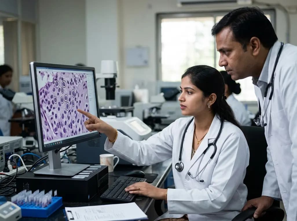
Accurate cellular nuclei detection represents a foundational pillar in computational pathology and modern oncological diagnostics. Digital pathology workflows increasingly rely on whole-slide imaging to evaluate tumor microenvironments, quantify cellular pleomorphism, and assess biomarker expression. However, traditional manual assessment of histological slides remains labor-intensive and prone to significant inter-observer variability. Automating this analytical pipeline provides standardized metrics that help oncologists and pathologists establish accurate prognostic stratifications. When automated algorithms analyze digital biopsies, detecting every single nucleus precisely determines tumor grading, mitotic indices, and lymphocytic infiltration patterns. Consequently, high-performance computational models are crucial for standardizing histopathological evaluation across diverse tissue types. In clinical practice, reliable detection directly influences therapeutic decision-making in breast, colorectal, and prostate malignancies. Nevertheless, conventional computer vision models often encounter major diagnostic bottlenecks when processing routine histological specimens. Variabilities in tissue fixation, staining intensity, and section thickness frequently obscure nuclear boundaries. Therefore, developing sophisticated machine learning frameworks capable of overcoming these technical hurdles has emerged as a top research priority in digital medicine and pathological diagnostics.
Despite substantial progress in whole-slide digitisation, achieving consistent diagnostic accuracy across varied hardware platforms remains challenging. Low-resolution digitized slides often suffer from pixelation artifacts, chromatic aberrations, and reduced contrast around delicate chromatin structures. When clinicians examine high-density tissue regions, closely packed or overlapping nuclei frequently blend into ambiguous morphological clusters. Standard deep learning detectors frequently misclassify these overlapping boundaries, leading to undercounting or false-positive segmentations. Furthermore, standard object detection networks typically struggle when forced to choose between fine cellular details and macro-level tissue architecture. Downsampling large histological gigapixel images to fit computational memory constraints inevitably sacrifices critical diagnostic nuances, such as nucleolar prominence or subtle nuclear grooves. Conversely, processing only localized, high-power image patches causes the detector to lose broader contextual awareness, such as architectural nesting and surrounding stroma. Consequently, isolated multi-scale algorithms or standard spatial enhancement filters fail to resolve these competing demands simultaneously. These persistent optical limitations emphasize the urgent need for integrated computational solutions that can enhance image clarity while preserving both local and global tissue features.
To overcome hardware limitations and compression degradation, biomedical engineers have introduced deep learning-based super-resolution into histopathology workflows. Super-resolution algorithms utilize deep convolutional neural networks to reconstruct high-frequency textural details from low-resolution inputs. In histological imaging, this transformation restores sharp nuclear membranes, intricate chromatin distributions, and precise inter-cellular margins. Rather than simply interpolating pixels, advanced generative and convolutional models infer lost structural data based on learned biological patterns. Consequently, applying super-resolution before object detection significantly reduces ambiguous edge artifacts that confuse bounding-box predictors. Furthermore, this computational enhancement allows laboratories with standard-resolution slide scanners to produce diagnostic outputs comparable to expensive, high-magnification systems. Importantly, modern super-resolution frameworks are optimized to prevent hallucinated artifacts that could otherwise mimic malignant atypia or atypical mitoses. By sharpening delicate nuclear contours without distorting diagnostic morphology, super-resolution serves as a powerful foundational layer for downstream analytical algorithms. As a result, subsequent detection models operate on clean, high-contrast visual representations, establishing optimal conditions for reliable cellular profiling across challenging hematoxylin and eosin preparations.
Standard computational pipelines often handle spatial scaling and image enhancement as disjointed tasks. In contrast, the recently proposed hybrid framework deploys a sophisticated dual-branch detection strategy that runs full-image and patch-based inference simultaneously. The full-image branch evaluates global architectural patterns, ensuring the network recognizes broader glandular structures and tissue orientation. Meanwhile, the patch-based branch zooms into localized super-resolved sub-regions, capturing intricate cellular morphology, nuclear folding, and subtle mitotic figures. Subsequently, the algorithm combines the outputs of both branches through a dedicated confidence-weighted fusion mechanism. This mechanism intelligently balances macro-contextual certainty with micro-level spatial precision. To eliminate redundant bounding boxes and resolve overlapping nuclear instances, the system executes non-maximum suppression followed by clustering-based spatial refinement. Additionally, this lightweight architecture preserves computational efficiency, avoiding the excessive memory overhead typically associated with large-scale vision transformers. Therefore, the coordinated dual-branch model achieves rapid, high-precision detection suitable for high-throughput laboratory settings, effectively solving the trade-off between panoramic context and microscopic detail.
Rigorous validation of deep learning models requires comprehensive, multi-institutional histological datasets. Researchers validated this hybrid super-resolution architecture using the widely recognized NuCLS benchmark, a curated dataset dedicated to cell nuclei classification and detection in breast cancer histopathology. The experimental findings demonstrated remarkable diagnostic performance, significantly outperforming baseline object detection models across multiple evaluation metrics. In particular, the combined workflow achieved up to a 20% increase in mean average precision (mAP@0.5-0.95) under stringent confidence thresholds. This significant improvement indicates that the model excels at distinguishing true nuclear boundaries even in densely crowded neoplastic fields. Furthermore, the hybrid detector maintained robust performance despite variations in tissue preparation, stain intensity, and optical illumination. The clustering-based refinement stage proved especially effective at resolving tightly clustered tumor-infiltrating lymphocytes and malignant epithelial cells. Consequently, these robust quantitative gains prove that coupling super-resolution with dual-branch fusion overcomes the traditional limitations of single-scale detectors, establishing a new state-of-the-art benchmark for computational pathology.
The integration of advanced deep learning algorithms into diagnostic pathology holds profound implications for everyday oncological care. Automated and reliable cellular nuclei detection streamlines labor-intensive prognostic assessments, such as evaluating Nottingham histologic scores in breast carcinoma or quantifying Ki-67 proliferation indices. Furthermore, standardized nuclear scoring minimizes diagnostic discordance between pathologists, ensuring more equitable and reproducible patient care across tertiary and community centers. By employing lightweight super-resolution architectures, clinical laboratories can process high-volume digital biopsy slides without requiring prohibitive supercomputing infrastructure. Additionally, these AI-driven systems assist pathologists by flagging subtle morphological abnormalities, thereby reducing diagnostic fatigue during prolonged slide reviews. In the future, combining these structural detection tools with multimodal patient data, including spatial transcriptomics and genomic profiling, will accelerate precision oncology. Clinicians and laboratory directors should actively monitor these technological developments, supporting the careful clinical validation and regulatory oversight necessary to integrate computer-assisted diagnostic tools safely into routine pathological workflows.
Super-resolution algorithms employ deep neural networks to reconstruct high-frequency microscopic details from lower-resolution digitized slides. In histopathology, this process sharpens nuclear borders, clarifies subtle chromatin patterns, and separates densely clustered cells. Consequently, downstream object detectors receive higher-contrast visual features, which significantly reduces false-positive detections and boundary inaccuracies without requiring costly high-magnification optical hardware upgrades in clinical laboratories.
Conventional single-scale detectors frequently struggle to balance broad tissue architecture with microscopic cellular details. In contrast, the dual-branch strategy processes both full-image global context and localized high-resolution image patches simultaneously. A confidence-weighted fusion mechanism then combines these predictions, refining overlapping boundaries through non-maximum suppression and spatial clustering. As a result, the framework maintains high detection sensitivity while preserving overall contextual comprehension across complex histological specimens.
Automating nuclei detection provides standardized, objective quantification of critical prognostic biomarkers, such as tumor-infiltrating lymphocytes, mitotic figures, and nuclear pleomorphism. This automation reduces inter-observer variability among pathologists, speeds up diagnostic turnaround times, and alleviates visual fatigue during microscopic examinations. Ultimately, highly accurate digital pathology algorithms ensure consistent tumor grading and support oncologists in formulating precise, individualized therapeutic strategies for cancer patients.
Disclaimer: This content is for informational and educational purposes only. It is not intended to substitute for professional medical advice, diagnosis, or treatment. Refer to the latest local and national guidelines for clinical practice.
References

Read summarized clinical updates, watch expert medical content, and earn CME certifications right from your smartphone.
Open your smartphone camera to scan and install.





A novel hybrid framework combines super-resolution imaging and dual-branch deep learning to dramatically enhance cellular nuclei detection in histopathology. Tested on the NuCLS dataset, it achieves up to a 20% increase in mAP, offering scalable precision for computational oncology and digital pathology.
Today

A comprehensive review of novel lipoprotein(a) therapies highlights RNA-based agents, oral inhibitors, and gene editing transforming residual cardiovascular risk management in clinical practice.
Today
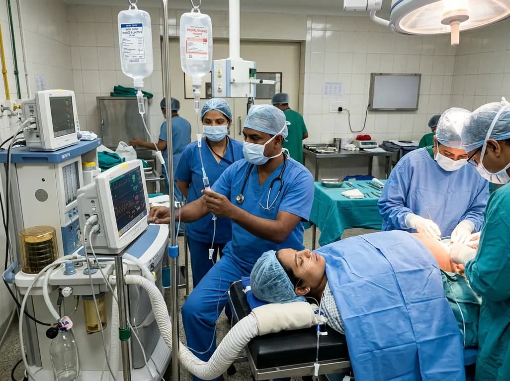
A multicenter MPOG database study of 289,047 cesarean delivery cases analyzed adherence to obstetric anesthesia best practices. General anesthesia avoidance reached 97.0%, while post-spinal vasopressor infusions (55.4%) and hypothermia prevention (56.7%) showed the lowest compliance, highlighting key targets.
Yesterday
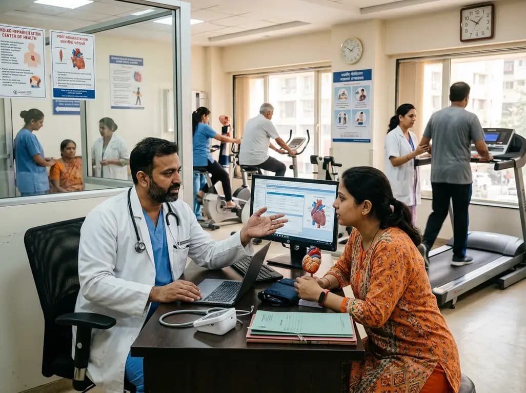
Post-COVID syndrome drives persistent vascular inflammation, endothelial dysfunction, and autonomic imbalance. Emerging evidence highlights structured exercise training as an essential non-pharmacological therapy to modulate long-term cardiovascular risk and accelerate clinical recovery.
Today
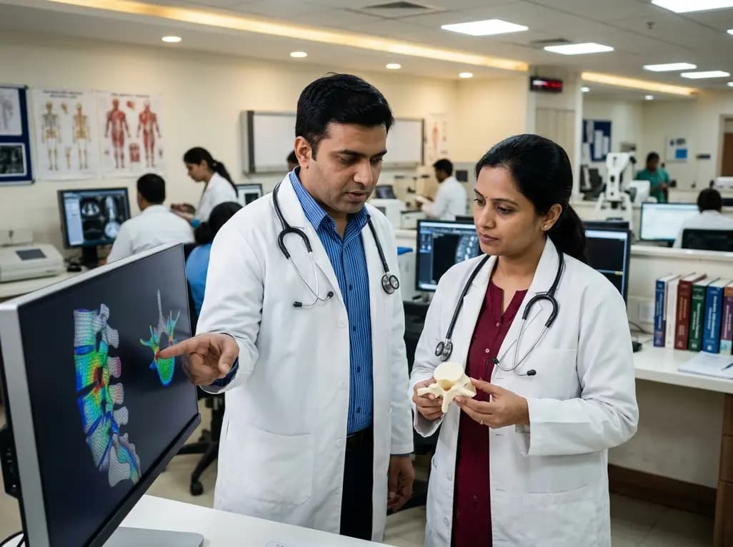
Recent research assesses an automated pipeline for CT-based vertebral finite element analysis, revealing how boundary condition components—especially load-point assignment—impact fracture-load accuracy in spinal biomechanics.
Today
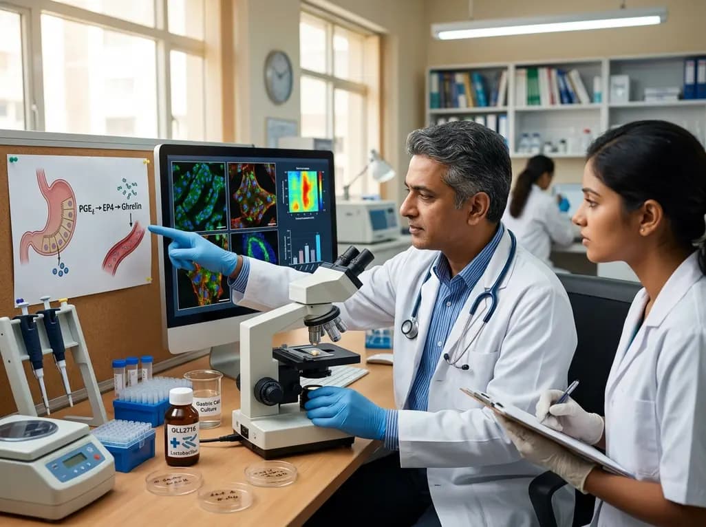
A study reveals that Lactobacillus paragasseri OLL2716 stimulates gastric cells to increase ghrelin expression and secretion via PGE2-EP4 signaling. This finding provides novel mechanistic insights into how specific probiotic strains improve delayed gastric emptying and modulate autonomic nervous activity.
Today
