.webp&w=384&q=75)

Loading, please wait...
Loading, please wait...
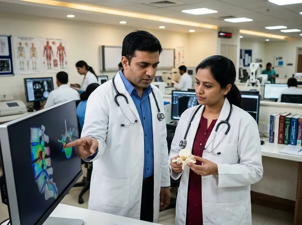
Assessment of bone strength is critical for evaluating osteoporotic collapse and spinal neoplastic instability. In recent years, vertebral finite element analysis has emerged as a state-of-the-art computational technique to quantify patient-specific fracture risk directly from clinical CT scans. However, traditional biomechanical modeling demands extensive manual configuration, creating a major barrier to routine clinical adoption. A groundbreaking study evaluated an automated pipeline to streamline single-vertebra simulations, revealing both the remarkable promise and key technical bottlenecks of automated boundary-condition workflows.
Conventional dual-energy X-ray absorptiometry measures areal bone mineral density, but it often fails to capture three-dimensional geometric variations and localized trabecular microarchitecture. Consequently, clinicians frequently encounter fragility fractures in patients categorized as having only moderate osteopenia. Biomechanical computed tomography overcomes these shortcomings by converting routine volumetric imaging into patient-specific structural models. Through calibrated material mapping, these simulations estimate failure thresholds under physiological compression. Furthermore, computational modeling accounts for cortical shell thickness, trabecular distribution, and complex spinal morphology. Therefore, non-invasive simulations offer superior diagnostic sensitivity compared to conventional densitometry alone. In clinical settings, objective failure predictions assist orthopedic surgeons and oncologists in deciding between conservative management and surgical stabilization. In addition, automated processing provides standardized strength indices across large patient cohorts. Nevertheless, the manual labor involved in segmenting vertebral bodies, establishing reference axes, and defining boundary conditions has historically restricted these tools to specialized academic research centers.
To transition biomechanical modeling from research laboratories into routine radiological practice, researchers designed a fully automated pipeline for clinical CT datasets. Specifically, the investigative team evaluated 113 vertebrae from 70 patients that had previously undergone manual reference modeling. The automated algorithm systematically identified superior and inferior endplates, constructed an intrinsic anatomical coordinate system, and positioned the axial load-application point. Subsequently, expert observers visually graded the automated configurations as good, acceptable, or bad according to rigid geometric and anatomical benchmarks. Among the 113 evaluated vertebrae, the automated workflow achieved good setups in 47% of cases, acceptable configurations in 35%, and bad setups in 18%. In the successfully processed cohort, the automated pipeline demonstrated excellent agreement with manual reference standards, achieving a high correlation coefficient of R = 0.950. Furthermore, the median percent difference in estimated fracture load was 13.0%. These findings demonstrate that automated pipelines can reliably reproduce manual configurations in nearly half of routine clinical cases without requiring human intervention.
Understanding which steps introduce numerical variance is essential for refining automated simulation algorithms. Therefore, investigators systematically isolated each boundary-condition component to quantify its independent effect on fracture-load estimates. Interestingly, automated endplate identification generated the smallest overall variation in failure load compared to manual reference models. Although minor deviations occurred during surface mesh generation, variations in calculated endplate surface area showed no significant statistical association with shifts in ultimate fracture-load predictions. Similarly, the automatic establishment of vertebra-specific anatomical coordinate systems introduced only modest deviations. When algorithms properly oriented the sagittal and coronal reference planes, axial compressive force vectors remained physiologically aligned with the principal trabecular trajectories. Consequently, standard segmentation discrepancies along the peripheral cortical margins did not substantially disrupt internal stress distributions. These observations confirm that contemporary machine learning and thresholding algorithms have attained adequate maturity for anatomical surface extraction and coordinate alignment in non-deformed vertebrae.
In contrast to endplate delineation, the assignment of the load-application point emerged as the most critical determinant of simulation accuracy. Specifically, automated shifts in the loading centroid produced the largest numerical discrepancies and the widest limits of agreement relative to manual baselines. Because compressive forces interact with the natural curvature and cross-sectional geometry of the vertebral body, even slight eccentricities in the load vector induce substantial bending moments. As a result, anterior or posterior displacement of the loading point dramatically alters tensile and shear strain concentrations across the cancellous core. The researchers observed a direct, pronounced association between load-point translation and shifts in computed fracture strength. When the automated algorithm miscalculated the geometric center of the superior endplate, the simulation either overpredicted or severely underpredicted structural capacity. Thus, boundary-condition definitions—specifically the precise point of mechanical contact—represent the most vulnerable component of automated biomechanical pipelines.
Although automated workflows significantly reduce labor and enable high-throughput data processing, the current failure rate precludes fully unsupervised clinical deployment. The fact that nearly one in five cases produced unsatisfactory boundary conditions highlights the need for robust quality-assurance checkpoints. Anatomical abnormalities, including severe osteophyte formation, degenerative scoliosis, Schmorl nodes, and prior compression deformities, frequently confound automated landmark algorithms. Furthermore, low-dose imaging protocols and beam-hardening artifacts from adjacent metallic implants can distort endplate boundary detection. Therefore, future developmental iterations must integrate hybrid approaches combining deep neural networks with physical plausibility checks. Until artificial intelligence algorithms achieve higher diagnostic reliability across abnormal anatomies, automated pipelines should incorporate rapid visual verification interfaces for radiologists and spine specialists. By maintaining an efficient human-in-the-loop oversight model, clinical institutions can leverage computational efficiency while preventing erroneous mechanical predictions in complex patient cases.
Despite current operational limitations, the standardization of automated simulations holds immense translational promise for modern medicine. In geriatric healthcare, opportunistic screening of routine abdominal and chest CT scans can identify individuals with occult skeletal fragility before catastrophic fractures occur. Moreover, longitudinal finite element tracking can evaluate patient-specific responses to novel anabolic and antiresorptive pharmacological agents. In spinal oncology, automated mechanical modeling provides objective metrics to refine the Spinal Instability Neoplastic Score. When metastatic osteolytic lesions compromise structural integrity, patient-specific simulations accurately pinpoint impending pathological collapse. Consequently, spine surgeons can optimize prophylactic stabilization strategies, select targeted cement augmentation volumes, and spare stable patients from unnecessary invasive interventions. As computational architectures become increasingly robust against anatomical deformities, automated finite element modeling will become an indispensable asset in individualized musculoskeletal precision care.
Vertebral finite element analysis integrates three-dimensional bone geometry, cortical shell thickness, and trabecular density distribution from clinical CT scans. Unlike two-dimensional areal bone mineral density testing, finite element modeling simulates actual mechanical responses under physiological loading conditions. Consequently, it provides superior precision in predicting bone strength and individualized fracture risk across diverse clinical populations.
The position of the applied load dictates the distribution of compressive, tensile, and shear stresses within the vertebral body. Even minor shifts in the loading point create unintended bending moments that drastically alter localized strain concentrations. Therefore, eccentric load placement significantly changes the calculated fracture load compared to an anatomically centered vector.
Currently, automated modeling requires human supervision because approximately 18% of automated setups generate unacceptable boundary errors. Degenerative changes, severe osteophytes, and anatomical variations frequently confuse automated landmark algorithms. Consequently, experts recommend a human-in-the-loop workflow where clinicians visually verify model alignment before utilizing the generated fracture predictions in clinical decision-making.
Disclaimer: This content is for informational and educational purposes only, and does not constitute medical advice, diagnosis, or treatment. Healthcare professionals should exercise their independent clinical judgment. Refer to the latest local and national guidelines for clinical practice.
References

Read summarized clinical updates, watch expert medical content, and earn CME certifications right from your smartphone.
Open your smartphone camera to scan and install.





Recent research assesses an automated pipeline for CT-based vertebral finite element analysis, revealing how boundary condition components—especially load-point assignment—impact fracture-load accuracy in spinal biomechanics.
last month
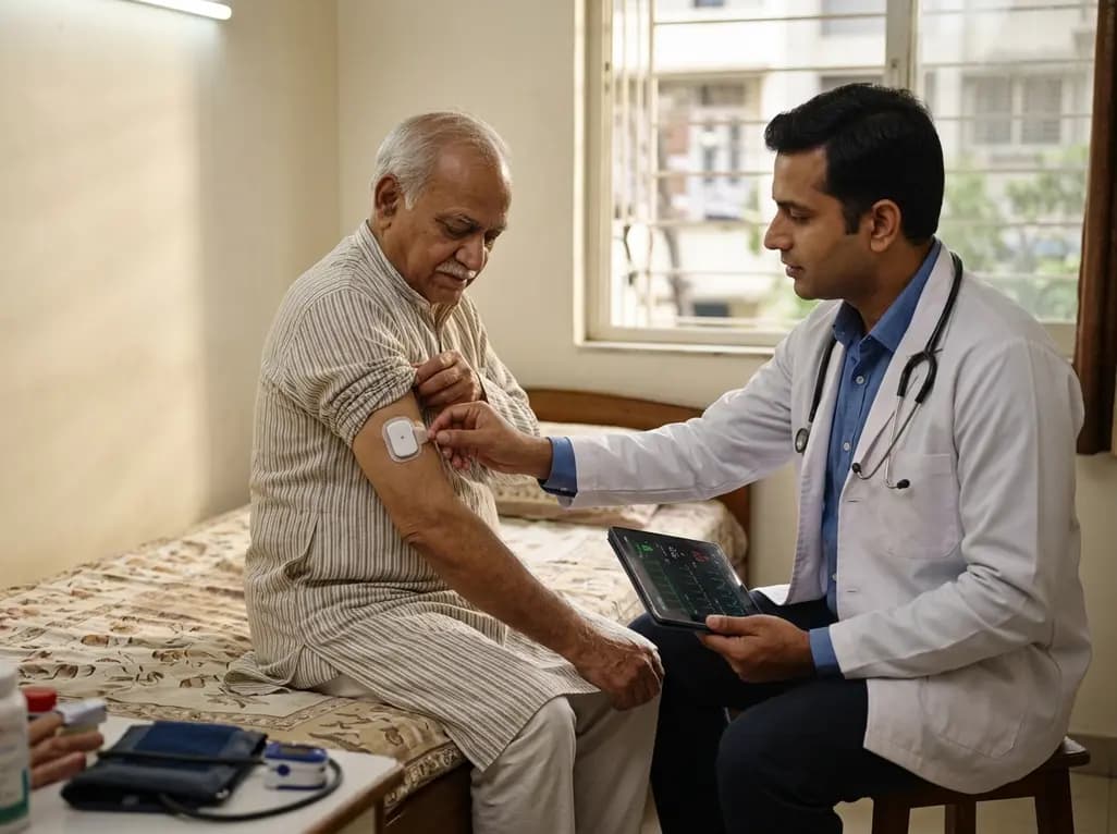
A feasibility study evaluates remote vital sign monitoring in acute hospital at home patients. Continuous wearable patches captured heart rate and respiratory rate with high adherence (89% and 75%), significantly exceeding manual spot checks for blood pressure, oxygen saturation, and temperature.
Today

A scoping review evaluates 116 studies on AI in women's reproductive health, finding that 83.6% carry high risk of bias. While ensemble models show promise, disparities and lack of validation hinder LMIC deployment.
Today

Differential oxygenation during peripheral TA-NRP can cause undetected coronary hypoxemia due to arch vessel clamping. A novel circuit with continuous aortic root monitoring detects desaturation early, facilitating left ventricular unloading and preventing allograft dysfunction and unnecessary heart discard.
Today

A longitudinal study reveals that patients undergoing staged bilateral hip arthroscopy experience progressive contralateral labral deterioration during surgical intervals. However, structural degradation does not compromise 2-year postoperative clinical outcomes or patient-reported satisfaction scores.
Today

A post hoc analysis of the PARM-T2D study shows pemafibrate exerts baseline-dependent effects on LDL-C and particle size in type 2 diabetes and hypertriglyceridemia. It lowers triglycerides and remnants while favorably remodeling atherogenic LDL particles based on initial lipid control status.
Today
