.webp&w=384&q=75)

Loading, please wait...
Loading, please wait...
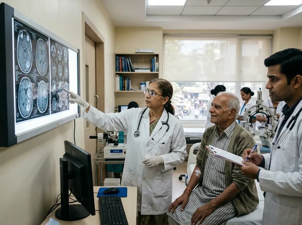
Stereotactic brain biopsy provides critical histopathological guidance for intracranial lesions, yet clinicians often hesitate to perform this procedure in octogenarians. Clinicians frequently fear high surgical morbidity, rapid functional decline, or limited therapeutic utility in elderly populations. Consequently, many elderly patients receive presumptive diagnoses or empirical palliative treatments without histological confirmation. However, a landmark retrospective study published in the Journal of Neurosurgery demonstrates that stereotactic brain biopsy is both safe and diagnostically reliable in patients aged 80 years and older. Obtaining definitive tissue diagnosis empowers oncology teams to tailor treatment strategies effectively while preserving functional independence.
As life expectancy expands worldwide, neurosurgeons encounter a rising incidence of brain tumors among geriatric patients. Historically, clinical teams approached elderly patients with intracranial tumors conservatively. Many practitioners assumed that advanced age precluded active oncological management. However, neuroimaging alone cannot reliably distinguish high-grade gliomas from primary central nervous system lymphoma, metastatic disease, or non-neoplastic inflammatory lesions. Relying exclusively on radiological features carries substantial risk of misdiagnosis and inappropriate treatment selection.
Therefore, establishing an accurate histological diagnosis remains critical for determining appropriate therapy. The 15-year retrospective cohort study analyzed 209 stereotactic biopsies performed in 208 patients aged 80 years and older. Importantly, the researchers showed that chronological age alone should not prevent diagnostic neurosurgery. Instead, modern stereotactic techniques offer high precision, minimal tissue trauma, and reliable tissue yields. Consequently, these findings encourage multidisciplinary teams to reconsider rigid age thresholds and offer individualized diagnostic evaluations to elderly candidates.
Achieving a conclusive histological diagnosis represents the primary goal of any diagnostic neurosurgical procedure. In this elderly cohort, stereotactic biopsy achieved a conclusive histological diagnosis in 96.2% of cases. This remarkably high success rate confirms that stereotactic sampling remains technically dependable in aged brain tissue.
Furthermore, the biopsy results diverged from the initial radiological diagnosis in 11.1% of patients. In these 23 individuals, tissue examination fundamentally altered clinical management. For instance, misidentifying a central nervous system lymphoma as a glioblastoma could expose patients to inappropriate surgical debulking while delaying life-saving systemic chemotherapy. Similarly, discovering non-neoplastic pathologies, such as vascular inflammation, localized infections, or demyelinating conditions, spares patients from cytotoxic chemotherapy. Thus, histological confirmation provides indispensable diagnostic clarity, protecting vulnerable octogenarians from inappropriate therapies and guiding targeted medical interventions.
Concerns regarding surgical hemorrhage and neurological deficits often discourage clinicians from recommending intracranial procedures in elderly patients. Geriatric individuals frequently present with vascular fragility, systemic hypertension, and concurrent anticoagulant therapy. Nevertheless, modern image-guided stereotactic surgery provides an exceptional safety profile.
In the analyzed study, only 1.9% of octogenarian patients experienced persistent postoperative neurological deficits. In addition, procedure-related mortality remained exceedingly low across the 15-year study period. When surgical teams adhere to meticulous trajectory planning and standardized perioperative coagulation protocols, hemorrhagic complications remain rare. Stereotactic techniques avoid the substantial physiological stress, extensive cortical disruption, and prolonged recovery associated with open craniotomy. Therefore, neurosurgeons can confidently inform families that stereotactic biopsy carries a minimal risk of disrupting baseline functional independence.
Although stereotactic biopsy is technically safe in octogenarians, clinical benefits depend heavily on careful patient selection. Multivariable analysis demonstrated that baseline functional status serves as the strongest predictor of clinical outcomes. Specifically, a prebiopsy Karnofsky Performance Status score of 70% or higher increased the likelihood of completing adjuvant therapy by more than twenty-four times (OR 24.3, 95% CI 7.0–84.1; p < 0.001).
Following histological confirmation, 80.7% of patients received adjuvant radiotherapy, chemotherapy, or combined regimens. However, patients with a baseline KPS score below 70% often suffered rapid decline and could not tolerate aggressive treatments. Furthermore, a KPS score under 70%, grade 4 glioma histology, and tumor contrast enhancement independently predicted shorter overall survival. Conversely, completing prescribed adjuvant therapy significantly extended survival. Consequently, neuro-oncologists must evaluate comprehensive functional reserve rather than chronological age when deciding on diagnostic biopsy.
Stereotactic tissue acquisition provides essential tumor material for modern molecular characterization. In patients diagnosed with grade 4 glioma, those with methylated MGMT promoters achieved significantly longer survival than those with unmethylated promoters. Molecular analysis transforms prognostic accuracy into actionable therapeutic pathways for geriatric patients.
This molecular distinction directly shapes therapeutic decisions in geriatric oncology. Elderly patients with methylated MGMT promoters derive meaningful survival benefits from temozolomide chemotherapy. Conversely, identifying unmethylated tumors prevents unnecessary chemotherapy toxicity, guiding teams toward hypofractionated radiotherapy alone or supportive care. In addition, molecular confirmation facilitates tailored treatments for primary central nervous system lymphoma and brain metastases, including targeted biologic agents. Therefore, stereotactic biopsy serves as a vital gateway to precision neuro-oncology, optimizing therapeutic efficacy while preserving quality of life.
Translating these findings into clinical practice requires a coordinated multidisciplinary approach tailored to elderly patients. Neurosurgeons, neuro-oncologists, radiation oncologists, and geriatricians must collaborate closely to evaluate both procedural candidacy and downstream therapeutic viability. Before scheduling a biopsy, clinicians should perform comprehensive geriatric assessments to evaluate baseline independence, cognitive reserve, and systemic frailty.
Furthermore, clinical teams must engage in transparent discussions with patients and family members regarding expected outcomes. When a patient demonstrates preserved functional performance (KPS ≥ 70%), stereotactic biopsy provides the definitive pathological data required to administer modern adjuvant therapies. Conversely, when severe baseline disability or profound medical comorbidities preclude active oncological intervention, teams can prioritize compassionate symptom control. Ultimately, this individualized framework balances surgical safety, diagnostic precision, and patient-centered goals of care.
Stereotactic brain biopsy demonstrates an excellent safety profile in octogenarians. Recent clinical data show that only 1.9% of patients aged 80 and older experienced persistent postoperative neurological deficits. Modern stereotactic precision and minimally invasive instrumentation allow neurosurgeons to obtain diagnostic tissue with minimal surgical morbidity and low complication rates.
Baseline Karnofsky Performance Status strongly predicts whether an elderly patient can tolerate subsequent oncological therapies. Patients with a KPS score of 70% or higher have twenty-four times greater odds of completing adjuvant treatment compared to those with lower functional scores, directly translating into significantly extended overall survival.
Histological confirmation accurately identifies tumor histology in over 96% of cases and alters presumptive radiological diagnoses in 11% of patients. Furthermore, obtaining tissue enables critical molecular profiling, such as MGMT promoter methylation testing, allowing oncologists to tailor hypofractionated radiation, chemotherapy, or targeted therapies to individual tumor biology.
Disclaimer: This content is for informational and educational purposes only and is not intended to substitute for professional medical advice, diagnosis, or treatment. Clinicians must exercise their independent clinical judgment. Refer to the latest local and national guidelines for clinical practice.
References

Read summarized clinical updates, watch expert medical content, and earn CME certifications right from your smartphone.
Open your smartphone camera to scan and install.





A 15-year retrospective study demonstrates that stereotactic brain biopsy in patients aged ≥80 years achieves a 96.2% diagnostic yield with only a 1.9% persistent deficit rate. Prebiopsy KPS ≥70% was the primary predictor of completing adjuvant therapy and prolonging survival.
4 weeks back

Left bundle branch pacing is an emerging physiological resynchronization strategy for heart failure. While observational data and some trials show benefits over biventricular pacing, divergent trial outcomes highlight the importance of confirmed conduction capture and specialized operator technique.
Today

Postoperative adhesions cause severe complications, including bowel obstruction and infertility. This review highlights functional biomaterials, evaluating polysaccharide films and hydrogels while addressing retention, tissue conformability, and translational hurdles in clinical surgery.
Today

Radiofrequency catheter ablation of the cavotricuspid isthmus successfully terminates typical atrial flutter. This article reviews the mechanisms driving coronary sinus electrogram changes, wavefront activation pathways, and clinical criteria to verify bidirectional block during electrophysiology studies.
Today
New biomechanical research reveals that foot-strike angle significantly influences knee energy absorption during cutting tasks in female soccer players. Understanding these kinematic patterns offers clinicians valuable pathways for targeted ACL injury prevention and athletic retraining.
Today

A cross-sectional study demonstrates that increased BMI in adolescents significantly enhances mandibular length and alters salivary adipokines like adiponectin and resistin, providing novel insights into craniofacial growth and orthodontic planning.
Today
