

Loading, please wait...
Loading, please wait...

Lumbar discectomy remains the definitive surgical standard for persistent lumbar radiculopathy caused by disc herniation. However, recurrent lumbar disc herniation presents a frustrating clinical setback for both spine surgeons and recovering patients. When herniated disc fragments re-emerge at the previously operated level, revision surgery frequently becomes mandatory. Consequently, spinal researchers actively seek to isolate reliable anatomical and biomechanical risk factors prior to initial operative intervention. Although surgical precision and patient lifestyle choices play substantial roles, global sagittal alignment profoundly dictates spinal load distribution. Preoperative spinopelvic alignment directly influences mechanical shear stresses across intervertebral disc spaces during everyday physical weight-bearing. Specifically, researchers demonstrate that reduced lordotic curvature transfers pathological compressive loads directly onto the posterior annular fibers. This abnormal load concentration accelerates degenerative breakdown and compromises annular tissue integrity over time. Moreover, natural pelvic compensation mechanisms cannot completely overcome a profound mismatch between pelvic anatomy and lumbar curvature. Understanding these biomechanical forces allows clinicians to identify vulnerable individuals before performing index discectomy procedures. Therefore, evaluating spinopelvic parameters on standing radiographs offers critical insights into postoperative mechanical stability and clinical durability.
To evaluate these biomechanical hypotheses, investigators conducted a rigorous retrospective comparative cohort study examining 117 surgical patients. All participants underwent single-level lumbar discectomy between 2015 and 2018 at a single academic medical center. The researchers divided the cohort into two distinct clinical groups based on their long-term postoperative outcomes. The recurrent lumbar disc herniation group consisted of 62 patients who required secondary surgical revision due to symptomatic recurrence. Conversely, the control cohort comprised 55 matched patients who maintained recurrence-free status for at least 24 months of verified follow-up. Clinicians obtained standardized standing lateral radiographs preoperatively for every patient across both study cohorts. From these standing radiographic images, investigators measured fundamental spinopelvic parameters using validated digital software tools. These essential radiographic measurements included lumbar lordosis, pelvic incidence, pelvic tilt, sacral slope, and the pelvic incidence-lumbar lordosis mismatch. Furthermore, the investigators conducted univariate logistic regression analyses to discover independent radiological risk markers for recurrence. They also performed receiver operating characteristic curve analysis to assess diagnostic performance and discriminatory power. Consequently, their protocol provides robust evidence regarding baseline sagittal anatomy and subsequent surgical failure.
The statistical analysis revealed striking differences in sagittal geometry between the recurrent and nonrecurrent cohorts. Most notably, patients in the recurrent group demonstrated significantly lower preoperative lumbar lordosis compared to recurrence-free controls. The average lumbar lordosis measured only 38 degrees in recurrent cases, whereas control patients averaged 53 degrees. In addition, the recurrent cohort exhibited a markedly greater pelvic incidence-lumbar lordosis mismatch, averaging 9 degrees versus minus 3 degrees in controls. This significant discrepancy underscores the deleterious role of hypolordotic alignment in destabilizing the posterior lumbar column. Interestingly, the two cohorts showed no statistically significant differences regarding pelvic incidence, pelvic tilt, or sacral slope. Thus, pelvic morphology alone does not dictate disc reherniation risk after surgical decompression. Rather, the dynamic adaptation of the lumbar spine to that fixed pelvic shape drives clinical failure. Logistic regression confirmed that lower lumbar lordosis independently increased recurrence odds with an odds ratio of 0.91. Similarly, greater mismatch increased recurrence risk with an odds ratio of 1.09. Therefore, uncorrected sagittal disharmony remains an influential determinant of mechanical discectomy failure.
Establishing precise diagnostic thresholds allows spine surgeons to implement these retrospective findings directly into clinical decision-making. Through receiver operating characteristic curve analysis, the investigators identified powerful diagnostic cutoffs for both parameters. Lumbar lordosis demonstrated strong predictive accuracy, achieving an area under the curve of 0.815. Specifically, a preoperative lumbar lordosis angle of 49 degrees or less strongly predicted subsequent disc reherniation. Furthermore, the pelvic incidence-lumbar lordosis mismatch showed an impressive area under the curve of 0.786. The optimal cutoff point for mismatch was 4 degrees or greater. When mismatch exceeded this threshold, patients faced a substantially elevated likelihood of surgical failure. In contrast, patients who maintained proportional lordosis relative to pelvic incidence avoided reherniation across the follow-up period. Because standard radiographs remain inexpensive and widely available, surgeons can calculate these metrics rapidly during routine outpatient consultations. Consequently, identifying high-risk sagittal alignment does not require expensive advanced imaging modalities. These actionable cutoffs equip clinicians to stratify surgical risk accurately, improve patient counseling, and guide informed consent before undertaking primary discectomy procedures. Moreover, adopting these thresholds helps multidisciplinary teams design tailored rehabilitation pathways to mitigate long-term postoperative mechanical failure.
Recognizing sagittal malalignment prior to primary discectomy fundamentally changes postoperative rehabilitation paradigms and long-term care plans. When clinicians identify a hypolordotic spine, they must address the underlying mechanical vulnerability promptly. Therefore, physical therapists should implement targeted exercise regimens that restore core stability and encourage natural lordotic posture. Strengthening the deep lumbar extensors and abdominal wall reduces excessive shear stress across vulnerable caudal motion segments. In addition, patients must practice proper ergonomic biomechanics during lifting, sitting, and repetitive occupational tasks. For patients with severe spinopelvic mismatch, simple fragmentectomy might prove biomechanically insufficient over prolonged follow-up. In such scenarios, surgeons might consider minimally invasive stabilization, interbody fusion, or lordosis-restoring techniques if secondary degeneration occurs. Moreover, patient counseling must highlight how uncorrected hypolordosis elevates revision risk over time. Clinicians should educate individuals on weight management, posture correction, and tailored strengthening exercises. Ultimately, integrating spinopelvic parameter evaluation into daily orthopaedic practice enhances surgical risk stratification. This proactive biomechanical approach safeguards surgical outcomes, optimizes long-term spinal function, and significantly minimizes future reoperation rates for vulnerable patients.
Reduced lumbar lordosis alters spinal biomechanics by flattening the natural lower back curve. Consequently, this hypolordotic posture shifts axial compressive loads anteriorly and concentrates excessive shear forces on posterior annular fibers. Over time, repeated mechanical stress damages the repaired annulus fibrosus, which substantially elevates the risk of disc material reherniating.
Preoperative standing lateral radiographs offer two dependable cutoffs for predicting recurrence. Specifically, a lumbar lordosis angle of 49 degrees or less strongly predicts surgical failure. Additionally, a pelvic incidence minus lumbar lordosis mismatch of 4 degrees or greater indicates elevated risk. Clinicians can evaluate these parameters easily to identify vulnerable surgical candidates.
Surgeons should incorporate spinopelvic assessment into preoperative counseling and rehabilitation planning. For patients with marked mismatch, clinicians must emphasize aggressive postoperative physical therapy focusing on core strengthening and lordosis maintenance. Furthermore, when persistent structural instability or recurrent herniation occurs, spine specialists should consider fusion procedures designed to restore physiological lordosis.
Disclaimer: This content is for informational and educational purposes only and is not intended as medical advice, diagnosis, or treatment. Always consult a qualified healthcare provider with any questions you may have regarding a medical condition. Clinicians must exercise their independent medical judgment when evaluating and treating patients. Refer to the latest local and national guidelines for clinical practice.
References

Read summarized clinical updates, watch expert medical content, and earn CME certifications right from your smartphone.
Open your smartphone camera to scan and install.





A comparative cohort study demonstrates that reduced lumbar lordosis and elevated pelvic incidence-lumbar lordosis mismatch independently predict recurrent lumbar disc herniation following discectomy, offering clear preoperative radiographic cutoffs for clinical risk assessment.
Today

An observational study from the NIH All of Us Research Program reveals that adding a multi-ancestry polygenic risk score to machine learning models provides only modest improvements in predicting adult weight gain, underscoring the enduring significance of routine clinical and lifestyle factors.
Today

A study in JAMIA Open shows lightweight open-source reasoning LLMs achieve up to 95% sensitivity and accelerate evidence synthesis screening by nearly twentyfold, presenting a cost-effective, scalable AI solution for biomedical research and systematic reviews.
Today
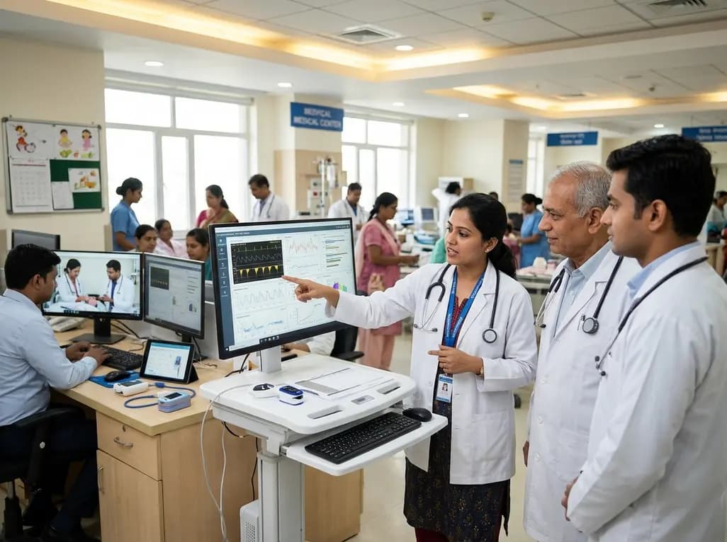
A landmark study details how an enterprise pediatric remote patient management model expanded from a single cardiology pilot to 16 diverse specialty programs by combining centralized digital operations with specialty clinical leadership.
Yesterday
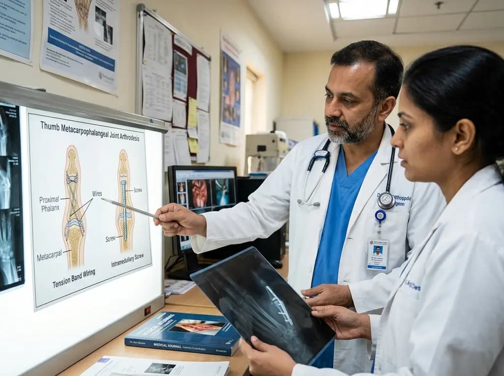
A comparative review of tension band wiring and intramedullary screw fixation in thumb metacarpophalangeal arthrodesis, evaluating union rates, hardware complications, and clinical outcomes.
Yesterday

A breakthrough ultrathin multimodal sensor decouples physical hemodynamic monitoring from disposable electrochemical calibration, achieving long-term wearable glucose prediction with 81.36% accuracy and a 9.12% MARD. This offers a robust, drift-free solution for outpatient metabolic surveillance.
Yesterday
