

Loading, please wait...
Loading, please wait...
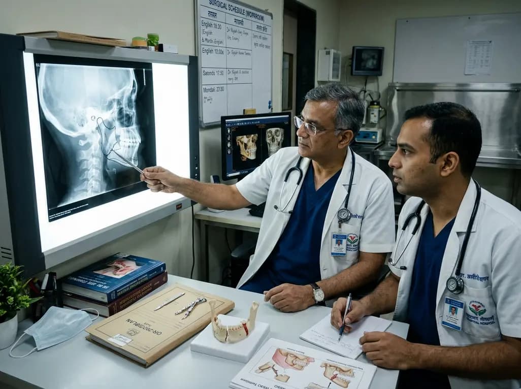
Orthognathic surgery corrects severe dentofacial discrepancies and restores harmonious masticatory function. Among mandibular interventions, bilateral sagittal split osteotomy serves as the premier surgical technique for correcting skeletal Class II retrognathia. A recent randomized clinical trial compared this conventional method with the anterior oblique osteotomy, evaluating their effects on bone contact area.
Surgical modifications of mandibular ramus procedures aim to maximize operative safety and bone healing. The traditional sagittal split osteotomy utilizes the classic Dal Pont modification refined by Epker. In this technique, the surgeon places a horizontal medial cut above the lingula and extends it along the anterior ramus border to the inferior mandibular margin. In contrast, the anterior oblique osteotomy modifies the ramus cut trajectory to alter split dynamics and minimize lingual soft tissue detachment.
Furthermore, clinicians created this oblique modification to protect the inferior alveolar neurovascular bundle during mobilization. However, altering cut angulations substantially changes how separated cortical surfaces align after advancement. When clinicians advance the distal tooth-bearing segment, proximal condylar stability relies entirely on the quality of intersegmental bony apposition. Therefore, surgeons must understand how distinct osteotomy designs influence osseous contact geometry. The double-blind trial analyzed 104 osteotomies across 52 skeletal Class II patients to evaluate these two techniques objectively. Consequently, their observations clarify essential biomechanical differences between conventional and modified ramal splits.
During mandibular advancement, fracture propagation along the medial cortex determines overall structural integrity. To classify lingual fracture lines accurately, researchers utilized the Lingual Split Scale developed by Plooij in 2009. This three-dimensional classification identifies four separate split configurations based on their anatomical relation to the mandibular foramen. A Type I split runs horizontally above the lingula toward the posterior border, representing an optimal cleavage path. In contrast, a Type II split descends behind the lingula toward the inferior ramal border.
Additionally, Type III splits propagate through the mandibular canal, exposing the inferior alveolar nerve to potential injury. Type IV fractures represent unfavorable comminution with severe cortical fragmentation. In this clinical trial, three-dimensional cone-beam imaging revealed notable variations between the comparative groups. The conventional osteotomy maintained predictable cleavage lines that favored structural preservation. Conversely, the anterior oblique design produced altered cortical separation planes. Thus, classifying lingual split patterns allows surgical teams to anticipate hardware placement needs and avoid inadvertent neurovascular entrapment.
Skeletal stability after orthognathic surgery requires intimate cortical contact between bone segments. When surgeons advance the distal mandible, spatial gaps inevitably appear along portions of the split interface. Therefore, maximizing bone contact area is critical for rapid revascularization and primary osteogenesis. The trial investigators utilized Dolphin Imaging software to quantify point B advancement and volumetric bone contact area across all osteotomy sites.
Importantly, the conventional sagittal split cohort demonstrated a significantly greater bone contact area than the anterior oblique group. Although both cohorts achieved equivalent linear advancements at point B, their surface interface measurements diverged substantially. The Dal Pont-Epker technique maintained broader bone overlap along the buccal and inferior borders. In contrast, the anterior oblique cut produced steeper interface angles that reduced mutual bony apposition. Consequently, the traditional technique provides a more substantial osseous substrate for rigid fixation. Furthermore, larger surface contact reduces dead space, accelerating secondary remodelling and ensuring robust osteosynthesis across the split.
Mandibular advancement in skeletal Class II malocclusion introduces considerable biomechanical challenges during bone healing. Forward repositioning of the distal segment increases tensile loads across the osteotomy site during mastication. If bone contact remains minimal, osteosynthesis hardware must withstand excessive bending forces without biological support. Consequently, compromised contact increases the clinical risk of plate fatigue, screw loosening, or skeletal relapse.
Furthermore, direct bone-to-bone apposition facilitates physiological load sharing between cortical plates and titanium hardware. Because the conventional sagittal technique yields a larger bone contact area, it disperses functional stresses more evenly across the healing interface. In contrast, limited contact areas generate localized stress concentrations that can impede osteoblast migration. Additionally, preserving extensive contact reduces the need for autogenous bone grafts in moderate to severe advancements. Clinicians can confidently proceed with early postoperative rehabilitation and stable orthodontic finishing. Therefore, selecting an osteotomy technique with proven surface area benefits directly enhances long-term skeletal stability.
Maxillofacial surgery in India addresses a wide spectrum of complex dentofacial deformities in tertiary centers across the country. Patients often present with severe retrognathia resulting from childhood temporomandibular joint ankylosis, trauma, or developmental hypoplasia. In these challenging situations, maxillofacial surgeons require reproducible techniques that ensure predictable osteosynthesis. While the anterior oblique osteotomy presents theoretical benefits for nerve handling, its diminished bone contact requires careful clinical deliberation.
Moreover, many Indian patients travel significant distances for surgical treatment, making long-term stability and minimal revision rates crucial. The greater bone contact observed with the conventional sagittal split osteotomy provides an added margin of biological safety. Furthermore, training curriculums in Indian institutions emphasize standardized osteotomy designs that junior surgeons can execute with confidence. Combining thorough virtual surgical planning with the classic Epker modification optimizes patient outcomes across diverse clinical settings. Ultimately, these evidence-based insights guide maxillofacial surgeons in selecting osteotomy configurations that maximize structural stability and minimize complications.
The conventional sagittal split osteotomy, modified by Dal Pont and Epker, employs a horizontal medial cut above the lingula extending along the external oblique ridge. Conversely, the anterior oblique osteotomy utilizes an oblique cut pattern designed to modify split vectors and reduce lingual manipulation. Although both approaches achieve successful mandibular movement, the conventional technique consistently generates a substantially broader bone contact surface between proximal and distal mandibular segments.
Adequate bone contact area ensures robust biological healing and enhances primary osseous union across the osteotomy gap. In addition, an expansive bony interface distributes occlusal and masticatory forces effectively, thereby reducing mechanical strain on internal fixation miniplates and monocortical screws. Consequently, optimizing bone contact significantly lowers the clinical incidence of postoperative skeletal relapse, nonunion, and hardware failure, particularly when surgeons perform large forward advancements in Class II retrognathic patients.
The Lingual Split Scale categorizes lingual fracture patterns into four distinct anatomical types based on post-split trajectory relative to the mandibular canal. By evaluating these patterns on three-dimensional computed tomography, clinicians can objectively identify whether the cleavage line preserved neurovascular structures or caused cortical comminution. Therefore, understanding these fracture types helps maxillofacial surgeons refine their osteotomy cuts, prevent unfavorable bad splits, and select ideal fixation configurations for individual mandibular anatomies.
Disclaimer: This content is for informational and educational purposes only... Refer to the latest local and national guidelines for clinical practice.
References

Read summarized clinical updates, watch expert medical content, and earn CME certifications right from your smartphone.
Open your smartphone camera to scan and install.





A double-blind RCT reveals that conventional sagittal split osteotomy yields significantly greater bone contact area than anterior oblique osteotomy during mandibular advancement, offering critical stability insights for maxillofacial surgeons.
Today

Maharashtra has launched a comprehensive initiative to halve anaemia prevalence across high-risk groups by 2030. The public health strategy integrates aggressive screening, point-of-care diagnostics, parenteral iron therapies, and targeted interventions for nutritional deficits and underlying hemoglobinopathies.
Today
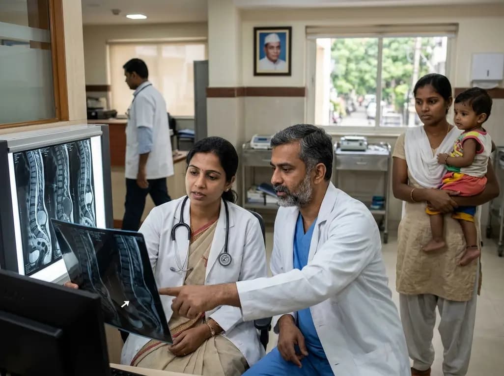
A rare presentation of late myelomeningocele sac rupture devoid of a neural placode demonstrates excellent 2-year functional outcomes, shedding light on the mechanical, chemical, and hydrostatic dynamics of congenital spinal dysraphism.
Today

Dynamic contrast 4DCT enables precise mapping of left atrial appendage blood transport in atrial fibrillation. By quantifying regional stasis, wash-in kinetics, and residence time, this imaging advance offers clinician-tailored thromboembolic risk stratification beyond standard clinical scores.
Today
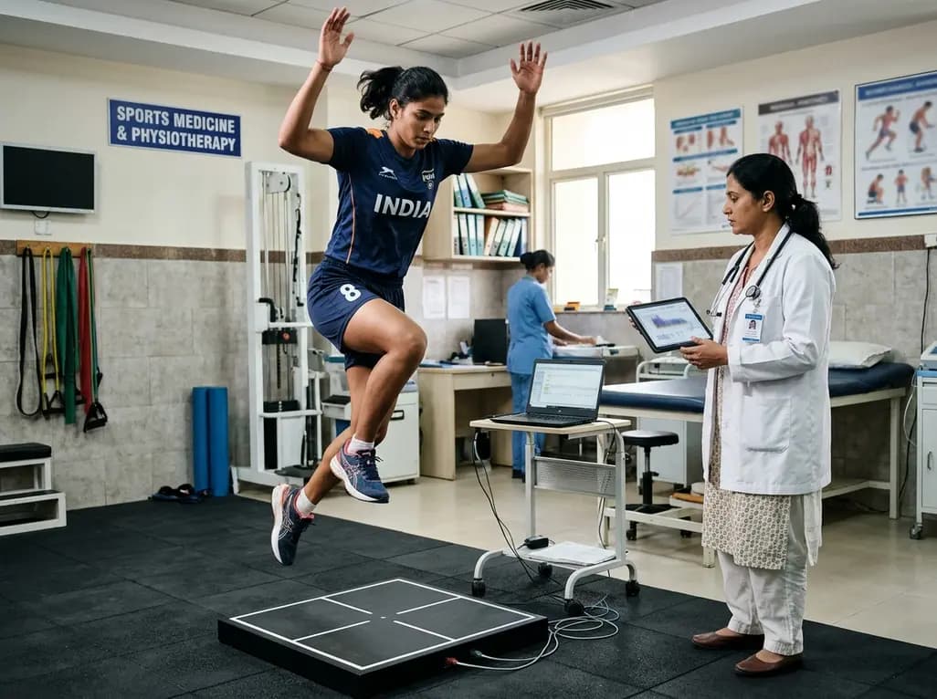
A systematic review and meta-analysis highlights the substantial impact of plyometric jump training on jump performance, reactive strength, and landing kinematics in female team-sport athletes, providing evidence-based programming guidelines for sports medicine and orthopedics.
Today
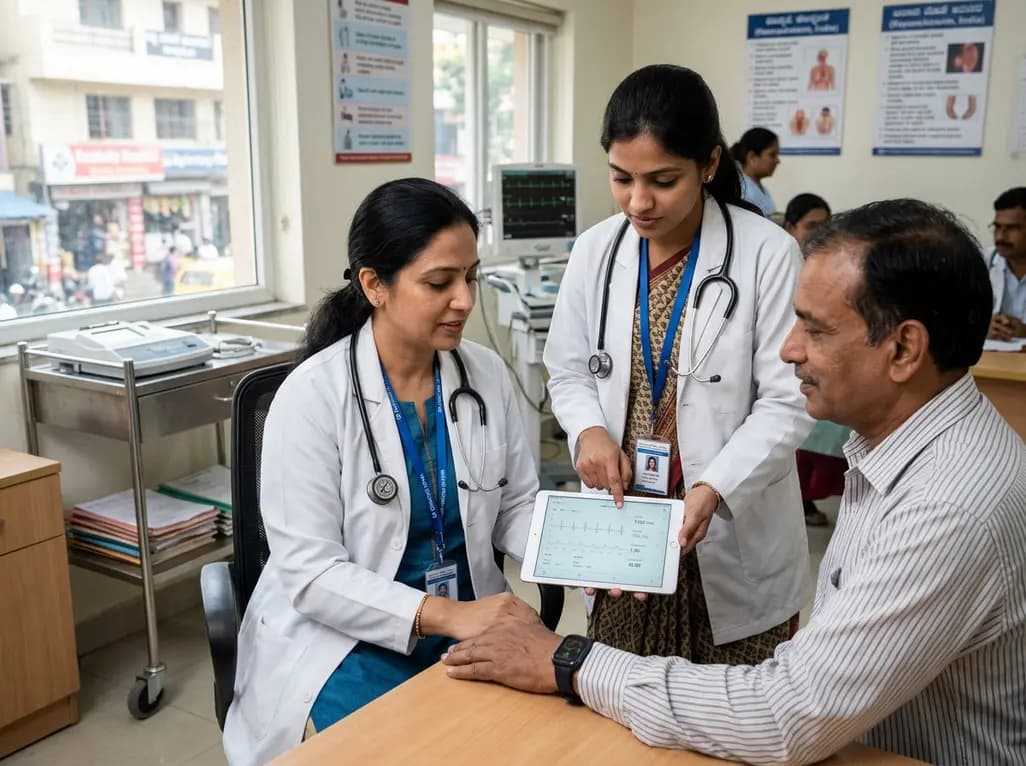
India is poised to transform chronic disease management through wearable health technology. Discover how continuous remote monitoring and predictive algorithms empower clinicians to detect early cardiovascular signals, personalize metabolic interventions, and deliver equitable preventive care across populations.
Today
