.webp&w=384&q=75)

Loading, please wait...
Loading, please wait...
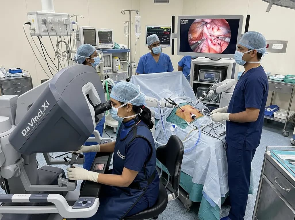
Adult congenital heart disease management continues to evolve rapidly with minimally invasive innovations. Specifically, robotic partial AVSD repair represents a major milestone in modern cardiac surgery. Partial atrioventricular septal defect combines an ostium primum atrial defect with left atrioventricular valve clefts. While conventional median sternotomy remains standard, endoscopic robotic platforms now deliver unmatched surgical visualization. Consequently, surgeons can execute intricate leaflet reconstruction and septal defect closure through keyhole incisions. This approach significantly shortens recovery times and optimizes cosmetic satisfaction for adult patients.
Partial atrioventricular septal defect presents unique anatomical challenges in adult patients. The malformation typically comprises a low-lying ostium primum defect and a zone of apposition, commonly termed a cleft, within the anterior mitral leaflet. Over time, left-to-right interatrial shunting causes right ventricular volume overload and progressive pulmonary vascular remodeling. Concurrently, mitral regurgitation exacerbates left atrial enlargement and promotes atrial arrhythmias. Therefore, timely surgical intervention is necessary to preserve long-term biventricular function.
Traditionally, surgeons corrected these defects through full median sternotomy. However, modern robotic technology allows skilled surgical teams to address complex intracardiac anomalies with minimal tissue disruption. Ideal candidates include adult patients with symptomatic left-to-right shunts, significant left atrioventricular valve regurgitation, and suitable peripheral vascular anatomy for femoral cannulation. Conversely, severe peripheral arterial disease or prior right thoracotomy represents important contraindications. By integrating high-resolution visualization with dexterous robotic instruments, surgeons can comprehensively evaluate the valvular apparatus and achieve durable anatomical correction.
Successful robotic cardiac procedures require rigorous positioning and standardized perfusion strategies. First, the surgical team places the patient in a supine position with slight elevation of the right hemithorax. Transesophageal echocardiography continuously guides line placement and monitors cardiac de-airing. Peripheral cardiopulmonary bypass is established through femoral arterial and venous cannulation under direct visual guidance.
Additionally, achieving complete bicaval isolation is critical for maintaining a bloodless intracardiac field. Perfusionists advance a superior vena cava cannula through the internal jugular or femoral vein. Surgeons achieve bicaval occlusion using endovascular balloons or robotic tourniquets inside the pericardium. Next, the team inserts robotic trocars through right thoracic intercostal spaces without rib spreading. A specialized transthoracic aortic cross-clamp, such as the Chitwood clamp, occludes the ascending aorta. Cold blood cardioplegia induces prompt diastolic cardiac arrest, ensuring optimal myocardial protection. Consequently, this meticulous setup guarantees complete circulatory control before atriotomy.
Intracardiac exposure represents a decisive step in adult congenital defect repair. Following cardiac arrest, the surgeon performs a right atriotomy or direct left atriotomy to visualize the anatomy. High-definition stereoscopic cameras provide incredible magnification of the delicate interatrial septum and conduction pathways. The surgeon clearly identifies the margins of the ostium primum atrial septal defect and the coronary sinus.
To close the septal communication, surgeons routinely select a glutaraldehyde-treated bovine pericardial patch. This material provides excellent handling characteristics, durability, and low thrombogenicity. The surgeon fixes the patch along the defect margins using continuous polypropylene sutures. During suturing near the atrioventricular node, the surgeon places superficial bites to avoid complete heart block. Specifically, the line of closure navigates carefully along the crest of the ventricular septum. Moreover, robotic instruments allow exceptional needle angles that prevent distortion of adjacent structures. Once secured, the surgeon confirms complete defect obliteration without residual shunt.
Mitral valve repair in partial AVSD requires meticulous reconstruction of the cleft and surrounding leaflets. The robotic console offers seven degrees of freedom, enabling precise micro-suturing inside the left atrium. First, the surgeon performs leaflet shaving to remove fibrous thickening along coaptation edges, restoring necessary leaflet pliability.
Subsequently, the surgeon approximates the edges of the zone of apposition using interrupted fine sutures. When tissue deficiency is pronounced, autologous pericardial augmentation helps expand the leaflet surface without generating tension. Avoiding excessive tension is vital, as overly tight cleft closure can cause iatrogenic mitral stenosis. Additionally, the surgeon inspects the subvalvular apparatus, identifying abnormal chordal attachments. To stabilize the repair and correct annular dilatation, the surgeon implants a flexible annuloplasty band. This flexible prosthesis supports annular remodeling while preserving physiological annular motion throughout the cardiac cycle. Finally, saline testing confirms excellent leaflet coaptation and trivial residual regurgitation.
Despite substantial technological advantages, robotic congenital heart repairs entail specific technical challenges. Inadequate visual exposure represents a major pitfall that can compromise accurate assessment of valvular structures. If the atrium is poorly retracted, cleft identification and chordal tension evaluation become extremely difficult. Therefore, dynamic atrial retractor placement is critical throughout every reconstructive step.
Furthermore, improper leaflet alignment during cleft approximation carries severe clinical consequences. Excessive traction can narrow the left ventricular inflow tract, causing progressive mitral stenosis. Conversely, incomplete cleft closure leads to significant residual regurgitation. Another critical risk involves conduction injuries. Because the atrioventricular conduction axis runs adjacent to the defect margin, deep suture placement can trigger permanent heart block. To mitigate this hazard, surgeons utilize shallow, precise suture trajectories along the nodal danger zone. Adhering strictly to these preventive strategies guarantees procedural safety and minimizes postoperative complications.
Clinical evidence demonstrates that totally endoscopic robotic congenital procedures provide outstanding outcomes. Patients undergoing robotic repairs experience significantly reduced surgical trauma compared to traditional sternotomy cohorts. Consequently, postoperative pain scores decrease, chest tube drainage remains minimal, and hospital stays are markedly shorter. Most patients return to full physical activities within two to three weeks.
Furthermore, avoiding a midline sternotomy prevents long-term skeletal instability and deep wound infections. Robotic approaches offer immense cosmetic and psychosocial benefits, especially for young adult patients. Longitudinal follow-up reveals stable mitral valve function, minimal recurrence of regurgitation, and preserved ventricular geometry. As robotic platforms continue to incorporate augmented reality and advanced tactile feedback, technical precision will increase further. In conclusion, robotic surgery transforms complex intracardiac repairs into safe, reproducible, and patient-centered therapies across modern surgical centers.
Robotic repair eliminates the need for median sternotomy, thereby significantly reducing surgical trauma, postoperative pain, and wound complications. Additionally, high-definition three-dimensional visualization enables precise micro-suturing of delicate valve leaflets. Consequently, patients experience minimal blood loss, shorter intensive care stays, rapid recovery, and superior cosmetic outcomes while achieving excellent long-term hemodynamic stability comparable to traditional open surgery.
Surgeons maintain optimal mitral valve exposure using dedicated robotic left atrial retractors and high-magnification stereoscopic cameras. These instruments provide exceptional direct views of the cleft and subvalvular structures through narrow access ports. Furthermore, surgeons perform delicate leaflet shaving, autologous pericardial patching, and flexible annuloplasty without applying excessive tension, which preserves natural leaflet mobility and prevents valve stenosis.
Adult patients with isolated partial atrioventricular septal defects, symptomatic left-to-right shunts, or moderate-to-severe mitral regurgitation benefit substantially from robotic intervention. Furthermore, active young adults seeking rapid recovery, early return to work, and minimal thoracic scarring represent ideal candidates. Patients must possess suitable peripheral femoral vessels to permit safe cardiopulmonary bypass cannulation and endovascular control.
Disclaimer: This content is for informational and educational purposes only. It is not intended to be a substitute for professional medical advice, diagnosis, or treatment. Always seek the advice of your physician or other qualified health provider with any questions you may have regarding a medical condition. Refer to the latest local and national guidelines for clinical practice.
References
Mazinani N et al. Totally Endoscopic Robotic Repair of Partial Atrioventricular Septal Defect. JACC Case Rep. 2026 Aug 25. doi: undefined. PMID: 42640240.
Gao C, Yang M, Xiao C, et al. Totally robotic repair of atrioventricular septal defect in the adult. J Cardiothorac Surg. 2015;10:153.
Tang Y, Wu Y, Zhu J, et al. Total endoscopic repair of atrial septal defect under on-pump beating heart. J Thorac Dis. 2018;10(12):6560-6566.

Read summarized clinical updates, watch expert medical content, and earn CME certifications right from your smartphone.
Open your smartphone camera to scan and install.





Explore the surgical technique of totally endoscopic robotic repair of partial atrioventricular septal defect with complex mitral valve reconstruction in adult patients.
last month

A meta-analysis demonstrates that Lille score-guided corticosteroid therapy significantly improves 28- and 90-day survival in severe alcohol-associated hepatitis compared to non-guided regimens, preventing fatal infections in non-responders.
Today

A cross-sectional study of over 33 million US live births from 2016 through 2024 reveals a 74% increase in hypertensive disorders of pregnancy. This clinical overview explores demographic drivers, maternal-fetal outcomes, and essential preventive strategies for obstetric practice.
Today

The phase II DAPA-AIC trial demonstrates that dapagliflozin 10 mg daily significantly preserves left ventricular ejection fraction compared to placebo over four months in adults undergoing anthracycline chemotherapy, highlighting a potential preventive strategy in cardio-oncology.
Today

A scoping review reveals severe gaps in long-term assistive technology provision following humanitarian emergencies. Addressing post-disaster rehabilitation requires systematic follow-up, workforce training, and integration into national health systems to sustain mobility and recovery.
Today

An interim propensity score-matched study comparing SASI vs OAGB demonstrates equivalent short- and mid-term outcomes. Both bariatric procedures achieved substantial excess weight loss and 100% medication-free blood glucose control at 18 months, underscoring the metabolic efficacy of both techniques.
Today
