

Loading, please wait...
Loading, please wait...
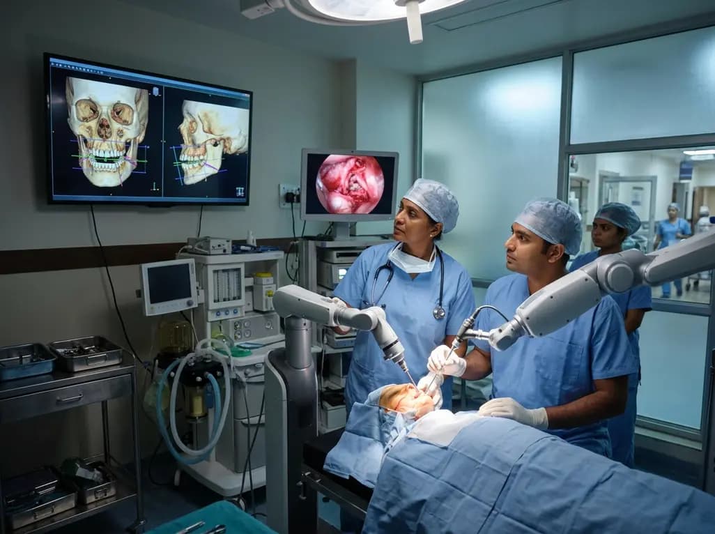
Orthognathic surgery restores facial harmony, optimal masticatory function, and airway stability in patients with severe dentofacial deformities. Over the past two decades, clinicians have transitioned from traditional mechanical dental articulators and manual cast surgery to three-dimensional virtual surgical planning. More recently, robotic orthognathic surgery has emerged as an innovative technological frontier that builds upon these digital planning foundations. Maxillofacial surgeons increasingly evaluate whether robotic platforms can translate virtual plans into surgical reality with superior precision. Although computer-aided design and three-dimensional splints have established reliable accuracy, robotic assistance promises dynamic control, robotic bone repositioning, and guided osteotomies. Consequently, surgeons must critically appraise the current scope of evidence, assessing technical performance, procedural safety, and pragmatic clinical feasibility.
Historically, orthognathic correction relied heavily on manual model surgery and acrylic occlusal splints. While these conventional methods offered functional correction, they frequently suffered from laboratory transfer errors and geometric discrepancies. Therefore, the introduction of cone-beam computed tomography and computer-assisted design substantially modernized craniofacial surgical planning. Virtual surgical planning allowed surgeons to simulate complex Le Fort I osteotomies and bilateral sagittal split osteotomies with remarkable detail. Furthermore, surgical navigation and patient-specific cutting guides significantly improved the transfer of digital simulations into the operative theater. However, manual positioning remained vulnerable to soft tissue resistance, intraoperative tool deflection, and unpredictable condylar torque during rigid fixation. To resolve these persistent mechanical challenges, researchers developed early passive robotic instruments and tracking systems. Initially, these devices merely guided bone positioning by tracking rigid optical markers attached to anatomical landmarks. Over time, investigators integrated active robotic manipulators equipped with multi-axis articulated arms and real-time navigation. Consequently, modern platforms can actively guide bone saws, position osteotomized maxillomandibular complexes, and verify spatial coordinates dynamically. This evolution marks a pivotal shift from static surgical guides toward intelligent, interactive surgical execution.
Contemporary research categorizes robotic applications into three main architectures: passive, semi-active, and autonomous systems. In passive systems, the surgeon retains complete manual control while the robotic arm acts as a rigid, spatial reference guide. In semi-active configurations, collaborative robotic arms restrict the surgeon's movements within predetermined safe anatomical boundaries using virtual fixtures and haptic feedback. For instance, the system prevents the osteotome from penetrating beyond pre-calculated depths, thereby protecting critical neurovascular bundles such as the inferior alveolar nerve. Conversely, fully task-autonomous prototypes execute specific linear osteotomies or drill sequences under continuous human supervision. Functionally, robotic platforms in orthognathic surgery execute two primary tasks: automated bone resection and fragment repositioning. For osteotomies, robotic arms hold oscillating saws, piezoelectric inserts, or cold ablative lasers to perform precise Le Fort I or ramus osteotomies. In fragment repositioning, robotic end-effectors physically grasp the osteotomized maxilla or mandible. They then move the bone segments through space to match three-dimensional coordinates established during virtual surgical planning. Moreover, advanced registration algorithms align preoperative imaging with intraoperative patient anatomy using optical tracking cameras and surface scanning. These combined mechanisms establish a reproducible bridge between virtual models and actual osseous movements.
A core objective of robotic integration is achieving sub-millimeter geometric accuracy during jaw realignment. Multiple preclinical investigations demonstrate that robotic systems achieve exceptional fidelity between preoperative virtual plans and final skeletal positions. Specifically, phantom and cadaveric trials evaluating robotic maxillary repositioning consistently report absolute spatial deviations well below one millimeter across translational axes. Similarly, rotational discrepancies along yaw, pitch, and roll frequently stay within acceptable thresholds of less than one degree. Controlled osteotomies guided by robotic manipulators exhibit significantly reduced linear and angular errors compared to freehand manual techniques. In addition, robotic guidance eliminates human tremor and minimizes unintended angular deviation during difficult ramus osteotomies. When compared with patient-specific cutting guides and 3D-printed intermediate splints, robotic platforms achieve comparable, and in specific experimental parameters, superior geometric precision. Nevertheless, the clinical advantage remains modest. Established digital workflows utilizing patient-specific implants already provide high clinical accuracy, often achieving translational discrepancies below 1.5 millimeters. Therefore, robotic platforms currently demonstrate equivalence to mature digital protocols rather than overwhelming superiority. While robotic arms reliably position rigid bone segments in laboratory simulations, biological factors such as soft tissue forces and muscle tone introduce unpredictable clinical resistance.
Despite encouraging engineering breakthroughs, robotic orthognathic surgery remains largely confined to experimental laboratories. Most published investigations rely entirely on synthetic sawbones, phantom models, and cadaveric heads. Clinical human trials are sparse, consisting primarily of single-case reports and small pilot feasibility series. Consequently, the literature lacks robust prospective comparative trials evaluating long-term functional and aesthetic outcomes. Several critical barriers impede rapid clinical translation. First, maxillofacial anatomy presents a highly constrained surgical corridor surrounded by vital vessels, cranial nerves, and dental roots. Bulky robotic arms and complex tracking frames can overcrowd the surgical field, impeding line-of-sight visibility and instrument manipulation. Second, intraoperative registration remains vulnerable to line-of-sight interruption and tracking marker displacement, which can degrade spatial accuracy. Furthermore, current platforms do not completely account for dynamic condylar sag, temporomandibular joint biomechanics, and soft-tissue rebound during skeletal fixation. Robotic systems also demand extensive operative setup times, steep technical learning curves, and significant capital expenditure. Finally, the absence of standardized reporting metrics across studies complicates meta-analytic evaluation. Until high-powered clinical trials validate procedural efficiency, complication rates, and long-term skeletal stability, robotics cannot routinely replace established virtual surgical planning workflows.
In India, the demand for orthognathic surgery is expanding rapidly due to rising aesthetic awareness and high prevalence of craniofacial malformations. Indian maxillofacial and plastic surgery centers have widely embraced digital virtual surgical planning, in-house three-dimensional printing, and custom surgical guides. These existing digital technologies offer highly cost-effective and accurate outcomes for diverse patient demographics. Introducing multi-million-rupee robotic suites, therefore, poses major economic and infrastructural hurdles. Public and private healthcare institutions must justify capital investments and maintenance contracts against real clinical dividends. However, tier-one academic institutes and premier tertiary surgical centers in India possess the surgical volume and engineering partnerships to pioneer this field. In high-volume centers, robotic platforms could serve as specialized adjuncts for intricate multi-piece maxillary asymmetries, syndromic craniosynostosis corrections, and severe hemifacial microsomia. In addition, local robotic development initiatives and collaborative healthcare technology programs could produce affordable, compact maxillofacial robotic solutions tailored for Indian operating theaters. Surgeons must balance clinical curiosity with rigorous evidence-based adoption. By prioritizing cost efficiency, surgical team training, and local clinical registries, Indian surgeons can strategically integrate robotic technologies while ensuring patient safety and healthcare affordability.
The future of robotic jaw surgery lies in the seamless convergence of robotics, artificial intelligence, and augmented reality. Rather than relying on rigid mechanical guides, future robotic platforms will incorporate real-time intraoperative imaging, such as cone-beam computed tomography and optical coherence tomography. Machine learning algorithms will analyze bone density variations in real time, enabling adaptive robotic force modulation during osteotomies to prevent osseous thermal necrosis and tool breakage. Furthermore, integrating augmented reality headsets with robotic consoles will allow surgeons to visualize hidden vascular channels and virtual osteotomy lines superimposed directly onto the patient's face. Haptic feedback mechanisms will also provide surgeons with tactile resistance cues, bridging the gap between autonomous precision and human tactile intuition. Collaborative robotics will become more compact, mountable directly onto the operating table or the patient's calvarium to eliminate line-of-sight constraints. Additionally, surgical societies must establish international multi-center registries to monitor complication rates, skeletal relapse, and nerve recovery over extended follow-up intervals. As these technologies mature, robotics will evolve from task-specific adjuncts into indispensable, comprehensive surgical partners in advanced maxillofacial reconstruction.
Robotic orthognathic surgery enhances precision by directly executing preprogrammed bone cuts and fragment repositions with sub-millimeter geometric accuracy. While conventional virtual planning relies on manual execution guided by static acrylic splints, robotic arms eliminate human hand tremor, prevent tool deflection, and restrict osteotomies within pre-calculated safety zones. Consequently, robotic systems reliably minimize translational and rotational transfer errors, ensuring that the postoperative skeletal alignment matches the digital simulation with high fidelity and reproducibility.
The primary obstacles include high financial costs, complex operating room logistics, and prolonged surgical setup times. Furthermore, the confined anatomy of the oral cavity restricts the movement of bulky robotic arms and tracking arrays. Current robotic platforms also struggle to compensate for unpredictable soft-tissue tension and dynamic temporomandibular joint displacement during rigid fixation. Finally, the lack of long-term prospective comparative clinical trials prevents surgeons from justifying routine clinical adoption over cheaper, proven digital guides.
Currently, robotic systems cannot independently perform complete orthognathic osteotomies in routine human surgery. Although autonomous cutting prototypes have demonstrated high geometric accuracy on phantom and animal models, live clinical procedures require direct surgeon control. Present systems operate in collaborative or semi-active modes, where the robot stabilizes instruments within virtual safety boundaries while the operating surgeon maintains oversight. This supervisory structure ensures absolute patient safety and prevents catastrophic damage to delicate adjacent neurovascular structures.
Disclaimer: This content is for informational and educational purposes only... Refer to the latest local and national guidelines for clinical practice.
References

Read summarized clinical updates, watch expert medical content, and earn CME certifications right from your smartphone.
Open your smartphone camera to scan and install.





A comprehensive scoping review evaluates robotic applications in orthognathic surgery, highlighting advances in osteotomies and bone positioning, clinical translation barriers, and surgical accuracy.
Today

Union Minister Pratap Jadhav highlights the pivotal role of pharmacists in fighting antibiotic misuse and antimicrobial resistance. By championing rational medicine use, patient counseling, and curriculum modernization, community pharmacists stand as vital guardians of public health across India.
Today
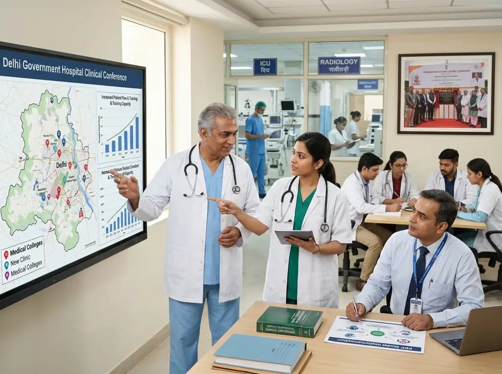
Delhi is undergoing a major healthcare overhaul with enhanced Ayushman Bharat insurance up to ₹10 lakh, increased MBBS and PG medical training seats, expanded primary network mandirs, new tertiary neuro-ICUs, and public-private reproductive technology initiatives to build a resilient, preventative healthcare model.
Today
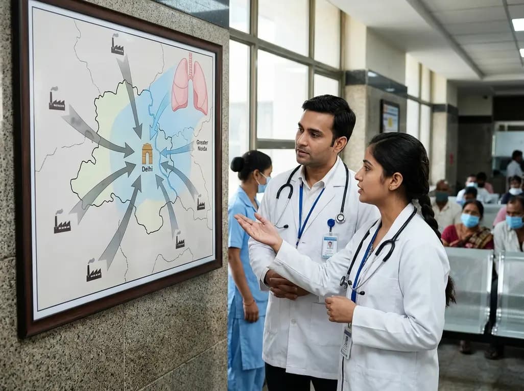
A landmark NIAS study reveals that regional airshed interventions can slash northern India's fine particulate levels by up to 60%. Discover the profound clinical implications of targeted emission curbs on cardiopulmonary health, chronic respiratory diseases, and population-level preventative strategies.
Today
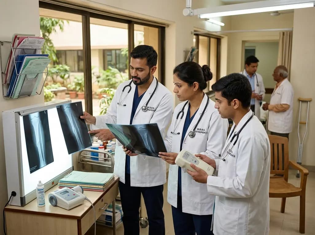
Non-union fractures occur in 2-5% of cases, causing significant disability and cost. Conservative modalities such as LIPUS, PEMF, and focused ESWT offer non-surgical healing options for defects up to 5-10 mm when adequate mechanical stability exists without infection.
Today
A Brazilian birth cohort reveals significant sex-specific associations between preterm birth and adolescent adiposity, underscoring the urgent need for targeted long-term cardiometabolic surveillance and tailored clinical monitoring.
Today
