

Loading, please wait...
Loading, please wait...

Open-heart surgery corrects structural defects in infants with congenital heart disease, but neurological complications remain a major concern. Notably, postoperative new brain injury occurs in a substantial proportion of young patients undergoing cardiopulmonary bypass. Understanding the underlying drivers of these cerebral insults enables clinical teams to implement tailored protective protocols. Consequently, identifying both modifiable and fixed determinants transforms surgical risk assessment and improves long-term neurodevelopmental outcomes.
Congenital heart disease alters fetal and neonatal cerebral hemodynamics, which predisposes vulnerable neural tissue to ischemic or hemorrhagic insults. In this cohort study of 172 infants and toddlers, 35.5% developed new brain lesions after corrective surgery. These acute postoperative injuries include white matter injury, cerebral infarction, intracranial hemorrhage, and cerebral venous thrombosis. Magnetic resonance imaging provides the gold standard modality to detect these subtle parenchymal abnormalities. Furthermore, early detection remains critical because subclinical injuries often escape immediate bedside diagnosis. In addition, white matter injury occurs frequently in this cohort due to the intrinsic vulnerability of developing pre-oligodendrocytes. These cells show exceptional susceptibility to oxidative stress and transient hypoperfusion. Therefore, surgical teams must anticipate neurological risks before initiating cardiopulmonary bypass. While modern surgical techniques achieve excellent survival rates, neurological morbidity impairs future cognitive, motor, and linguistic development. Pediatric specialists must actively monitor these children through standardized imaging before and after cardiopulmonary intervention. Ultimately, recognizing this significant 35.5% incidence highlights the urgent need for targeted neuroprotection during pediatric open-heart procedures. Consequently, multidisciplinary teams must unite to identify high-risk candidates prior to surgical exposure and tailor their intraoperative support accordingly.
Multivariable logistic regression reveals that prolonged cardiopulmonary bypass substantially increases the likelihood of brain injury. Specifically, each additional minute on bypass elevates the risk by nearly three percent. Extended pump duration triggers systemic inflammatory cascades, creates microemboli, and compromises dynamic cerebral autoregulation. Consequently, prolonged bypass times exacerbate microvascular hypoperfusion during critical phases of operative repair. In addition, anatomical anomalies directly influence this susceptibility. A smaller main pulmonary artery diameter emerged as an independent predictor of adverse cerebral events. Patients presenting with smaller pulmonary arteries often have complex hemodynamics, hypoplastic pulmonary vascular beds, or reduced systemic-to-pulmonary collaterals. Consequently, these structural limitations reduce baseline oxygen reserve and complicate intraoperative cannula placement. Furthermore, restricted arterial anatomy frequently necessitates protracted surgical dissection and extended circulatory support. Thus, surgeons encounter challenging technical demands that indirectly elevate cardiopulmonary bypass time. Surgical teams should therefore prioritize streamlined operative strategies that minimize total bypass exposure. Moreover, preoperative echocardiography and advanced vascular mapping help cardiologists anticipate anatomical hurdles. By recognizing the additive risk of small vascular dimensions and lengthy pump runs, clinicians can refine perfusion protocols to protect cerebral autoregulation.
Neurological vulnerability in pediatric patients does not follow a uniform trajectory across all diagnostic groups. Instead, multivariable analysis demonstrates that risk varies according to patient age and cyanotic status. In the overall cohort, older age independently increased injury odds, reflecting accumulated hemodynamic stress and complex late repairs. However, stratified subgroup analyses uncovered distinct, age-cyanosis interactions. Among noncyanotic infants under one year of age, younger age actually amplified neurological risk. In this infant cohort, developing neural substrates exhibit profound sensitivity to hemodynamic instability and immature cerebrovascular autoregulation. Conversely, in noncyanotic children older than one year, prolonged bypass duration represented the dominant independent determinant of parenchymal damage. Cyanotic children, meanwhile, demonstrated unique pathophysiological patterns with borderline age and sex effects. Chronic cyanosis induces cerebral capillary proliferation and altered hematocrit, which modifies blood viscosity and oxygen delivery. Therefore, chronic hypoxemia primes the central nervous system differently than acyanotic cardiac lesions. As a result, clinicians cannot adopt a single neuroprotective algorithm for all pediatric patients. Rather, teams must stratify monitoring protocols based on whether the infant has cyanotic anatomy and whether surgery occurs during early infancy.
The study highlights two critical hematological predictors: lower postoperative platelet counts and lower mean corpuscular hemoglobin concentration. Both factors significantly correlated with increased brain injury odds. Cardiopulmonary bypass circuits consume platelets and dilute clotting factors through hemodilution and mechanical sheer stress. Consequently, profound thrombocytopenia impairs primary hemostasis and promotes microvascular intracranial hemorrhage. Furthermore, dysfunctional platelet aggregation destabilizes the blood-brain barrier under conditions of altered perfusion. Lower postoperative mean corpuscular hemoglobin concentration similarly compounds cerebral ischemia. Mean corpuscular hemoglobin concentration reflects intra-erythrocytic hemoglobin density, which directly governs oxygen-carrying capacity. When this index declines, oxygen unloading at the microcapillary level becomes inefficient. Consequently, vulnerable cerebral territories experience tissue hypoxia despite acceptable arterial saturation readings. In noncyanotic infants under one year, this hematological vulnerability proved particularly pronounced. Pediatric perfusionists and intensivists must therefore monitor red blood cell indices and coagulation parameters with rigorous precision. Maintaining adequate platelet levels and optimizing red blood cell quality during hemodilution minimizes ischemic and hemorrhagic events. Therefore, aggressive hematological optimization represents an actionable, highly modifiable pathway for clinical teams aiming to preserve neurodevelopmental integrity.
Translating these prognostic indicators into practical clinical workflows offers an effective means of safeguarding neurodevelopment. Because cardiopulmonary bypass duration and hematological derangements are modifiable, perioperative teams can intervene proactively. First, perfusionists should implement customized prime volumes and ultrafiltration techniques to prevent excessive hemodilution and platelet loss. In addition, near-infrared spectroscopy provides continuous noninvasive assessment of regional cerebral oxygen saturation. When cerebral desaturation occurs, clinicians can promptly adjust pump flow, mean arterial pressure, or arterial blood gas targets. Furthermore, anesthesia protocols should emphasize cerebral metabolic suppression and temperature management during critical stages of cardiopulmonary bypass. Preventing hyperthermia during rewarming reduces secondary ischemic injury and free radical generation. In the intensive care unit, clinicians must maintain tight glycemic control and preserve cerebral perfusion pressure. Managing mechanical ventilation to prevent hypocapnia avoids undesirable cerebral vasoconstriction. Moreover, standardized postoperative neuroimaging protocols facilitate rapid identification of subclinical lesions, prompting prompt medical or antithrombotic management. Through synchronized collaboration among cardiac surgeons, pediatric intensivists, neurologists, and perfusionists, surgical centers can substantially reduce the burden of perioperative brain lesions.
Postoperative new brain injury results from a complex combination of prolonged cardiopulmonary bypass exposure, microembolism, ischemia-reperfusion injury, and impaired cerebral autoregulation. In addition, severe hemodilution, acute platelet consumption, and decreased hemoglobin concentration compromise microvascular oxygen delivery. Together, these intraoperative stressors damage immature neural tissues and cause white matter injury, stroke, or hemorrhage.
Extended cardiopulmonary bypass duration increases the generation of gaseous and particulate microemboli while activating systemic inflammatory pathways. Consequently, prolonged perfusion damages endothelial integrity and alters cerebral autoregulation. Minimizing bypass time through streamlined surgical techniques directly reduces cellular ischemic stress and preserves microvascular perfusion, thereby lowering the probability of postoperative neurological lesions.
Platelets maintain microvascular seal integrity and prevent intracranial bleeding during postoperative coagulopathy, meaning that severe thrombocytopenia increases hemorrhage risks. Meanwhile, mean corpuscular hemoglobin concentration reflects cellular hemoglobin density. A lower concentration reduces oxygen carrying capacity and capillary tissue oxygen delivery, exacerbating ischemic injury across sensitive white matter regions during hemodynamic transitions.
Disclaimer: This content is for informational and educational purposes only and does not constitute medical advice, diagnosis, or treatment. Healthcare professionals must rely on their clinical judgment and confirm all clinical findings with current evidence and institutional protocols. Treatments, dosages, and procedures should follow approved local product labels and guidelines. The authors and publishers are not liable for any injuries, losses, or damages arising from the use or misuse of this information. Refer to the latest local and national guidelines for clinical practice.
References

Read summarized clinical updates, watch expert medical content, and earn CME certifications right from your smartphone.
Open your smartphone camera to scan and install.





Postoperative new brain injury affects 35.5% of infants and toddlers undergoing cardiac surgery. Key independent predictors include bypass duration, smaller pulmonary artery diameter, low platelets, and low MCHC, revealing vital targets for age- and cyanosis-stratified neuroprotection.
Today

Ambient digital scribes promise effortless medical documentation, but a critical sociotechnical commentary reveals that AI alters rather than eliminates charting work. Discover how shifting cognitive burdens, oversight risks, and workflow redesign impact clinical safety and healthcare practice.
Today

A new comparative study demonstrates that intraoperative imprint cytology achieves 96.6% diagnostic accuracy in ovarian tumours, outperforming frozen section in sensitivity for borderline lesions and offering an accessible, rapid alternative for resource-constrained surgical centres.
Today

Thoracic aortic aneurysm lacks etiologic medical therapy. A new study reveals that targeting specific αV integrins with Cilengitide attenuates TGF-β and mechanotransduction pathways, effectively limiting pro-fibrotic remodeling and aortic bulb dilation in both syndromic and sporadic disease models.
Today
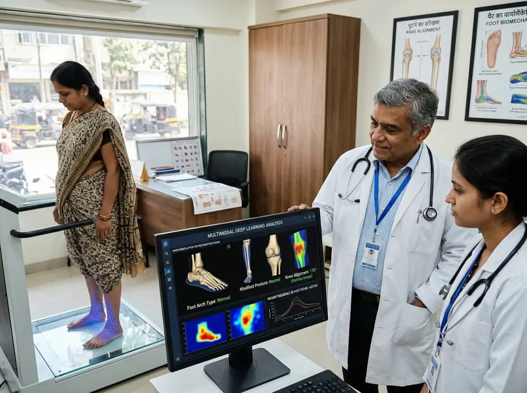
A multimodal deep learning framework utilizing weight-bearing 3D foot scans accurately screens lower-limb alignment, including arch type, hindfoot posture, and knee alignment, offering an accessible, radiation-free alternative to radiography for early biomechanical triage.
Today
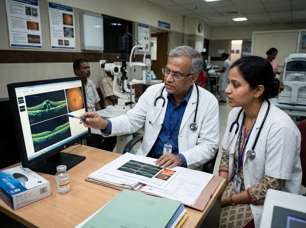
A real-world retrospective study demonstrates that combining intravitreal ranibizumab with focal/grid macular laser significantly improves visual acuity and central macular thickness compared to laser monotherapy in patients with center-involved diabetic macular edema.
Today
