

Loading, please wait...
Loading, please wait...

Evaluating suspicious adnexal masses during surgery presents a complex challenge for gynecologists and oncosurgeons. Clinicians must quickly distinguish benign cysts from invasive malignancies to determine the appropriate surgical extent. Although standard frozen section histopathology remains common, intraoperative imprint cytology provides an agile, accurate alternative. Recent clinical evidence highlights its remarkable diagnostic fidelity when compared against final paraffin histology. Therefore, surgical teams can leverage touch imprint preparations to obtain rapid microscopic assessments without relying on costly cryosection hardware.
Intraoperative decision-making profoundly influences patient survival and quality of life in ovarian neoplasia. For instance, young reproductive-age women presenting with complex adnexal cysts frequently desire fertility preservation. In these patients, an erroneous intraoperative diagnosis of malignancy triggers unnecessary bilateral salpingo-oophorectomy and premature surgical menopause. Conversely, underestimating a malignant ovarian tumor leads to incomplete cytoreductive staging. As a result, the patient faces subsequent repeat operations or suboptimal oncologic outcomes.
Surgeons traditionally depend on intraoperative frozen sections to establish immediate diagnostic clarity. However, frozen sections demand expensive cryostat instruments, stable power supply, and specialized technicians. These technical resources remain scarce in many community hospitals and district healthcare facilities across India. In contrast, imprint cytology requires only clean glass slides, gentle tissue contact, and rapid routine staining. Pathologists can review cytologic smears within ten minutes of tumor excision. Consequently, this rapid technique bridges an essential diagnostic gap during urgent gynecological procedures.
A recent tertiary hospital study by Miladinović and colleagues systematically evaluated 29 consecutive ovarian mass consultations. Pathologists reviewed both imprint cytology smears and frozen sections against final paraffin histopathology. Interestingly, intraoperative imprint cytology achieved a remarkable overall diagnostic accuracy of 96.6%, correctly classifying 28 of 29 cases. In comparison, frozen section analysis demonstrated an overall accuracy of 89.7%, correctly categorizing 26 cases.
Furthermore, statistical concordance with final histopathology was notably higher for imprint cytology than for frozen sections. Imprint cytology demonstrated near-perfect agreement with a kappa coefficient of 0.907. Meanwhile, frozen section yielded substantial agreement with a kappa coefficient of 0.704. McNemar testing revealed no statistically significant divergence in overall accuracy between the two diagnostic methods. Notably, imprint cytology achieved 100% sensitivity for benign neoplasms and borderline tumors, alongside 80% sensitivity for malignant lesions. In contrast, frozen section reached only 50% sensitivity for borderline neoplasms and 60% for malignant lesions.
Borderline ovarian tumors represent a notorious diagnostic trap during intraoperative consultations. These neoplasms frequently feature extensive cystic regions interspersed with subtle epithelial proliferations. When pathologists freeze large mucinous or serous tumors, freezing artifacts often distort cellular features and mask cellular stratification. Furthermore, tissue sampling errors during gross sectioning frequently trigger diagnostic discrepancies on frozen sections. In the Montenegro cohort, frozen sections missed half of the borderline neoplasms.
Conversely, touch imprint cytology extracts discohesive cells directly from suspicious papillae and solid mural nodules. Cellular smears highlight nuclear enlargement, hyperchromasia, and prominent nucleoli without freeze distortion. Pathologists readily recognize cellular atypia while correlating smear findings with gross examination. Similarly, in high-grade carcinomas, cytology clearly reveals marked pleomorphism, atypical mitoses, and tumor diathesis. However, cytologists must distinguish reactive mesothelial proliferations from low-grade serous neoplasms. Because cytologic preparations lack architectural depth, careful gross pathological examination remains vital during intraoperative review.
In India, access to advanced histopathology laboratories varies widely between metropolitan centers and rural districts. Many peripheral medical institutions perform complex gynecological surgeries without an on-site cryostat or histology technician. Consequently, surgeons must choose between deferred paraffin histology or transporting frozen specimens to distant laboratories. Intraoperative imprint cytology presents an accessible and transformative diagnostic solution for these resource-constrained settings.
The procedure requires minimal consumables, using standard glass slides and rapid staining solutions such as Field stain or Toluidine blue. Alternatively, rapid hematoxylin and eosin protocols yield clear diagnostic smears within eight minutes. Additionally, imprint cytology eliminates the heavy capital expense and ongoing maintenance required for microtome cryostats. Pathologists can review cytologic preparations rapidly and communicate findings directly to operating surgeons. Therefore, adopting this cost-effective technique in Indian surgical oncology centers can prevent unnecessary second-look laparotomies. Furthermore, this approach avoids both overtreatment in young women with benign cysts and undertreatment in epithelial ovarian cancers.
Despite its high accuracy, imprint cytology presents specific diagnostic limitations that surgical teams must understand. Cytology evaluates individual cell morphology but cannot directly evaluate tissue architectural patterns such as capsular breach or microinvasion. For instance, distinguishing a benign mucinous cystadenoma from an early borderline tumor requires extensive histologic sectioning. Similarly, large cystic masses with copious mucinous fluid may yield hypocellular smears, causing potential false-negative interpretations.
To minimize sampling errors, surgical teams should establish standardized protocols combining meticulous gross evaluation with multiple touch smears. Pathologists should systematically open multilocular cysts to identify solid areas, internal papillary projections, and necrotic zones. Touching or scraping these focal areas ensures high cellular yield and reduces diagnostic discrepancies. Furthermore, in tertiary centers with cryostats, combining imprint cytology with frozen sections provides optimal diagnostic accuracy. Cytology provides crisp nuclear details, while frozen sections evaluate architectural growth patterns. Consequently, this combined strategy delivers maximal safety during complex pelvic operations.
Imprint cytology involves pressing a freshly cut tissue surface directly onto a clean glass slide to transfer cellular layers for rapid staining. In contrast, frozen section requires embedding the tissue in a freezing medium, sectioning it with a cryostat microtome at subzero temperatures, and staining the thin slice. Cytology preserves fine nuclear details in minutes, whereas frozen sections evaluate tissue architecture at the cost of freezing artifacts.
Cytology accurately detects epithelial atypia, nuclear pleomorphism, and proliferation, demonstrating high sensitivity for borderline and malignant cases. However, imprint cytology cannot directly visualize basement membrane disruption or stromal invasion due to the absence of three-dimensional tissue architecture. Therefore, pathologists must correlate cytological smears with thorough gross examination of the tumor capsule and mural nodules. When ambiguity persists, pathologists recommend performing concurrent frozen section or awaiting permanent paraffin histopathology.
In many rural and semi-urban Indian medical centres, hospitals lack expensive cryostat equipment and subspecialized pathology staff. Imprint cytology offers an affordable, rapid alternative using standard light microscopy and inexpensive staining solutions like rapid hematoxylin or Field stain. Consequently, surgical teams can achieve accurate intraoperative diagnosis without costly infrastructure. This immediate diagnostic feedback guides appropriate surgical extent, preventing unnecessary organ removal in young women or inadequate cancer staging in malignant disease.
Disclaimer: This content is for informational and educational purposes only and should not be taken as professional medical advice. Always seek the guidance of your physician or other qualified health provider with any questions you may have regarding a medical condition or treatment. Refer to the latest local and national guidelines for clinical practice.
References

Read summarized clinical updates, watch expert medical content, and earn CME certifications right from your smartphone.
Open your smartphone camera to scan and install.





A new comparative study demonstrates that intraoperative imprint cytology achieves 96.6% diagnostic accuracy in ovarian tumours, outperforming frozen section in sensitivity for borderline lesions and offering an accessible, rapid alternative for resource-constrained surgical centres.
Today

A study reveals that extracellular vesicle-derived miR-144-5p is a highly predictive diagnostic biomarker for carotid artery plaques (AUC 0.907). Correlating with IL-1β and TNF-α, it opens new avenues for liquid biopsy-based stroke risk assessment and vascular monitoring.
Today
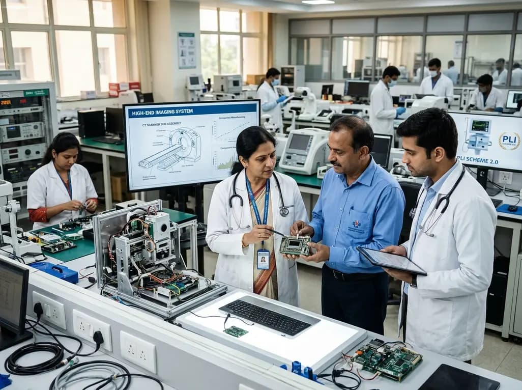
The Department of Pharmaceuticals has extended the medical devices PLI scheme to 2035-36 with a Rs 2,700 crore outlay. Featuring relaxed investment barriers and eliminated sales thresholds, the revised framework targets deep manufacturing, high-end diagnostics, critical MedTech components, and supply resilience.
Today

A metabolomic study reveals that aging delays glutathione synthesis and gamma-glutamylcysteine accumulation following muscle injury. Although precursor supplementation targets this enzymatic bottleneck, in vitro tests show no immediate effect on myogenesis, highlighting the complexity of muscle repair in older adults.
Today
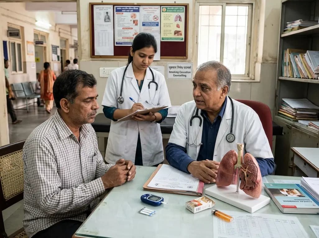
A systematic review evaluates psychological interventions for smoking cessation in diabetes, showing short-term success but highlighting challenges in long-term abstinence.
Today
