

Loading, please wait...
Loading, please wait...

Lower-limb alignment directly dictates musculoskeletal load transmission across the entire human kinetic chain. Biomechanical malalignment alters joint contact stress and accelerates debilitating musculoskeletal disorders. For instance, abnormal frontal-plane knee alignment and severe hindfoot deviations contribute to knee osteoarthritis, patellofemoral pain syndrome, and chronic plantar fasciitis. Currently, clinicians evaluate lower-limb alignment through full-leg standing radiographs or comprehensive physical examinations. However, routine radiography requires costly specialised imaging suites and exposes individuals to ionising radiation. In addition, manual orthopedic examinations consume considerable clinical time and introduce observer subjectivity. These practical constraints severely limit the availability of early community screening programs. Consequently, many patients present only after irreversible articular cartilage damage or chronic tissue inflammation has already occurred. Scalable lower-limb alignment screening offers an effective solution to bridge this diagnostic bottleneck. Non-invasive optical assessments allow clinical teams to identify vulnerable structural profiles rapidly. By detecting alignment variations during preliminary consultations, healthcare providers can introduce timely conservative therapies. Therefore, implementing accessible screening tools in primary care and sports medicine can protect long-term joint integrity.
Weight-bearing three-dimensional foot scanning captures dynamic anatomical geometry under genuine physiological loads. Distal foot posture closely reflects proximal joint alignment through interconnected lower-limb kinetic mechanics. For example, excessive subtalar joint pronation triggers internal tibial torsion and compensatory knee valgus angles. Traditional footprint assessments rely on flat two-dimensional surface contact outlines. Consequently, conventional ink or pressure prints fail to capture vital volumetric features and arch collapse patterns. Modern optical scanners, in contrast, generate detailed digital surface meshes within a few seconds. These volumetric scans accurately document subtle spatial elevations across the plantar vault and calcaneal structures. Recently, researchers compiled a standardised dataset of 167 participants aged 6 to 60 years to explore this relationship. Clinicians labeled each subject for medial arch type, hindfoot posture, and frontal-plane knee alignment. The investigators concurrently extracted 16 geometric morphological parameters alongside the full 3D surface images. Furthermore, this broad age demographic ensured diverse structural profiles across pediatric and adult cohorts. Thus, non-invasive plantar surface digitization supplies objective morphological markers that reflect mechanical alignment across the lower limb.
Automated biomechanical screening requires artificial intelligence systems that synthesize heterogeneous medical data modalities seamlessly. To address this computational challenge, researchers engineered a multimodal deep learning framework that integrates image representations with discrete geometric measurements. Specifically, the neural network processes high-resolution plantar scan images alongside 16 discrete morphological features simultaneously. The investigators implemented a feature-wise attention mechanism to prioritize alignment-relevant cues across both data streams. This attention module dynamically weighs regional surface contours against linear anthropometric ratios. Consequently, the network focuses on predictive structural features while suppressing non-informative anatomical variance. Moreover, the researchers benchmarked diverse computer vision backbones, including Convolutional Neural Networks and advanced Vision Transformers. They also completed systematic ablation studies to evaluate standalone image encoders, morphology-only models, and unweighted fusion layers. In doing so, the investigators observed how multimodal integration influences specific classification tasks. Therefore, this computational framework combines structural morphology with spatial vision models to establish an objective diagnostic pipeline.
The attention-based multimodal deep learning model achieved competitive but distinct performance across the three clinical classification tasks. Internal validation on a held-out test cohort revealed an accuracy of 89 percent for arch type identification. Additionally, the network achieved an 81 percent accuracy for hindfoot alignment and 76 percent accuracy for frontal-plane knee alignment. These empirical metrics confirm that distal foot morphology reliably predicts local and adjacent segment posture. However, comparative ablation experiments uncovered fascinating task-dependent model behaviors. For instance, the image-only Vision Transformer achieved the single highest classification accuracy for arch type categorization. Conversely, a Vision Transformer operating without attention fusion attained slightly superior accuracy and macro-F1 scores for knee alignment. These findings illustrate that multimodal attention mechanisms do not provide uniform benefits across every biomechanical task. Rather, proximal joint prediction may rely on broader visual features rather than fine-grained geometric parameters. Nevertheless, the model reliably captured functional mechanical variations across the pediatric and adult study cohort.
Translating artificial intelligence systems into routine clinical orthopedics requires high algorithmic interpretability and physician trust. To provide transparent diagnostic explanations, the research team implemented class activation mapping across the trained models. Notably, these activation heatmaps consistently highlighted clinically meaningful plantar territories associated with specific structural alignments. For example, the networks focused intensely on the medial longitudinal arch perimeter and the calcaneal contact zone during prediction. This anatomical attention corroborates the distal-to-proximal kinetic chain hypothesis familiar to orthopaedic specialists. Furthermore, explainable visual overlays enable clinicians to verify algorithmic reasoning during outpatient consultations. Primary care physicians and sports medicine clinicians can utilize these objective outputs to guide early physical therapy and custom orthotic prescription. However, the authors emphasize that multicenter external validation remains necessary prior to widespread clinical adoption. Future studies must test diverse patient populations, varying foot types, and distinct clinical environments. Ultimately, integrating explainable multimodal algorithms into non-invasive scanning devices could revolutionize screening-level musculoskeletal healthcare delivery across modern healthcare systems.
The human lower extremity operates as an integrated closed kinetic chain during weight-bearing activities. Consequently, structural alterations in the foot, such as severe pes planus, induce internal tibial rotation and alter frontal knee moments. By capturing these morphological adaptations with high-resolution scans, deep learning algorithms detect subtle structural markers that correlate with abnormal knee stress. Therefore, screening identifies individuals predisposed to joint wear before structural osteoarthritis manifests clinically.
Standard long-leg radiographs require dedicated imaging suites, deliver ionising radiation, and incur substantial institutional costs. In contrast, weight-bearing three-dimensional foot scanners are non-invasive, fast, and completely radiation-free. They capture dynamic structural deformation under real body weight, providing rich volumetric and geometric data within seconds. As a result, 3D optical scanning enables scalable population triage in community and outpatient settings where expensive radiographic equipment is unavailable.
Although internal validation demonstrated impressive diagnostic performance, the initial study examined a modest cohort of 167 individuals. Furthermore, accuracy for knee alignment reached 76 percent, meaning the tool serves as a preliminary triage aid rather than a definitive diagnostic replacement. External multicenter validation across diverse patient demographics, body mass indices, and athletic populations remains necessary before clinicians can reliably implement this artificial intelligence system in routine practice.
Disclaimer: This content is for informational and educational purposes only. It is not intended to provide medical advice, diagnosis, or treatment. Always seek the advice of a qualified healthcare provider with any questions you may have regarding a medical condition or treatment. Refer to the latest local and national guidelines for clinical practice.
References

Read summarized clinical updates, watch expert medical content, and earn CME certifications right from your smartphone.
Open your smartphone camera to scan and install.





A multimodal deep learning framework utilizing weight-bearing 3D foot scans accurately screens lower-limb alignment, including arch type, hindfoot posture, and knee alignment, offering an accessible, radiation-free alternative to radiography for early biomechanical triage.
Today

Targeting bacterial signaling pathways marks a major shift in antimicrobial strategies. By disrupting quorum sensing and biofilm formation, anti-virulence tools suppress foodborne pathogens without driving antimicrobial resistance, protecting vulnerable patients and the broader food ecosystem.
Today
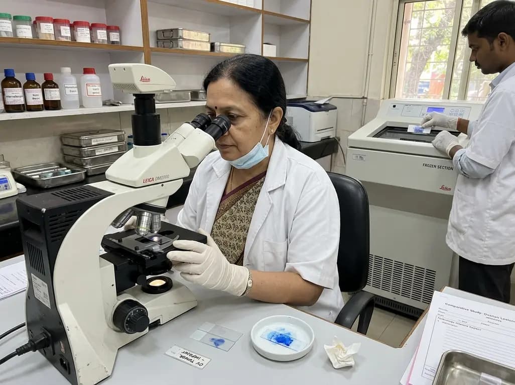
A new comparative study demonstrates that intraoperative imprint cytology achieves 96.6% diagnostic accuracy in ovarian tumours, outperforming frozen section in sensitivity for borderline lesions and offering an accessible, rapid alternative for resource-constrained surgical centres.
Today
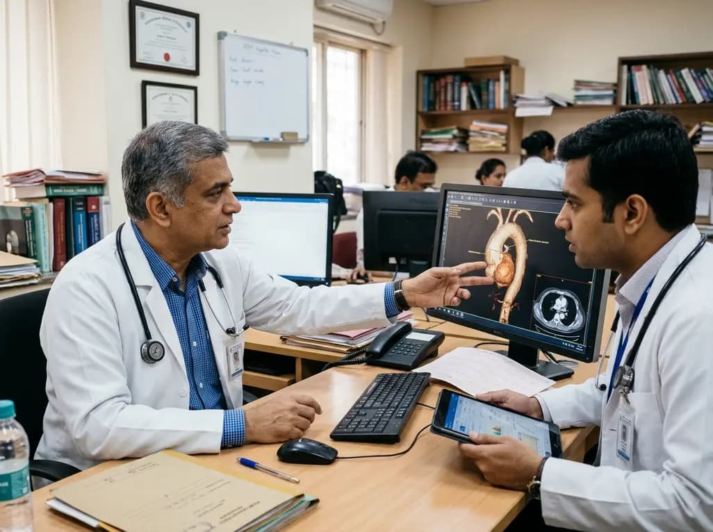
Thoracic aortic aneurysm lacks etiologic medical therapy. A new study reveals that targeting specific αV integrins with Cilengitide attenuates TGF-β and mechanotransduction pathways, effectively limiting pro-fibrotic remodeling and aortic bulb dilation in both syndromic and sporadic disease models.
Today
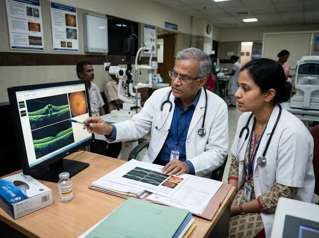
A real-world retrospective study demonstrates that combining intravitreal ranibizumab with focal/grid macular laser significantly improves visual acuity and central macular thickness compared to laser monotherapy in patients with center-involved diabetic macular edema.
Today
