

Loading, please wait...
Loading, please wait...

Digital workflows continue to transform modern prosthodontic practice across the globe. However, capturing passive fit across edentulous arches remains a notable technical hurdle for many dental surgeons. Traditional intraoral scanning often accumulates stitching errors over long spans of edentulous ridge. Therefore, clinicians frequently experience distortion when fabricating complete-arch implant-supported prostheses. To solve these errors, innovative researchers have explored reverse scanning techniques alongside advanced optical technologies. Digital dentistry demands exceptional trueness to prevent biological complications and mechanical screw loosening. Furthermore, minor deviations in implant positions can create substantial stress within restorative frameworks. Stereophotogrammetry has emerged as a dedicated optical solution for recording multiple implant coordinates. Meanwhile, intraoral scanners have received major hardware upgrades and algorithmic refinements. Consequently, clinicians must understand which digital recording modality delivers the highest trueness and reproducibility. Evaluating new scanning protocols provides vital clarity for full-arch implant rehabilitations. This comparative investigation addresses that fundamental knowledge gap by examining linear and angular deviations directly across multiple systems.
The investigation compared three distinct digital recording methods under standardized in vitro laboratory conditions. Specifically, investigators evaluated direct intraoral scanning, stereophotogrammetry, and reverse scanning techniques using an edentulous maxillary master cast. This definitive cast featured six multi-unit abutment replicas simulating an immediate complete-arch restoration in daily practice. Furthermore, the operators completed fifteen recordings for each experimental group, generating forty-five total datasets. For direct intraoral scanning and reverse scanning, the operators utilized an advanced intraoral scanner, the Primescan system. In contrast, they captured stereophotogrammetric measurements using the specialized Oxo Core device. To establish an uncompromising benchmark, an industrial-grade desktop optical scanner digitized the master cast. Reverse-engineering software then aligned each test scan against this high-resolution reference dataset. Consequently, the researchers calculated linear trueness by measuring the deviation from the master model. Similarly, they assessed precision by quantifying measurement repeatability within each experimental cohort. Through robust non-parametric testing, the authors analyzed spatial coordinates across all three dimensions. This rigorous experimental setup allowed an objective comparison without confounding biological variables.
The experimental findings revealed statistically significant differences in linear trueness among the three digital modalities. Notably, reverse scanning demonstrated the highest linear trueness, exhibiting a remarkably low deviation of 39.1 micrometers. Intraoral scanning followed with a mean linear deviation of 69.7 micrometers across the complete arch. Meanwhile, stereophotogrammetry recorded the largest linear deviation at 102.8 micrometers. Post hoc statistical comparisons confirmed that reverse scanning significantly outperformed stereophotogrammetry in linear trueness. However, the difference between direct intraoral scanning and reverse scanning did not reach statistical significance. In terms of linear precision, all three digital workflows displayed excellent consistency. Thus, repeated scans within each group produced tightly clustered measurements without substantial scattering. In addition, the high precision values indicate that each device maintains reliable internal calibration during complete-arch scanning. Nevertheless, trueness represents the decisive parameter because systematic displacement compromises prosthetic framework seating. These linear data suggest that reverse scanning avoids cumulative error propagation effectively. Consequently, the protocol offers an exceptionally accurate method for digitizing full-arch multi-unit abutment locations in complex edentulous cases.
Prosthetic longevity requires precise angular orientation alongside accurate linear distances. In this study, angular trueness also exhibited statistically significant variance among the test cohorts. Intraoral scanning achieved outstanding angular trueness, showing minimal angular discrepancy relative to the reference standard. Similarly, reverse scanning maintained strict angular fidelity across all six multi-unit abutments. In contrast, stereophotogrammetry exhibited greater angular variability during complete-arch recording. Post hoc testing demonstrated that intraoral scanning generated significantly lower angular errors than stereophotogrammetry. Moreover, severe angular tilt creates localized strain around implant necks and retaining screws. Therefore, maintaining angular accuracy prevents mechanical screw loosening and prosthetic screw fracture under functional mastication. The superior angular performance of intraoral scanning highlights the sophistication of modern optical triangulation algorithms. Furthermore, reverse scanning successfully preserved these angular advantages by utilizing the same intraoral scanner platform. Consequently, both intraoral scanning and reverse scanning provide the angular precision necessary for rigid prosthetic frameworks. These findings reassure clinicians seeking predictable passive fit for complex screw-retained restorations in completely edentulous arches.
These laboratory findings offer immediate practical relevance for dental surgeons and restorative specialists. Complete edentulism presents severe anatomical challenges, including mobile oral mucosa and lack of distinct landmarks. Traditional direct scanning often struggles across extensive edentulous spans due to image stitching errors. In contrast, reverse scanning bridges this gap by digitizing an interim prosthesis or verification structure extraorally. As a result, clinicians capture critical tissue contours and precise implant coordinates simultaneously without patient interference. Furthermore, reverse scanning eliminates the need for expensive stereophotogrammetry camera systems in private practice. Clinicians can achieve superior linear trueness using their existing intraoral scanners and standard laboratory analogs. In addition, minimizing linear error to approximately 39 micrometers safely falls within the threshold for clinical passivity. Consequently, dental technicians can mill monolithic zirconia or titanium frameworks with supreme confidence. However, practitioners must maintain meticulous technique during abutment tightening and analog seating. Ultimately, integrating reverse scanning streamlines full-arch workflows, shortens chairside appointments, and delivers exceptional prosthetic outcomes for edentulous patients undergoing full mouth rehabilitation.
Reverse scanning techniques involve recording the positions of dental implants by scanning an existing provisional prosthesis, verification jig, or stone cast extraorally. Clinicians attach scannable implant analogs to the prosthesis intaglio surface before using an intraoral or laboratory scanner. Consequently, this indirect approach bypasses intraoral optical challenges such as soft tissue mobility, limited mouth opening, and saliva. The technique accurately transfers verified clinical contours, aesthetics, and implant coordinates directly to CAD software.
Stereophotogrammetry relies on optical triangulation between specialized extraoral targets and high-speed multi-camera sensors. In this specific investigation, stereophotogrammetry demonstrated higher linear deviation than reverse scanning due to hardware calibration parameters and target geometry. Furthermore, intraoral scanning technology has improved substantially, narrowing performance differences. While stereophotogrammetry remains valuable for avoiding soft tissue errors in vivo, benchtop comparisons reveal that high-precision intraoral scanners with reverse protocols achieve superior absolute trueness under controlled laboratory conditions.
Yes, clinicians can adopt reverse scanning protocols without investing in expensive stereophotogrammetry systems. Practitioners only require their current intraoral scanner, compatible scan bodies, and standard multi-unit abutment analogs. By scanning the patient's verified interim prosthesis extraorally on the benchtop, the clinician obtains accurate 3D implant positions and anatomical contours seamlessly. Therefore, this workflow provides an accessible, cost-effective alternative for general dental practitioners and prosthodontists performing full-arch rehabilitations.
Disclaimer: This content is for informational and educational purposes only... Refer to the latest local and national guidelines for clinical practice.
References

Read summarized clinical updates, watch expert medical content, and earn CME certifications right from your smartphone.
Open your smartphone camera to scan and install.





A comparative in vitro study evaluated reverse scanning techniques, intraoral scanning, and stereophotogrammetry for full-arch implant impressions. Reverse scanning achieved the highest linear trueness at 39.1 µm, offering clinicians an accurate, accessible alternative for edentulous rehabilitation.
Today
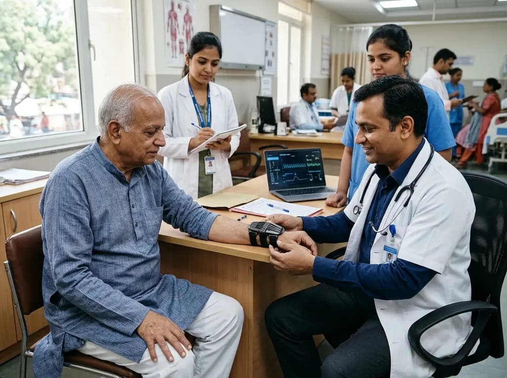
The TREM-AB open-label pilot study evaluated a wearable passive nonlinear vibration absorber for essential tremor. The Nonlinear Energy Sink bracelet reduced clinical Fahn-Tolosa-Marin tremor scores by 32.1% and electrophysiological tremor power by 49.2% with no adverse effects reported.
Today
A population-based study of 20,928 children reveals that small vulnerable newborns face sex-divergent mental capital outcomes at ages 6-7: term SGA girls show higher behavioral difficulties and lower resilience, while preterm non-SGA boys experience increased behavioral problems.
Today

A landmark study details how an enterprise pediatric remote patient management model expanded from a single cardiology pilot to 16 diverse specialty programs by combining centralized digital operations with specialty clinical leadership.
Today

A comparative review of tension band wiring and intramedullary screw fixation in thumb metacarpophalangeal arthrodesis, evaluating union rates, hardware complications, and clinical outcomes.
Today
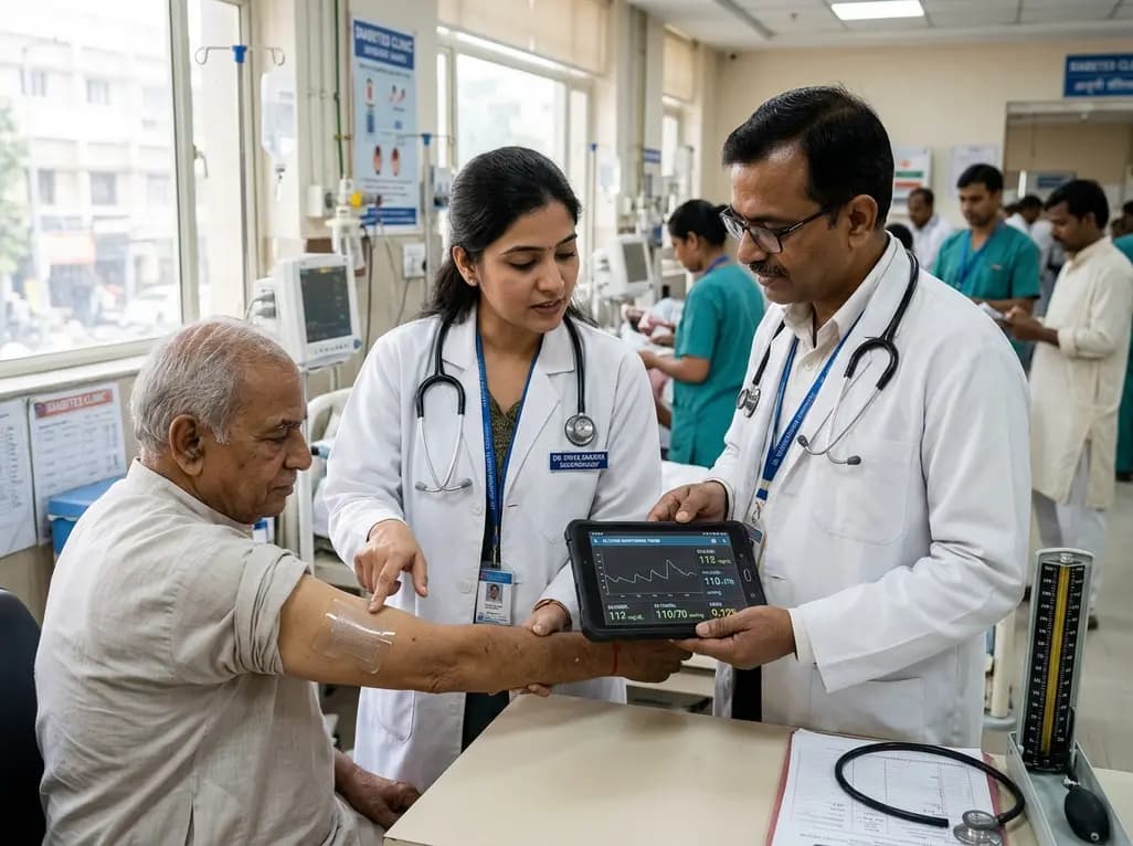
A breakthrough ultrathin multimodal sensor decouples physical hemodynamic monitoring from disposable electrochemical calibration, achieving long-term wearable glucose prediction with 81.36% accuracy and a 9.12% MARD. This offers a robust, drift-free solution for outpatient metabolic surveillance.
Today
