.webp&w=384&q=75)

Loading, please wait...
Loading, please wait...

Medical experts recognize central retinal artery occlusion (CRAO) as a severe vascular event that mirrors a cerebral stroke. While clinicians often prioritize treating the affected eye, the clinically normal fellow eye frequently harbors subclinical signs of damage. A new study highlights the significant prevalence of Retinal Ischemic Perivascular Lesions (RIPLs) in these seemingly unaffected eyes. These findings suggest that the underlying vascular pathology affects both eyes simultaneously, regardless of acute symptoms.
RIPLs represent permanent markers of previous focal ischemia within the inner nuclear layer of the retina. These lesions appear on spectral-domain optical coherence tomography (SD-OCT) as localized areas of thinning. Researchers recently compared 39 fellow eyes of CRAO patients with 57 healthy controls to assess these markers. Results demonstrated that RIPLs occurred in 28.2% of fellow eyes compared to only 3.5% in healthy individuals. Furthermore, the CRAO group exhibited a significantly higher total number of lesions.
The morphology of these lesions provides critical clues regarding the severity of systemic vascular compromise. The study categorized the lesions into narrow types and larger wedge-shaped types. Notably, wedge-shaped lesions occurred exclusively in the eyes of CRAO patients, never in healthy controls. Since the presence of these lesions correlates with hypertension and cardiovascular disease, they serve as vital biomarkers for future ischemic events. Consequently, ophthalmologists should meticulously screen the fellow eye to identify patients at high risk for systemic complications.
Moreover, the presence of these ischemic markers correlates with advanced conditions like carotid artery stenosis and coronary artery disease. Therefore, identifying these lesions in an otherwise healthy eye warrants a comprehensive systemic workup. This proactive approach ensures that patients receive timely interventions to prevent myocardial infarction or secondary strokes. Additionally, identifying subclinical ischemia helps clinicians tailor long-term management strategies for these high-risk patients.
RIPLs are subclinical, focal areas of atrophy in the inner nuclear layer of the retina. They serve as permanent indicators of past ischemic events and clinicians can easily detect them using SD-OCT imaging.
Screening is vital because nearly 30% of unaffected fellow eyes in CRAO patients harbor these lesions. Their presence indicates a high burden of systemic vascular disease and a potential risk for future strokes or cardiac events.
Disclaimer: This content is for informational and educational purposes only. It does not constitute medical advice or a professional relationship. Always seek the advice of a qualified healthcare provider for any medical condition. Refer to the latest local and national guidelines for clinical practice.
References
Adıyeke SK et al. Retinal Ischemic Perivascular Lesions in the Fellow Eyes of Patients with Central Retinal Artery Occlusion. Retina. 2026 May 06. doi: 10.1097/IAE.0000000000004876. PMID: 42090196.
Bousquet E, Santina A, Au A, et al. Retinal ischemic perivascular lesions are associated with myocardial infarction in patients with coronary artery disease. Am J Ophthalmol. 2024. doi: 10.1016/j.ajo.2024.03.027.
Colcombe J, et al. Retinal Ischemic Perivascular Lesions: An Exploratory Study of Their Potential as Biomarkers for Cardiovascular Disease. J. Pers. Med. 2023;13(10):1564. doi: 10.3390/jpm13101564.

Read summarized clinical updates, watch expert medical content, and earn CME certifications right from your smartphone.
Open your smartphone camera to scan and install.





A study reveals that 28.2% of fellow eyes in CRAO patients have Retinal Ischemic Perivascular Lesions, acting as subclinical markers for cardiovascular risk...
5 months ago

An observational study from the NIH All of Us Research Program reveals that adding a multi-ancestry polygenic risk score to machine learning models provides only modest improvements in predicting adult weight gain, underscoring the enduring significance of routine clinical and lifestyle factors.
Today

A study in JAMIA Open shows lightweight open-source reasoning LLMs achieve up to 95% sensitivity and accelerate evidence synthesis screening by nearly twentyfold, presenting a cost-effective, scalable AI solution for biomedical research and systematic reviews.
Today
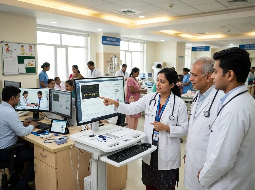
A landmark study details how an enterprise pediatric remote patient management model expanded from a single cardiology pilot to 16 diverse specialty programs by combining centralized digital operations with specialty clinical leadership.
Today

A comparative review of tension band wiring and intramedullary screw fixation in thumb metacarpophalangeal arthrodesis, evaluating union rates, hardware complications, and clinical outcomes.
Yesterday

A breakthrough ultrathin multimodal sensor decouples physical hemodynamic monitoring from disposable electrochemical calibration, achieving long-term wearable glucose prediction with 81.36% accuracy and a 9.12% MARD. This offers a robust, drift-free solution for outpatient metabolic surveillance.
Yesterday
