.webp&w=384&q=75)

Loading, please wait...
Loading, please wait...

Radiomics for LSCC metastasis is currently transforming head and neck oncology by offering a more precise staging method. Specifically, predicting cervical lymph node metastasis (LNM) in laryngeal squamous cell carcinoma (LSCC) is vital for determining surgical extent. However, traditional imaging often struggles with occult metastases. Therefore, recent research demonstrates that integrating artificial intelligence and radiomics can significantly enhance diagnostic accuracy before surgery. Notably, this advancement aligns with the global shift toward precision medicine. Consequently, clinicians can now leverage advanced imaging data more effectively. Furthermore, the use of multi-sequence MRI provides a detailed map of the tumor microenvironment. Indeed, this approach allows for more informed decision-making.
Traditionally, radiologists rely on size and morphology to identify metastatic nodes. However, these criteria often lack the sensitivity required for early-stage detection. Specifically, radiomics identifies patterns invisible to the human eye by extracting quantitative features. For instance, in a major study, researchers analyzed over 2,300 features from T1-enhanced and T2-weighted images. Furthermore, they used LASSO regression to select the most stable biomarkers for prediction. Additionally, this process ensures that only the most relevant features inform the final model. Similarly, the integration of these features improves predictive power. Consequently, this method offers a significant leap over conventional techniques. Moreover, it provides a non-invasive way to assess node status.
The study found that the Random Forest algorithm performed exceptionally well as a standalone radiomics model. Nevertheless, the real breakthrough occurred when researchers combined these digital biomarkers with clinical features. Because of this, the integrated nomogram achieved an impressive AUC of 0.908 in the testing phase. Therefore, the combined model outperformed standalone clinical or radiomic approaches. In fact, the synergy between clinical data and digital features is clear. Consequently, clinicians can now use this data to refine their surgical strategies. In other words, the nomogram acts as a superior decision-support tool. Furthermore, it helps in tailoring therapy for individual patients.
In addition, this approach provides a robust tool for personalized treatment planning. As a result, surgeons can better identify patients who require radical neck dissection. Alternatively, they can spare low-risk patients from unnecessary morbidity. Moreover, this non-invasive technique minimizes the risk of overtreatment while ensuring high-risk patients receive comprehensive care. Above all, the integration of radiomics into routine workflows could redefine the standard of care. With this in mind, future studies should focus on larger cohorts. Finally, the role of AI in oncology continues to expand, offering hope for better patient outcomes. Specifically, these tools will likely become standard in clinical practice soon.
Traditional interpretation relies on a radiologist\'s visual assessment of node size and shape. In contrast, radiomics uses algorithms to extract thousands of quantitative data points from the image, revealing microscopic textures and patterns associated with malignancy.
No, the nomogram is a preoperative tool intended to guide clinical decision-making and surgical planning. Histopathology remains the gold standard for final diagnosis and staging.
Disclaimer: This content is for informational and educational purposes only. It does not constitute professional medical advice, diagnosis, or treatment. Always seek the advice of your physician or other qualified health provider with any questions you may have regarding a medical condition. Refer to the latest local and national guidelines for clinical practice.
References
Li B et al. Radiomics analysis of MRI improves prediction of lymph node metastasis in laryngeal squamous cell carcinoma. Future Oncol. 2026 Feb 13. doi: 10.1080/14796694.2026.2630630. PMID: 41686503.
Zhao X et al. Radiomics analysis of CT imaging improves preoperative prediction of cervical lymph node metastasis in laryngeal squamous cell carcinoma. Eur Radiol. 2023; 33:1121–31. doi: 10.1007/s00330-022-09051-4.
Lin P et al. Intratumoral and peritumoral radiomics of MRIs predicts pathologic complete response to neoadjuvant chemoimmunotherapy in head and neck squamous cell carcinoma. J Immunother Cancer. 2024; 12(4):e008561. doi: 10.1136/jitc-2023-008561.

Read summarized clinical updates, watch expert medical content, and earn CME certifications right from your smartphone.
Open your smartphone camera to scan and install.





Recent research highlights how a radiomic-clinical nomogram using MRI features significantly improves the preoperative prediction of lymph node metastasis i...
8 months ago

Following regulatory scrutiny from FSSAI regarding promotional representations and lean muscle preservation claims, Dr. Reddy's Laboratories and Nestlé Health Science are currently reviewing the branding and marketing strategy for their joint nutritional supplement Celevida GLP+.
Today
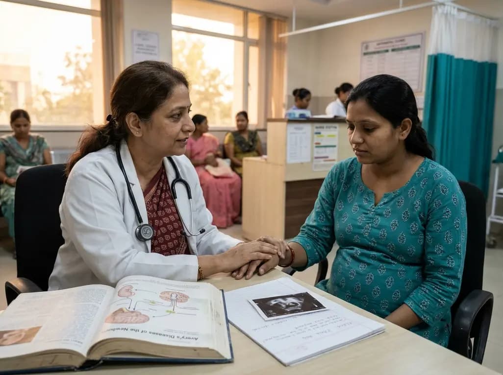
A comprehensive scoping review synthesises evidence linking prenatal maternal mental health to long-term offspring neurodevelopment, evaluating neuroendocrine, inflammatory, and epigenetic pathways while highlighting clinical screening and future research priorities.
Today
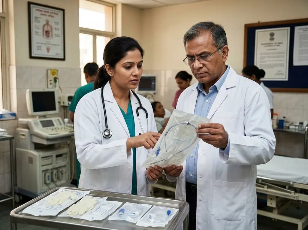
The Directorate General of Health Services directs Indian states to halt the hazardous reuse and reprocessing of single-use medical devices. Discover regulatory implications under the Drugs and Cosmetics Act, critical infection risks, legal liabilities, and operational changes required across clinical specialties.
Today

A 25-year multi-institutional cohort reveals high recurrence rates in pediatric giant cell tumor of bone, particularly in patients with open physes and bone marrow edema on preoperative MRI. Early surveillance and careful balance between growth plate preservation and tumor clearance are essential.
Today

India is advancing preventive healthcare by integrating genomics, artificial intelligence, and quantum technologies. With the SANGAM programme sequencing one million Indian genomes, clinicians can unlock personalized therapies, detect cardiometabolic risks early, and curb the burden of diabetes and heart disease.
Today
