

Loading, please wait...
Loading, please wait...

Diagnosing and managing primary bone neoplasms in children requires distinct oncologic expertise. Although relatively common in young adults, pediatric giant cell tumor of bone remains exceptionally rare and represents less than five percent of all primary bone neoplasms in skeletally immature individuals. Because clinicians encounter this condition infrequently, standard treatment algorithms often rely on adult paradigms that fail to account for open physes. Consequently, surgeons face an ongoing dilemma between achieving wide tumor clearance and preserving long-term limb growth.
Giant cell tumors originate as locally osteolytic neoplasms characterized by multinucleated, osteoclast-like giant cells and neoplastic mononuclear stromal cells. In adults, these lesions characteristically expand into the epiphysis and reach the subchondral bone. However, skeletally immature children frequently display lesions situated within the metaphysis or showing combined metaphyseal-epiphyseal architecture. The open physis functions as a dynamic barrier, yet tumor growth can disturb physeal vascularization and cellular development. Consequently, children face significant risks of angular limb deformities and leg-length discrepancy if tumor expansion or surgical instrumentation violates the cartilaginous growth plate. Histopathological confirmation remains vital because mimickers such as aneurysmal bone cysts, chondroblastoma, and osteosarcoma occur frequently in pediatric populations. Clinicians frequently utilize molecular immunohistochemistry, particularly targeting H3.3 G34W mutations, to distinguish authentic giant cell tumors from secondary reactive giant cell lesions. Furthermore, younger patients exhibit heightened bone turnover and altered local cytokine signaling, which may facilitate aggressive local biology. Therefore, orthopedic oncology teams must evaluate skeletally immature patients with meticulous precision before deciding on definitive surgical intervention.
A landmark 25-year multi-institutional study led by Werenski and colleagues evaluated forty-two patients younger than nineteen years treated surgically across three tertiary academic centers. The study revealed a median patient age of sixteen years, alongside a notable female predominance of sixty-two percent. Tumors predominantly arose in the appendicular skeleton, with seventy-six percent involving long tubular bones, most frequently the distal or proximal femur. Strikingly, the overall local recurrence rate reached forty-five percent, with a median recurrence interval of only seven months postoperatively. This timeline highlights an aggressive early recurrence pattern that sharply contrasts with the more indolent courses seen in mature adults. In addition, when researchers stratified patients by skeletal maturity, individuals with open physes showed a fifty-five percent local recurrence rate compared to thirty-eight percent in patients with fused growth plates. Although this numerical disparity did not achieve statistical significance due to cohort size, it strongly indicates that younger skeletal biology poses distinct therapeutic vulnerabilities. Furthermore, systemic metastatic disease occurred in five percent of patients, demonstrating that these histologically benign lesions still harbor genuine potential for pulmonary dissemination.
One of the most consequential findings in the Werenski multi-institutional study involves preoperative imaging analysis. Among twenty-seven patients with complete baseline magnetic resonance imaging, fifty-six percent exhibited prominent peritumoral bone marrow edema. Notably, patients demonstrating bone marrow edema experienced a dramatic sixty-seven percent local recurrence rate, whereas patients without edema suffered only a seventeen percent recurrence rate. This difference reached strong statistical significance and identified bone marrow edema as a vital prognostic biomarker. Pathophysiologically, extensive bone marrow edema reflects heightened mechanical stress, localized hypervascularity, osteoclastic hyperactivity, and microvascular infiltration extending beyond the visible osteolytic lesion margins. Therefore, conventional mechanical curettage may leave behind microscopic neoplastic cell clusters embedded within inflamed marrow compartments. In contrast, standard radiographic margins frequently underestimate the true biological extent of neoplastic invasion in immature bone. Surgeons must consequently interpret preoperative fluid-sensitive MRI sequences with great scrutiny. Incorporating comprehensive multiplanar imaging helps multidisciplinary teams define accurate curettage margins or consider more radical resection planes whenever high-risk edema patterns appear.
Surgical management in growing children requires a careful balance between oncologic eradication and functional preservation. Intralesional extended curettage remains the primary surgical modality because it preserves native joint architecture and protects articular cartilage. However, high-speed burring, pulsatile lavage, and local chemical or thermal adjuvants—such as liquid nitrogen, phenol, or polymethylmethacrylate cement—carry inherent risks to the nearby growth plate. Exposure to extreme thermal or chemical injury can prematurely fuse open physes, triggering progressive angular deformities and limb-shortening deformities. Conversely, conservative curettage aimed solely at avoiding physeal trauma increases the risk of microscopic residual disease, fostering rapid local recurrence. When extensive subchondral destruction threatens joint integrity, surgeons often employ subchondral bone grafting or structural allografts alongside osteosynthesis plates. Systemic medical therapies, such as the RANK-ligand inhibitor denosumab, provide valuable cytoreduction in unresectable or sacral lesions. Nevertheless, surgeons must approach denosumab cautiously in growing pediatric cohorts due to risks of rebound hypercalcemia upon cessation and post-treatment osteosclerosis, which can obscure surgical margins. Thus, surgeons must tailor every operative plan to the child's precise skeletal maturity and anatomical geometry.
Given that the median time to tumor recurrence was merely seven months, postoperative follow-up in pediatric cohorts must follow an aggressive, structured schedule. Clinical teams should avoid extending surveillance intervals during the critical first two years following index surgery. Instead, clinicians must obtain serial high-resolution radiographs and MRI scans every three to four months during the initial twenty-four months. Furthermore, because pulmonary metastases developed in five percent of cohort patients, routine baseline and follow-up chest computed tomography remains mandatory. Optimal patient outcomes depend on close collaboration within a dedicated sarcoma multidisciplinary tumor board. Pediatric oncologists, musculoskeletal radiologists, specialized orthopedic oncologists, and bone pathologists must jointly evaluate each case from initial biopsy to long-term reconstruction. Moreover, physical therapists and rehabilitation teams play a vital role in restoring joint mobility and strengthening adjacent muscular structures after bone reconstruction. By aligning meticulous surgical technique with intensified surveillance and interdisciplinary care, clinicians can achieve successful local control while optimizing functional and developmental outcomes for pediatric patients.
Skeletally immature patients often present with tumors in close proximity to active growth plates. Consequently, surgeons frequently adopt conservative curettage margins to protect the delicate open physis and prevent growth arrest. This conservative approach increases the likelihood of leaving microscopic neoplastic tissue behind. Furthermore, heightened pediatric bone turnover and altered local cytokines promote rapid proliferation, driving recurrence rates up to fifty-five percent in open physes.
Extensive peritumoral bone marrow edema on fluid-sensitive MRI indicates active hypervascularity, osteolysis, and microvascular infiltration beyond the macroscopic tumor border. Clinical research demonstrates that patients with preoperative bone marrow edema face a sixty-seven percent recurrence risk compared to seventeen percent in those without edema. Therefore, identifying bone marrow edema prompts surgeons to extend adjuvant curettage, use chemical or thermal adjuncts, and execute vigilant post-resection follow-up.
Because the median time to recurrence in pediatric patients is only seven months, clinicians recommend rigorous early surveillance. Orthopedic oncology guidelines advise conducting physical examinations, localized MRI, and plain radiography every three to four months during the first two postoperative years. Additionally, teams must perform regular chest imaging, such as low-dose computed tomography, every six to twelve months to detect rare pulmonary metastases promptly.
Disclaimer: This content is for informational and educational purposes only and should not be considered medical advice. Always consult a qualified healthcare professional regarding any medical condition or before making healthcare decisions. Refer to the latest local and national guidelines for clinical practice.
References

Read summarized clinical updates, watch expert medical content, and earn CME certifications right from your smartphone.
Open your smartphone camera to scan and install.





A 25-year multi-institutional cohort reveals high recurrence rates in pediatric giant cell tumor of bone, particularly in patients with open physes and bone marrow edema on preoperative MRI. Early surveillance and careful balance between growth plate preservation and tumor clearance are essential.
Today
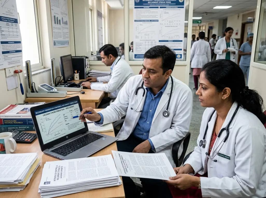
Explore vital recurrent stroke predictors in lacunar stroke patients managed with intensive blood pressure lowering. This analysis examines trial findings from SPS3 to uncover residual vascular risks, metabolic factors, and targeted secondary prevention strategies for high-risk neurological patients.
Today
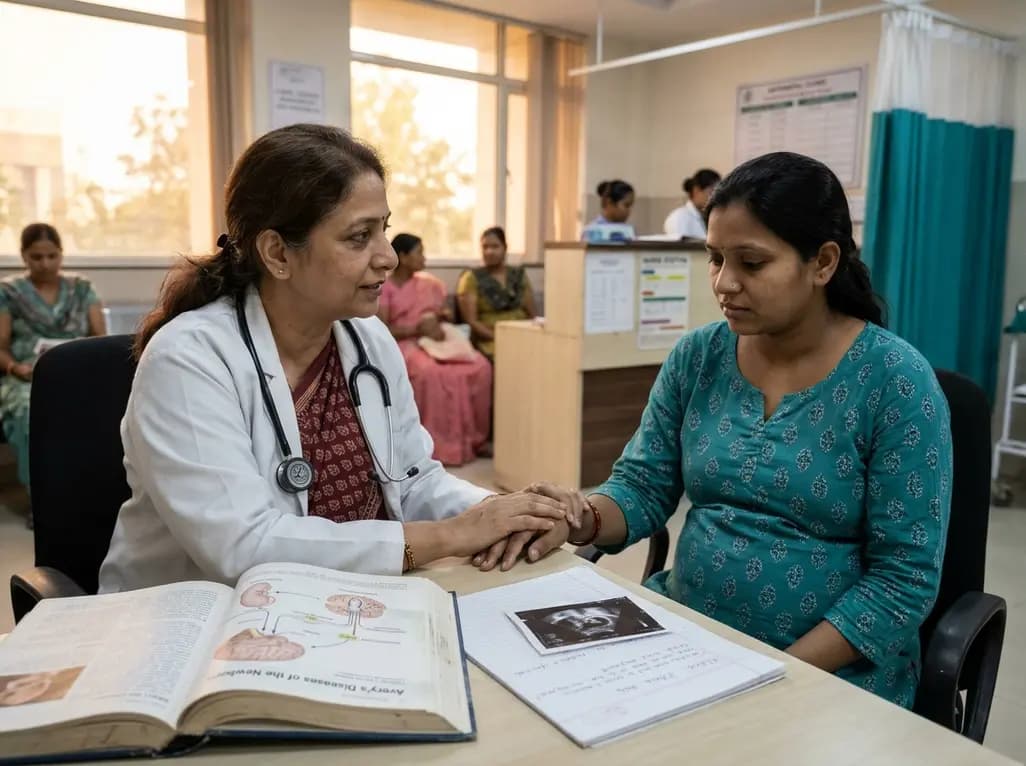
A comprehensive scoping review synthesises evidence linking prenatal maternal mental health to long-term offspring neurodevelopment, evaluating neuroendocrine, inflammatory, and epigenetic pathways while highlighting clinical screening and future research priorities.
Today
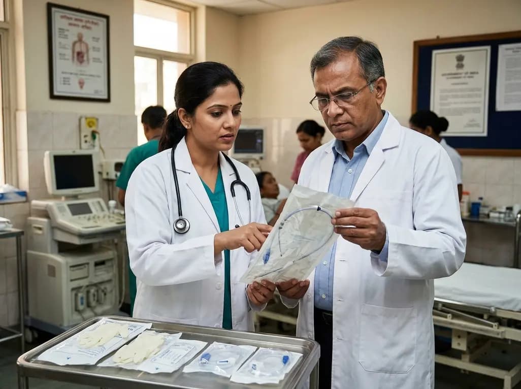
The Directorate General of Health Services directs Indian states to halt the hazardous reuse and reprocessing of single-use medical devices. Discover regulatory implications under the Drugs and Cosmetics Act, critical infection risks, legal liabilities, and operational changes required across clinical specialties.
Today
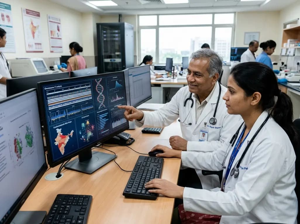
India is advancing preventive healthcare by integrating genomics, artificial intelligence, and quantum technologies. With the SANGAM programme sequencing one million Indian genomes, clinicians can unlock personalized therapies, detect cardiometabolic risks early, and curb the burden of diabetes and heart disease.
Today
