.webp&w=384&q=75)

Loading, please wait...
Loading, please wait...

Adolescent idiopathic scoliosis represents a complex three-dimensional deformity characterized by coronal deviation, sagittal alterations, and rotational abnormalities. Surgical intervention aims to achieve balanced three-dimensional alignment while halting curve progression. Historically, surgical techniques prioritized coronal plane alignment, often leaving significant axial torsion unaddressed. However, the introduction of modern segmental pedicle screw instrumentation and advanced correction maneuvers has shifted this paradigm. Currently, posterior spinal fusion derotation techniques permit surgeons to actively correct true vertebral rotation alongside coronal and sagittal profiles. Despite widespread adoption, questions remain regarding how stable these axial plane corrections remain across long-term postoperative recovery.
Axial plane deformity in scoliosis drives much of the clinical cosmetic deformity, specifically thoracic rib humps and lumbar prominences. Consequently, spine surgeons utilize en-bloc derotation maneuvers during posterior spinal fusion derotation procedures. This technique applies simultaneous corrective torque across multiple contiguous pedicle screws to untwist the spinal column. Therefore, surgeons can mobilize rigid periapical segments and restore physiological chest wall symmetry. Biomechanical studies confirm that en-bloc derotation distributes mechanical forces evenly across multiple screw purchase points. Thus, the maneuver reduces localized bone-screw interface failure while optimizing derotation. However, achieving acute intraoperative correction represents only the initial clinical hurdle. Clinicians must understand how these rotational corrections behave longitudinally during musculoskeletal remodeling.
To characterize these long-term rotational adjustments, investigators conducted a single-center retrospective study using a prospective surface topographic registry. The cohort comprised 49 adolescents aged 11 to 20 years who underwent posterior spinal fusion derotation. Notably, the cohort had a mean age of 14.6 years, 73.4% female distribution, and an average preoperative coronal Cobb angle of 57.9 degrees. Furthermore, major thoracic curves accounted for 67% of cases. The researchers excluded any patients with prior spinal procedures to avoid confounding variables. To capture the full spectrum of axial geometry, the study tracked three distinct parameters across six time points: preoperative, six weeks, three months, six months, one year, and two years. First, clinicians measured the angle of trunk rotation using a standardized scoliometer. Second, radiologists assessed true skeletal axial vertebral rotation via low-dose biplanar EOS imaging. Third, investigators recorded external body surface rotation using high-resolution surface topographic scanning.
The study yielded encouraging findings regarding the durability of structural corrections. Specifically, the angle of trunk rotation showed significant clinical improvement at every postoperative visit compared to baseline. Most importantly, the cohort demonstrated no statistically significant loss of trunk rotation correction throughout the entire two-year follow-up period. Similarly, maximum axial vertebral rotation and apical axial vertebral rotation showed robust correction on biplanar radiographs. Both parameters remained significantly improved at two years compared to preoperative values. However, researchers noted a slight but statistically significant loss of apical vertebral rotation between the six-week and one-year visits. After one year, this minor vertebral recalibration stabilized completely. Therefore, the internal bony correction achieved through posterior spinal fusion derotation remains fundamentally durable and resilient over time.
In contrast to internal skeletal alignment, external body surface rotation exhibited a surprisingly divergent longitudinal pattern. Surface topography initially revealed significant improvements in maximum body surface rotation at three months postoperatively. In addition, apical body surface rotation showed significant improvement at both three months and one year. However, by the two-year postoperative evaluation, body surface rotation failed to maintain statistically significant improvement relative to baseline. This intriguing mismatch demonstrates that skeletal derotation does not translate permanently into proportional external surface symmetry. Several soft-tissue factors likely account for this gradual dissipation. As adolescent patients mature, postoperative chest wall remodeling, paraspinal muscle scarring, and asymmetrical muscle tension evolve continuously. Furthermore, secondary rib cage deformation may persist despite rigid realignment of the vertebral column.
These findings provide essential guidance for orthopedic surgeons managing adolescent idiopathic scoliosis. First, posterior spinal fusion derotation reliably delivers lasting skeletal correction and clinical scoliometer improvement up to two years. Second, clinicians must practice nuanced preoperative counseling regarding aesthetic surface expectations. Patients and their families frequently equate internal spinal straightening directly with external torso symmetry. Therefore, surgeons must clarify that external surface contouring involves dynamic muscular and soft-tissue factors beyond bone realignment. Highlighting that cosmetic surface changes may fluctuate helps establish realistic expectations and reduces postoperative dissatisfaction. Moreover, objective surface topographic scanning serves as an exceptional radiation-free tool during surveillance. By combining surface topography with selective EOS radiography, clinicians can comprehensively track physical outcomes while minimizing cumulative radiation exposure.
Direct vertebral derotation manipulates individual vertebrae independently, which concentrates mechanical stress on single screws. In contrast, en-bloc derotation links multiple segmental screws together using rigid bridges. This distributes rotational corrective forces across several vertebral levels simultaneously, enhancing safety, mechanical purchase, and spinal correction efficiency.
Skeletal instrumentation permanently fuses and stabilizes the vertebral column. However, external surface contours depend heavily on ribs, paraspinal musculature, and subcutaneous fat. Over two years, continuing adolescent growth, soft-tissue remodeling, postural adaptation, and asymmetric muscle tone can gradually alter superficial surface aesthetics despite stable bony alignment.
Clinicians effectively monitor clinical axial deformity using a handheld scoliometer to measure the angle of trunk rotation. In addition, low-dose biplanar EOS radiographs quantify true axial vertebral rotation. Non-invasive surface topographic scanning accurately tracks superficial contours, reducing repetitive radiographic radiation exposure during routine postoperative follow-up.
Disclaimer: This content is for informational and educational purposes only. It is not intended to be a substitute for professional medical advice, diagnosis, or treatment. Always seek the advice of your physician or other qualified health provider with any questions you may have regarding a medical condition. Refer to the latest local and national guidelines for clinical practice.
References
Linden GS et al. The Longitudinal Effects of Posterior Spinal Fusion With Derotation on Axial Deformity in Adolescent Idiopathic Scoliosis. Spine (Phila Pa 1976). 2026 Oct 15. doi: 10.1097/BRS.0000000000005021. PMID: 38709012.
Upadhyay N. Posterior Instrumented Adolescent Idiopathic Scoliosis Correction and Fusion (T4 to L4). OrthOracle. 2019. doi: 10.1234/orthoracle.114.
Sung PS. Lumbar spine coordination during axial trunk rotation in adolescents with and without right thoracic idiopathic scoliosis. Hum Mov Sci. 2020;74:102680. doi: 10.1016/j.humov.2020.102680.

Read summarized clinical updates, watch expert medical content, and earn CME certifications right from your smartphone.
Open your smartphone camera to scan and install.





This study evaluates 2-year axial correction outcomes in adolescent idiopathic scoliosis following posterior spinal fusion with en-bloc derotation. While internal vertebral and clinical trunk rotation maintain correction, body surface improvements diminish over time, offering vital guidance for surgical expectations.
Today
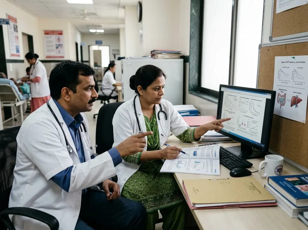
Recent research reveals that subclinical cholestasis, marked by elevated serum gamma-glutamyl transferase, reflects gut dysbiosis and predicts resistance to immune checkpoint blockade. Bile acid remodeling and mucosal MAdCAM-1 loss impair antitumor immunity, highlighting novel therapeutic targets.
Today

Maharashtra has initiated the appointment of two dedicated special public prosecutors in every district alongside a joint police-health SOP to fast-track PCPNDT and MTP cases. Medical imaging centers, radiologists, and obstetricians must strengthen statutory documentation and audit preparedness.
Today
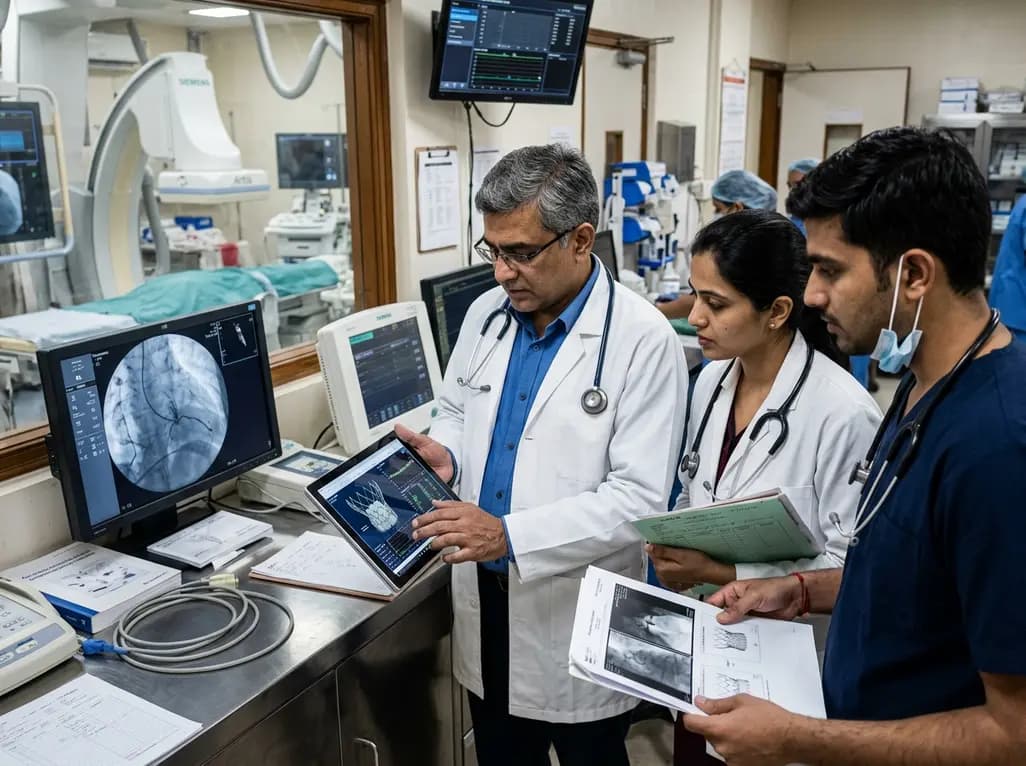
The US FDA has released an early safety alert regarding Edwards Lifesciences Commander M delivery catheters used in transcatheter mitral valve replacement. Reports of balloon bursting during deployment led to one death and one injury, prompting a recall of specific lots and urgent clinical inventory reviews.
Today
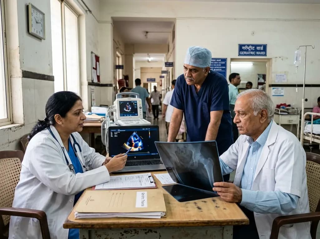
A meta-analysis evaluates the impact of preoperative transthoracic echocardiography on surgical delay and hospital stay in geriatric hip fracture patients. Evidence emphasizes balancing cardiac workups against operative urgency and adhering to clinical guidelines.
Today

An exploratory pilot study evaluates metoprolol pharmacokinetics across cisgender males, cisgender females, and transgender women on gender-affirming hormone therapy. Findings reveal distinct drug disposition trends, emphasizing the critical need for genotype-stratified research in transgender cardiovascular care.
Today
