.webp&w=384&q=75)

Loading, please wait...
Loading, please wait...

Elderly patients who sustain fragility hip fractures present complex medical challenges to acute surgical services worldwide. Historically, clinicians routinely obtain preoperative transthoracic echocardiography to assess structural heart disease before taking patients to the operating theater. However, fragile geriatric patients often suffer from acute physiological decompensation when immobilized. Prolonged bed rest increases the risks of pneumonia, pressure injuries, deep vein thrombosis, and delirium. Consequently, global orthopedic and geriatric guidelines strongly advocate for definitive surgical repair within 24 to 48 hours of injury. When physicians order non-invasive cardiac imaging, obtaining and interpreting scans can create operational bottlenecks. Orthopedic surgeons and anesthesiologists frequently debate whether routine echocardiograms confer tangible safety benefits or merely postpone urgent mechanical stabilization. Furthermore, indiscriminate testing often reveals incidental, chronic valvular abnormalities that do not alter the planned anesthetic technique. Therefore, surgical teams must determine whether specialized cardiac investigations genuinely optimize patient outcomes. Balancing cardiac risk stratification against surgical expediency remains a continuous perioperative challenge in acute geriatric trauma care. Consequently, establishing clear institutional protocols helps multidisciplinary teams resolve these diagnostic dilemmas without jeopardizing timely patient management.
A recent comprehensive meta-analysis pooled data from five relevant studies encompassing 2,581 geriatric hip fracture patients. Among this cohort, 865 patients underwent preoperative echocardiography, whereas 1,716 patients proceeded to surgical intervention without it. Baseline demographic characteristics, including mean age, sex distribution, fracture patterns, and surgical fixation techniques, remained comparable across both cohorts. Nevertheless, patients selected for cardiac imaging demonstrated higher baseline rates of coronary artery disease, congestive cardiac failure, and cardiac arrhythmias. When investigators examined operative timing, the mean time to surgery was 9.4 hours shorter in the uninvestigated group. However, this numerical difference did not achieve statistical significance, likely due to substantial heterogeneity across centers. In contrast, the analysis revealed a statistically significant reduction in overall hospital stay. Specifically, patients who bypassed preoperative scanning experienced a mean hospital stay that was 0.6 days shorter. The researchers noted that existing retrospective data feature considerable residual confounding and insufficient statistical power. Consequently, while imaging may lengthen logistical processes, definitive proof of independent surgical delay requires further rigorous investigation.
Current consensus from the American College of Cardiology emphasizes targeted cardiovascular risk assessment rather than routine screening. Specifically, guidelines discourage non-invasive imaging in asymptomatic patients with known, stable cardiac conditions. Preoperative imaging provides high clinical utility when patients exhibit active cardiac symptoms, including decompensated heart failure, severe aortic stenosis, or unstable hemodynamic parameters. However, in routine geriatric trauma practice, clinicians frequently request consultations out of defensive medicine or rigid institutional habits. Many clinicians assume that finding reduced ejection fraction will guide safer anesthetic administration. In reality, modern intraoperative monitoring and careful hemodynamic titration already accommodate moderate ventricular dysfunction. Furthermore, studies indicate that fewer than 15 percent of preoperative scans actually change perioperative medical management. When clinicians strictly apply established cardiovascular guidelines, they eliminate up to one-third of unnecessary scans without missing severe pathology. Therefore, orthogeriatric services should enforce strict adherence to American College of Cardiology recommendations. By distinguishing stable baseline comorbidities from decompensated disease, multidisciplinary teams avoid non-contributory diagnostic workups and prioritize rapid surgical stabilization.
The logistical burden of preoperative cardiac evaluation is particularly evident in public hospitals and high-volume trauma centers. In many resource-limited healthcare environments, access to trained sonographers and certified cardiologists remains restricted during weekends and evenings. Consequently, an urgent request for imaging often pushes surgery into the subsequent daytime operating lists. This prolonged delay directly compromises patient physiology and escalates hospital bed occupancy costs. Furthermore, delayed surgical fixation increases the requirement for supplemental parenteral analgesia, which elevates the incidence of postoperative confusion and urinary retention. In addition, family members and hospital administrators face financial strain from prolonged preoperative inpatient days. Therefore, optimizing turnaround times for essential diagnostic evaluations is critical for maintaining patient flow. Hospital administrators must streamline cardiac consultation pathways to ensure on-call imaging services respond promptly. When institutions optimize internal communication between cardiology, anesthesiology, and orthopedic departments, they prevent unnecessary cancellations. Ultimately, establishing swift and reliable diagnostic pipelines safeguards both clinical safety and healthcare resource stewardship in overburdened trauma environments.
To resolve persistent diagnostic bottlenecks, forward-thinking medical centers are redesigning their acute orthogeriatric intake pathways. Many institutions successfully implement focused cardiac ultrasound protocols conducted directly by trained anesthesiologists or emergency physicians. Point-of-care cardiac ultrasound rapidly evaluates left ventricular gross systolic function, detects pericardial effusion, and identifies severe aortic stenosis within minutes. Consequently, clinicians obtain actionable physiological insights at the bedside without waiting for formal department-level echocardiographic reporting. Furthermore, establishing dedicated orthogeriatric co-management units aligns clinical objectives between medical and surgical teams. Under these collaborative care models, geriatricians promptly optimize fluid status and manage chronic medications while preparing the patient for surgery. Moreover, hospital systems should implement reflex-testing criteria based on functional capacity and validated risk scores. If an echocardiogram is genuinely indicated, the order receives high-priority triage to minimize scheduling friction. By uniting point-of-care diagnostics with structured risk algorithms, centers can safely expedite operative intervention. Thus, fragile fracture patients receive timely, high-quality interventions that optimize functional recovery and reduce mortality.
According to established cardiovascular guidelines, preoperative echocardiography is indicated for patients presenting with active cardiac conditions or unexplained cardiorespiratory symptoms. These conditions include acute coronary syndrome, decompensated heart failure, significant new murmurs suggestive of severe aortic stenosis, and unexplained syncope. However, stable patients with established, compensated comorbidities do not require routine screening, as excessive testing delays surgery without altering anesthetic management or improving overall clinical outcomes.
Delaying hip fracture fixation beyond 24 hours significantly increases post-trauma morbidity, hospital length of stay, and long-term mortality. Extended preoperative immobilization promotes venous thromboembolism, hypostatic pneumonia, skin breakdown, and severe delirium in frail individuals. Furthermore, surgical delays prolong severe bone pain, increasing reliance on opioid medications that further depress mental status. Therefore, clinical teams strive to stabilize physiological parameters rapidly to ensure surgical repair occurs within the first day of hospital admission.
Point-of-care ultrasound performed by trained anesthesiologists or acute care clinicians effectively bridges the diagnostic gap during urgent evaluations. This targeted examination quickly assesses severe ventricular dysfunction, hypovolemia, pericardial effusion, and gross valvular pathology right at the bedside. Consequently, point-of-care protocols provide immediate physiological clarification without incurring scheduling delays. However, patients with complex, ambiguous cardiac findings still require formal comprehensive echocardiography and multidisciplinary consultation to guide safe perioperative care.
Disclaimer: This content is for informational and educational purposes only and does not constitute formal medical advice, diagnosis, or treatment. Healthcare providers must exercise independent clinical judgment when evaluating individual patients. Refer to the latest local and national guidelines for clinical practice.
References

Read summarized clinical updates, watch expert medical content, and earn CME certifications right from your smartphone.
Open your smartphone camera to scan and install.





A meta-analysis evaluates the impact of preoperative transthoracic echocardiography on surgical delay and hospital stay in geriatric hip fracture patients. Evidence emphasizes balancing cardiac workups against operative urgency and adhering to clinical guidelines.
Today
Recent research reveals that subclinical cholestasis, marked by elevated serum gamma-glutamyl transferase, reflects gut dysbiosis and predicts resistance to immune checkpoint blockade. Bile acid remodeling and mucosal MAdCAM-1 loss impair antitumor immunity, highlighting novel therapeutic targets.
Today
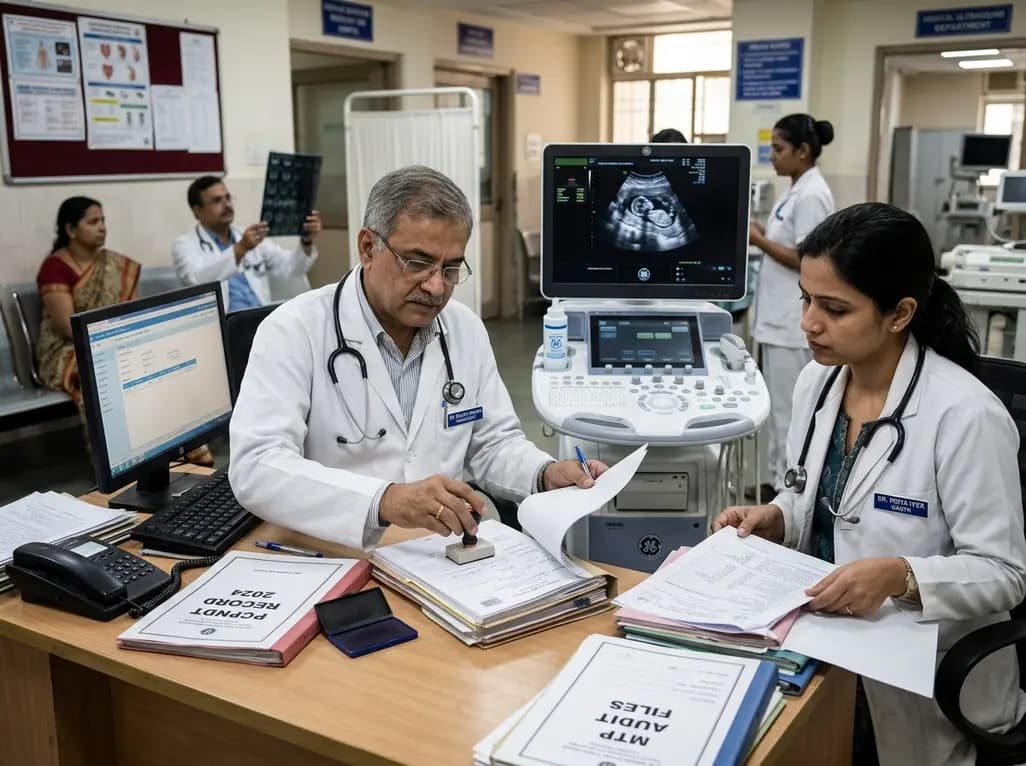
Maharashtra has initiated the appointment of two dedicated special public prosecutors in every district alongside a joint police-health SOP to fast-track PCPNDT and MTP cases. Medical imaging centers, radiologists, and obstetricians must strengthen statutory documentation and audit preparedness.
Today
Higher LACE+ scores independently predict non-home discharge and 90-day readmissions following intradural spinal tumor surgery. Utilizing this validated scoring index enables neurosurgical teams to stratify perioperative risk, improve transitional care, and optimize resource allocation.
Today

An exploratory pilot study evaluates metoprolol pharmacokinetics across cisgender males, cisgender females, and transgender women on gender-affirming hormone therapy. Findings reveal distinct drug disposition trends, emphasizing the critical need for genotype-stratified research in transgender cardiovascular care.
Today
