.webp&w=384&q=75)

Loading, please wait...
Loading, please wait...
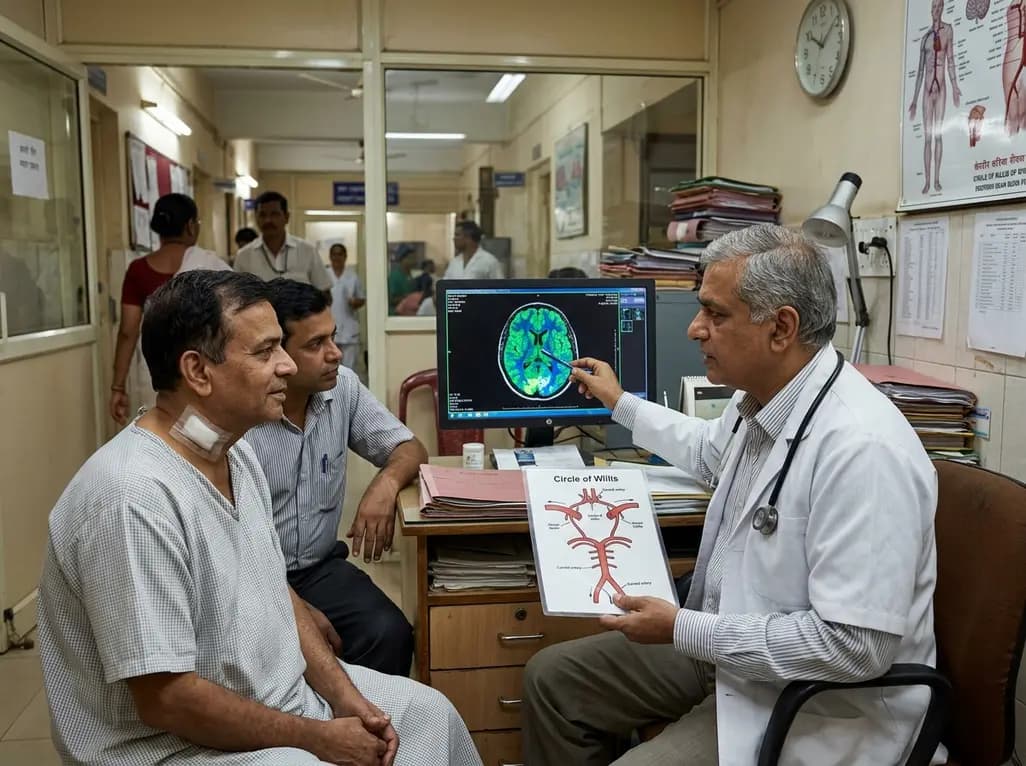
Asymptomatic carotid stenosis affects a substantial portion of the aging population globally. Clinicians frequently encounter severe stenosis in individuals who display no overt focal neurological deficits. However, cumulative neuroimaging evidence indicates that chronic hypoperfusion significantly impairs baseline neurocognitive capabilities. Whether surgical intervention restores cognitive ability remains a complex clinical debate. Recent scientific findings emphasize the crucial role of posterior cerebral perfusion in modulating cognitive recovery following surgical revascularization. By tracking cerebral hemodynamics preoperatively and postoperatively, researchers have identified key perfusion characteristics that dictate neurocognitive trajectories. Specifically, posterior cerebral perfusion represents a primary hemodynamic determinant of cognitive resilience. Understanding these physiological mechanisms allows vascular surgeons and neurologists to identify candidates who will truly benefit from carotid endarterectomy beyond mere primary stroke prevention.
Patients with unilateral severe asymptomatic carotid stenosis frequently present with subtle, unrecognized cognitive decline. Computed tomographic perfusion imaging demonstrates widespread hemodynamic alterations across major vascular territories. Although tissue hypoperfusion occurs most frequently within the anterior cerebral artery territory, severe functional deficits correlate more strongly with hemodynamic changes in secondary vascular networks. Consequently, researchers evaluated regional perfusion impairment across the anterior, middle, and posterior cerebral vascular zones. Patients exhibiting impaired posterior cerebral perfusion showed significantly lower baseline scores across global cognitive batteries, memory assessments, and executive functioning tests. Furthermore, quantitative electroencephalogram monitoring revealed reduced ipsilateral occipital gamma-band power in these specific patients. This objective electrophysiological finding confirms that posterior hemodynamic deficits directly compromise functional cortical activity prior to revascularization.
Carotid endarterectomy restores luminal diameter and enhances distal hemodynamic pressure. Following successful surgical intervention, a significant majority of preoperatively impaired cerebral regions show marked perfusion recovery. However, evaluating cognitive outcomes at an overall cohort level often masks significant subgroup variations. While aggregate cognitive scores across unselected patient cohorts show modest overall changes, specific physiological subgroups exhibit dramatic cognitive recovery. Quantitative electroencephalogram assessments after six months confirm that successful restoration of posterior cerebral perfusion directly normalizes cortical oscillations. Specifically, surgical revascularization restored occipital gamma-band power symmetry between hemispheres. This neurophysiological restoration strongly correlates with microvascular recovery and enhanced functional neuronal connectivity across posterior cortical structures.
Targeted analyses reveal that six-month post-operative cognitive gains depend heavily on baseline perfusion status. Patients who achieved measurable restoration of posterior cerebral perfusion demonstrated superior baseline-adjusted global cognitive scores compared to non-restored peers. Furthermore, these patients demonstrated profound improvements in executive function and visual-spatial memory tests. Because the posterior cerebral territory supplies crucial visual processing and associative integration centers, restoring hemodynamic flow directly resolves persistent ischemic suppression. Therefore, baseline posterior cerebral perfusion status serves as an effective physiological marker for identifying patients with reversible neurocognitive dysfunction. Clinicians must recognize that anterior revascularization positively influences posterior hemodynamics through collateral circulatory pathways.
Multivariate analytical models provide valuable clinical insights regarding predictive factors for postoperative neurocognitive improvement. Researchers identified concomitant vertebrobasilar stenosis as an independent predictor of favorable cognitive outcomes following carotid revascularization. Additionally, preoperative hypoperfusion localized to the cuneus region at the lateral ventricular plane strongly predicted post-operative global cognitive gains. These specific anatomical parameters highlight the intricate interdependence between the anterior and posterior intracranial arterial networks. Recognizing these imaging biomarkers assists multidisciplinary medical teams in refining patient selection algorithms. By integrating quantitative perfusion metrics into routine preoperative evaluations, clinicians can accurately identify asymptomatic individuals who are most likely to experience cognitive recovery.
These empirical insights redefine how clinicians evaluate asymptomatic carotid stenosis in daily practice. Relying solely on luminal narrowing percentages often fails to capture individual neurocognitive risk profiles. Incorporating computed tomographic perfusion parameters into clinical workflows provides a more nuanced understanding of microvascular reserve. Moreover, recognizing posterior cerebral perfusion as a mediator of cognitive outcomes offers clear practical guidance for patient counseling. Clinicians can confidently discuss potential cognitive benefits when specific posterior perfusion deficits are identified preoperatively. Future prospective trials will continue validating these physiological biomarkers to further standardize care guidelines across vascular surgery and interventional neurology specialties.
Carotid endarterectomy restores proper blood flow to hypoperfused brain regions. Although unselected patient groups show variable overall cognitive changes, individuals with baseline posterior cerebral perfusion deficits frequently experience substantial cognitive improvement. Restoring microvascular circulation improves global cognition, executive function, and memory performance after surgical revascularization.
The posterior cerebral territory supplies vital cortical structures involved in visual processing and memory integration. Chronic hypoperfusion in this territory severely reduces baseline cognitive reserve and alters cortical electrophysiological activity. Restoring posterior perfusion resolves underlying functional ischemia, leading to restored occipital gamma-band symmetry and measurable cognitive recovery.
Multivariate analyses demonstrate that concomitant vertebrobasilar stenosis and severe preoperative hypoperfusion in the cuneus region at the lateral ventricular plane serve as strong predictors. Identifying these specific hemodynamic and anatomical biomarkers allows clinicians to accurately select patients who will derive significant cognitive benefits from carotid revascularization.
Disclaimer: This content is for informational and educational purposes only and does not constitute medical advice, diagnosis, or treatment. Always consult a qualified healthcare provider regarding any clinical condition or procedure. Refer to the latest local and national guidelines for clinical practice.
References
Teng L et al. Posterior Cerebral Perfusion Associates With Cognitive Improvement After Carotid Endarterectomy in Asymptomatic Stenosis. J Am Heart Assoc. 2026 Aug 07. doi: 10.1161/JAHA.126.050735. PMID: 42568096.

Read summarized clinical updates, watch expert medical content, and earn CME certifications right from your smartphone.
Open your smartphone camera to scan and install.





A prospective cohort study demonstrates that posterior cerebral perfusion impairment and its post-operative restoration are strongly associated with cognitive recovery following carotid endarterectomy in asymptomatic severe carotid stenosis, identifying key anatomical predictors for patient selection.
last month

A retrospective cohort study demonstrates that comprehensive pharmacy transitions of care—including admission medication history, discharge review, and 72-hour telephone follow-up—significantly reduced 30-day readmission rates in acute care patients, highlighting the value of pharmacist-led clinical interventions.
Today

Meghalaya recorded a historic 55% reduction in maternal deaths, dropping from 244 to 107 over five years. Through the MOTHER app, dedicated transit homes, and expanded infrastructure like the 150-bed Tura Maternity Hospital with its first public NICU, the state demonstrates an effective public health model.
Today

Explore modern perioperative management protocols for clinical xenotransplantation. Learn how costimulation blockade, anti-inflammatory perfusion, and coagulopathy rescue overcome interspecies hurdles.
Today
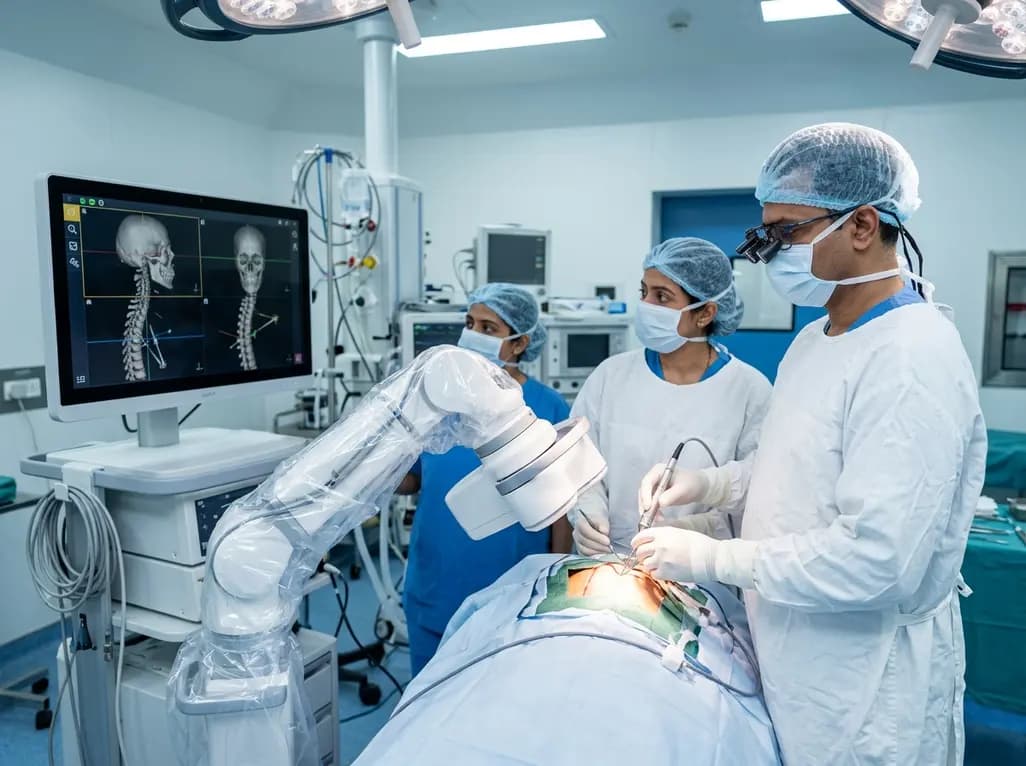
North India's first Centre of Excellence for Brain and Spine at Amrita Hospital Faridabad integrates Brainlab's Loop-X mobile robot. This landmark facility combines cone-beam computed tomography with surgical navigation to elevate precision, optimize operative workflows, and enhance specialized clinician training.
Today

At Homa CME 2026 at Hyderabad's T-Hub, healthcare leaders emphasized moving beyond routine metrics through digital calculators and precision stratification. Discover why characterizing individual insulin secretion, resistance patterns, and vascular risks transforms chronic metabolic care in India.
Today
