.webp&w=384&q=75)

Loading, please wait...
Loading, please wait...
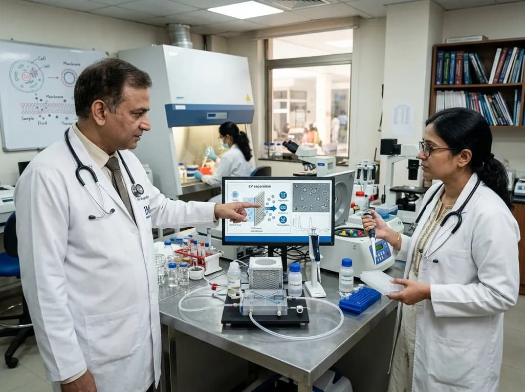
Extracellular vesicles have emerged as pivotal biological messengers in intercellular communication, liquid biopsies, and regenerative therapeutics. However, conventional isolation methods such as differential ultracentrifugation frequently face substantial bottlenecks in yield, purity, throughput, and operational complexity. Consequently, modern biomedical engineering is actively prioritizing next-generation extracellular vesicle processing through advanced porous membrane architectures. These sophisticated platforms move far beyond rudimentary size-exclusion filtration, serving as multifunctional interfaces for precise biomarker discovery and therapeutic interventions. By integrating tailored nanotechnology and surface modifications, researchers are fundamentally transforming how clinicians profile disease progression and manipulate pathological vesicles.
Historically, laboratory researchers and clinicians utilized standard porous membranes purely as physical sieves to isolate vesicles. These legacy systems separated vesicular structures from surrounding cellular debris based strictly on mechanical size cutoffs. While functional, standard filtration often caused severe membrane fouling, unpredictable clogging, shear-induced vesicle deformation, and reduced recovery rates. To overcome these persistent limitations, material scientists have engineered next-generation porous membranes featuring customized pore geometries and ordered nanoarchitectures. For instance, micro-machined and self-assembled membranes now provide exceptionally uniform pore dimensions and distribution. Therefore, these systems substantially optimize fluid dynamics and vesicle throughput during routine processing workflows. Additionally, novel surface chemistries minimize non-specific protein adsorption, ensuring high structural and molecular integrity of the isolated cargo. Consequently, clinical laboratories can process diverse physiological biofluids, including blood plasma, urine, and cerebrospinal fluid, with remarkable speed and reproducibility. Furthermore, advanced membrane platforms streamline diagnostic sample preparation, effectively bridging the gap between benchtop discovery and clinical implementation.
Recent breakthroughs in materials science have introduced highly functionalized membranes equipped with smart surface modifications. Notably, engineers have developed surface-enhanced membranes that enable direct plasmonic detection of intact vesicles on the membrane matrix. When coupled with localized surface plasmon resonance or Raman spectroscopy, these substrates facilitate ultra-sensitive, label-free optical quantification of disease-specific surface markers. Moreover, researchers can selectively interrogate and manipulate the biomolecular protein corona that naturally forms around vesicles in biological fluids. This corona characterization provides vital diagnostic insights regarding disease staging, immune interactions, and cellular origins. In addition, innovative membrane designs incorporate self-cleaning properties that dynamically mitigate biofouling during prolonged filtration procedures. Self-cleaning interfaces preserve constant permeation flux and eliminate the need for frequent, expensive filter replacements. By deploying patterned nanomembranes, manufacturers ensure high batch-to-batch consistency across mass-produced microfluidic cartridges. Ultimately, these structural refinements deliver exceptional analytical sensitivity while significantly lowering operational costs across high-throughput clinical workflows.
Beyond diagnostic sample isolation, next-generation porous membranes now serve as robust scaffolds for sophisticated biological modeling. Specifically, researchers incorporate these semi-permeable membranes into dynamic three-dimensional cell culture environments and organ-on-a-chip platforms. By establishing physiologically realistic cellular interfaces, these models accurately recreate complex biological barriers, such as the blood-brain barrier and alveolar-capillary interfaces. Consequently, scientists can track horizontal extracellular vesicle transfer between adjacent cell layers in real time under precise microfluidic conditions. Furthermore, these biomimetic barrier models elucidate how malignant cells shed oncogenic vesicles to remodel distant tissues and establish pre-metastatic niches. In addition, environmental monitoring systems utilize functionalized porous membranes to capture aerosolized or waterborne vesicles, expanding our understanding of environmental exposures and toxicology. Thus, porous membranes function as versatile biological platforms that deepen our mechanistic insights into intercellular communication during health, infection, and malignant transformation.
In contemporary oncology, liquid biopsy offers a minimally invasive paradigm for early cancer detection and dynamic longitudinal surveillance. Extracellular vesicles protect fragile microRNAs, mutated oncogenic DNA, signaling lipids, and transmembrane proteins from rapid enzymatic degradation in circulating blood. However, isolating these low-abundance disease biomarkers previously required cumbersome, labor-intensive workflows. Fortunately, next-generation membrane systems capture tumor-derived vesicles with high specificity, enabling immediate downstream molecular profiling. Clinicians can rapidly extract preserved oncogenic payloads to guide individualized chemotherapy regimens and targeted immunotherapies. In addition, serial vesicle profiling allows oncologists to monitor therapeutic responses, detect minimal residual disease, and recognize emergent drug resistance months before radiographic manifestation. Because porous membrane-based assays require minute sample volumes, they are particularly advantageous for fragile oncology patients and pediatric populations. Consequently, these point-of-care membrane assays will accelerate the widespread adoption of precision medicine across diverse healthcare settings.
The clinical potential of extracellular vesicles extends far beyond diagnostic biomarkers into direct therapeutic applications. For instance, bioengineers utilize porous membranes to harvest and purify therapeutic vesicles secreted by mesenchymal stem cells for regenerative tissue repair. Concurrently, membrane purification protocols ensure that engineered vesicles loaded with synthetic drugs or therapeutic RNA remain structurally intact and contaminant-free. Conversely, oncologists are actively investigating selective membrane-based apheresis devices for targeted vesicle removal. Malignant tumors frequently release immunosuppressive vesicles carrying programmed death-ligand 1 (PD-L1) to neutralize host cytotoxic T lymphocytes. By integrating affinity-functionalized porous membranes into extracorporeal circuits, clinicians can selectively filter out these immunosuppressive vesicles from the patient's bloodstream. Therefore, this targeted depletion therapy could restore systemic anti-tumor immunity and synergize effectively with immune checkpoint inhibitors. As a result, advanced membrane engineering provides promising therapeutic avenues for managing treatment-refractory malignancies and systemic inflammatory disorders.
The successful translation of advanced porous membrane platforms into routine clinical practice requires rigorous standardization and regulatory validation. Currently, international scientific consortia are establishing universal quality control parameters for membrane integrity, pore uniformity, and vesicle elution purity. Moreover, integrating automated membrane cartridges with microfluidic systems will minimize human technical error and facilitate high-volume sample processing in commercial pathology laboratories. In developing healthcare environments, cost-effective membrane chips could decentralize molecular diagnostics, making early cancer screening accessible outside major tertiary hospitals. Furthermore, interdisciplinary collaboration among oncologists, pathologists, materials scientists, and biomedical engineers will accelerate the commercialization of validated bedside platforms. As fabrication costs decrease and microfabrication improves, membrane-driven vesicle technologies will become indispensable instruments in everyday clinical practice. Ultimately, these membrane innovations will revolutionize cancer management, therapeutic delivery, and personalized clinical decision-making.
Traditional ultracentrifugation requires extensive processing time, expensive machinery, and high gravitational forces that frequently damage fragile vesicle membranes and cause protein aggregation. In contrast, next-generation porous membranes provide gentle, rapid, size-selective, and high-throughput separation. Moreover, customized pore architectures and anti-fouling surface chemistries preserve vesicle structural integrity while significantly enhancing purity and recovery yield. Consequently, membrane-based platforms offer a standardized, scalable approach suitable for routine clinical laboratories and point-of-care diagnostics.
Functionalized porous membranes enable ultra-sensitive capture and real-time detection of tumor-derived extracellular vesicles from circulating biofluids like blood or urine. By incorporating surface-enhanced plasmonic nanostructures and specific affinity ligands, these membranes isolate disease-specific vesicles without extensive sample pre-treatment. Therefore, clinicians can rapidly extract preserved oncogenic RNA, mutated DNA, and surface proteins. This capability facilitates early cancer detection, dynamic monitoring of treatment efficacy, and timely identification of therapeutic resistance mechanisms.
Malignant tumors release abundant extracellular vesicles that suppress immune surveillance, promote angiogenesis, and facilitate distant metastasis by transferring immunosuppressive factors like PD-L1. By employing specialized porous membranes within extracorporeal blood circuits, clinicians can selectively filter and deplete these pathogenic vesicles from circulation. Consequently, this targeted clearance eliminates tumor-mediated immunosuppression and restores cytotoxic T-cell activity. Furthermore, this therapeutic filtration strategy complements systemic immunotherapies, potentially overcoming drug resistance and improving patient survival outcomes.
Disclaimer: This content is for informational and educational purposes only and should not be considered medical advice. Always consult a qualified healthcare professional regarding any medical condition or treatment. Refer to the latest local and national guidelines for clinical practice.
References

Read summarized clinical updates, watch expert medical content, and earn CME certifications right from your smartphone.
Open your smartphone camera to scan and install.





Explore how next-generation porous membrane technologies are transforming extracellular vesicle processing, advancing cancer diagnostics, liquid biopsies, tissue barrier modeling, and targeted therapeutics.
4 weeks back
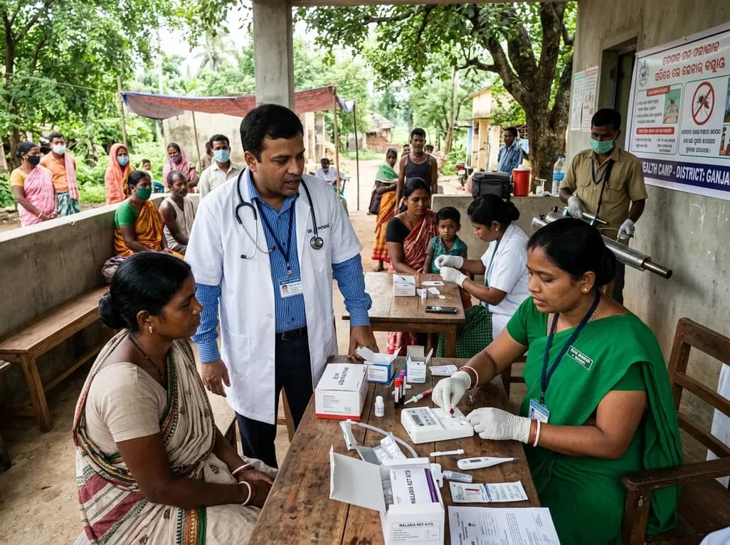
Following 37,899 reported cases and 9 deaths, Odisha has scaled up active case detection, rapid diagnostic testing, and vector control across high-burden districts. Here is a comprehensive clinical breakdown of the state's aggressive containment protocols, diagnostic pathways, and targeted field management.
Today
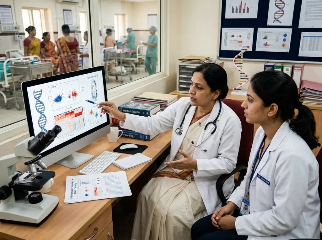
Endometriosis affects millions of women across India, yet diagnostic delays frequently exceed five years. Recent multi-centre genomic research has identified 21 novel genetic regions, notably on chromosome 13, establishing crucial population-specific insights to transform clinical screening and therapeutic strategies.
Today

Radiofrequency catheter ablation of the cavotricuspid isthmus successfully terminates typical atrial flutter. This article reviews the mechanisms driving coronary sinus electrogram changes, wavefront activation pathways, and clinical criteria to verify bidirectional block during electrophysiology studies.
Today
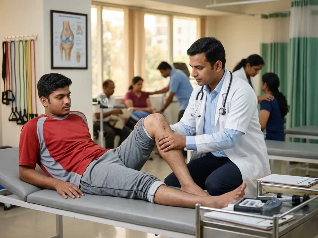
A study comparing four knee flexion strength tests after ACL reconstruction using hamstring tendon autografts reveals marked variability in limb symmetry indices. Clinicians must recognize that different strength testing modalities yield divergent return-to-sport clearance rates.
Today
Discover how to identify superimposed entrapment neuropathy in diabetic patients. This review details clinical evaluation, surgical decompression evidence, and a structured algorithm for primary care referrals to prevent ulcers and preserve limb function.
Yesterday
