.webp&w=384&q=75)

Loading, please wait...
Loading, please wait...
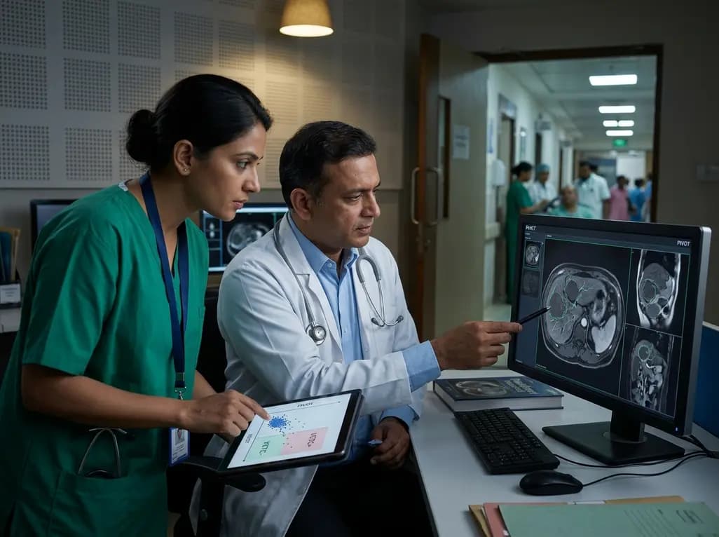
Primary liver cancer remains a major cause of cancer-related mortality across the globe, with hepatocellular carcinoma representing the overwhelming majority of cases. Despite surgical resections offering curative intent, disease recurrence frequently impairs long-term survival. Pathologists recognize vessels encapsulating tumor clusters as a distinct, highly aggressive vascular pattern linked to rapid metastatic spread. However, clinical teams historically identified this phenotype only through postoperative pathology. A landmark multicenter study now introduces a foundation model-enabled platform designed for accurate preoperative VETC prediction using routine clinical magnetic resonance imaging.
Vessels encapsulating tumor clusters represent a unique microvascular architecture in hepatocellular carcinoma. Unlike conventional microvascular invasion where isolated cancer cells invade endothelial walls, this phenotype envelops entire cohesive tumor clusters within continuous endothelial networks. Consequently, intact tumor clusters enter circulation without undergoing epithelial-mesenchymal transition. These aggregates evade host immune defenses and establish secondary metastatic foci with remarkable efficiency.
Multiple clinical studies confirm that this aggressive vascular structure drives early recurrence and reduces survival after liver resection. Nevertheless, confirming this morphology traditionally requires postoperative tissue analysis using CD34 immunohistochemistry. Because preoperative needle biopsies carry hemorrhage risks and suffer from sampling error, assessing this morphology before surgery has remained difficult. Therefore, noninvasive imaging biomarkers that reliably predict this aggressive signature address a vital clinical need.
To address this clinical barrier, researchers engineered PIVOT, an artificial intelligence framework powered by multimodal foundation models. Specifically, this platform bridges the gap between macroscopic imaging and microscopic cellular pathology. During model training, the team utilized paired preoperative multiparametric MRI scans, whole-slide histology images, and CD34 immunohistochemistry stains.
By aligning morphologic and vascular latent embeddings, the system identifies subtle radiological features corresponding to microscopic vessel encapsulation. Furthermore, ablation experiments proved that cross-modal alignment is critical. Indeed, removing histopathologic alignment branches caused the steepest performance decline.
Crucially, while development requires histological inputs, prospective inference relies solely on standard preoperative MRI scans. Consequently, clinicians obtain predictive scores without invasive biopsies, enabling rapid integration into routine surgical workflows.
The investigators demonstrated clinical reliability by evaluating the architecture across 2,052 patients with resectable hepatocellular carcinoma. This comprehensive multicenter retrospective study included an internal training cohort, an internal validation set, and four external validation cohorts. Across all evaluation subsets, the PIVOT-MRI model exhibited outstanding diagnostic precision.
Specifically, PIVOT-MRI achieved an area under the receiver operating characteristic curve of 0.887 in the internal test cohort. Moreover, it maintained strong accuracy across the four external cohorts, with values ranging between 0.839 and 0.861. These resilient metrics demonstrate robust generalizability across diverse imaging equipment and patient populations.
To enhance risk assessment, researchers built PIVOT-Clinical by combining imaging scores with serum alpha-fetoprotein and tumor size. This multimodal tool achieved an internal test area under the curve of 0.904, reliably stratifying two-year recurrence risks.
Beyond predictive accuracy, artificial intelligence models require biological plausibility to earn clinical adoption. Therefore, the investigators performed exploratory radiotranscriptomic analyses to investigate the genetic pathways underlying high PIVOT scores. Sequencing data revealed compelling biological concordance between noninvasive imaging predictions and aggressive tumor biology.
Importantly, tumors exhibiting elevated PIVOT-MRI scores demonstrated significant enrichment of transcriptional programs governing angiogenesis, endothelial cell activation, and cell proliferation. These pathways mirror the classic histological appearance of vessel-encapsulated clusters, where proliferating endothelial loops enclose tumor cell aggregates.
Additionally, the transcriptomic profile demonstrated activation of pro-metastatic signaling networks. In contrast, low-scoring tumors showed quiescent vascular programs. Consequently, these findings verify that PIVOT captures genuine underlying vascular biology rather than non-specific radiological artifacts, reinforcing confidence in its clinical validity.
Preoperative risk stratification fundamentally guides surgical planning and multidisciplinary care in liver cancer. Currently, hepatic resection provides curative potential, but early recurrence threatens long-term disease control. By delivering accurate vascular risk profiles before laparotomy, PIVOT empowers surgeons to personalize operative strategies effectively.
For example, identifying high-risk vascular phenotypes preoperatively could guide surgeons to choose anatomical resections with wider margins rather than limited wedge resections. Similarly, clinicians can prioritize intense postoperative surveillance protocols for high-risk patients. Moreover, these individuals may benefit substantially from neoadjuvant or adjuvant systemic regimens, including anti-angiogenic agents and checkpoint inhibitors.
Ultimately, integrating foundation model-based predictions into routine hepatology and surgical workflows represents a transformative advancement in oncology. As prospective studies validate these tools, preoperative computational pathology will guide risk-adapted surgical oncology decisions.
While these retrospective results show substantial promise, practical considerations will dictate clinical implementation. First, prospective clinical trials must evaluate whether treatment modifications based on algorithmic predictions improve patient survival. Multidisciplinary teams must determine whether altering resection margins or initiating adjuvant therapy based on model scores reduces recurrence rates.
Furthermore, standardizing image acquisition protocols across diverse MRI platforms remains essential. Health institutions must verify that variations in magnetic field strength and contrast timing do not impair diagnostic precision. Fortunately, foundation models display strong domain adaptability, which helps mitigate discrepancies across different hospital scanners.
In addition, combining radiomic assessments with circulating tumor cell assays and liquid biopsies could enhance prognostic accuracy. By integrating computational imaging, clinical biomarkers, and molecular testing, clinicians can achieve precise, personalized care for patients with hepatocellular carcinoma.
Vessels encapsulating tumor clusters represent a distinctive, highly aggressive vascular pattern seen in hepatocellular carcinoma. In this histological architecture, continuous endothelial cells wrap around cohesive tumor clusters instead of single invading cells. Consequently, intact tumor clusters enter circulation without undergoing epithelial-mesenchymal transition. This collective dissemination protects malignant cells against immune surveillance and shear stress, markedly accelerating early recurrence and compromising overall postoperative survival.
The PIVOT architecture incorporates multimodal foundation models trained on paired preoperative magnetic resonance imaging, whole-slide histology, and CD34 immunohistochemistry. By cross-aligning morphologic and vascular latent embeddings during model development, the network maps microscopic histological structures directly to macroscopic imaging features. During routine prospective clinical application, the system requires only standard multiparametric preoperative magnetic resonance scans. Thus, clinicians achieve accurate vascular phenotype predictions noninvasively without performing risky pre-resection biopsies.
Preoperative identification of aggressive vascular encapsulation empowers multidisciplinary teams to personalize hepatocellular carcinoma treatment. Surgeons encountering high-risk scores can optimize resection margins by selecting anatomical hepatectomies over narrow margin resections. Furthermore, oncologists can consider neoadjuvant or adjuvant systemic regimens, such as tyrosine kinase inhibitors or immunotherapy, for high-risk patients. Finally, clinical teams can schedule more frequent postoperative surveillance imaging to detect and treat early recurrences promptly.
Disclaimer: This content is for informational and educational purposes only. It is not intended to substitute professional medical advice, diagnosis, or treatment. Clinicians must exercise their independent judgment when evaluating clinical conditions. Refer to the latest local and national guidelines for clinical practice.
References

Read summarized clinical updates, watch expert medical content, and earn CME certifications right from your smartphone.
Open your smartphone camera to scan and install.





A multicenter retrospective study of 2,052 patients validates PIVOT, an AI foundation model that predicts vessels encapsulating tumor clusters (VETC) from preoperative MRI in hepatocellular carcinoma, enhancing risk stratification for recurrence.
Today
Health authorities in the Democratic Republic of Congo confirmed the spread of Ebola to North Ubangi province. With cross-border infections and high case counts, healthcare professionals worldwide must review early clinical recognition, differential diagnosis, infection control measures, and isolation protocols.
Today
Surgical oncologists at AIIMS Delhi have mapped two critical anatomical landmarks: Ray's Zone in the deep pelvis and Ray's Hepatocaval Triangle near the liver. These predefined hazard boundaries guide dissection, dramatically reduce severe intraoperative bleeding, and preserve vital nerves and visceral organs.
Today
A study reveals that the MYLK c.4079G>A, p.(Gly1360Asp) variant is linked to high-penetrance heritable thoracic aortic disease and early-onset aortic dissection, showing a founder effect in Finnish cohorts. Findings emphasize early cascade screening and vigilant surveillance.
Today
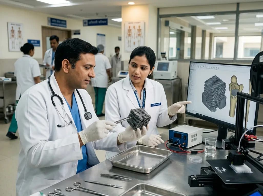
A novel sacrificial-template technique combines FDM polymer 3D printing and micro-arc oxidation to fabricate orientation-controlled porous titanium scaffolds. These bioactive constructs offer tailored stiffness, exceptional multidirectional load resistance, and robust osseointegration for bone defect repair.
Today
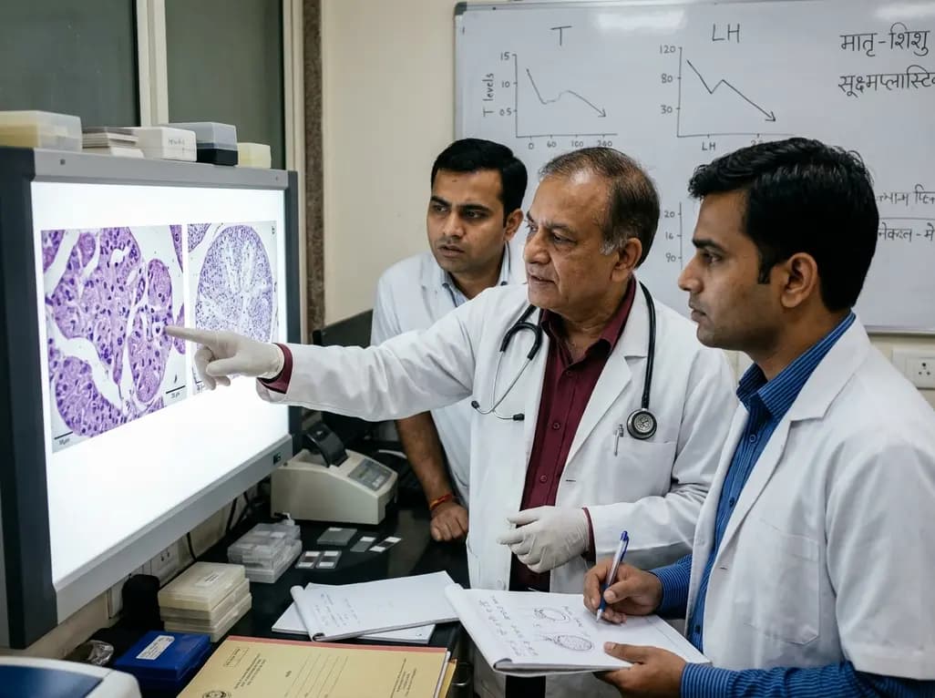
An animal model study reveals that gestational exposure to polystyrene microplastics causes persistent intrauterine growth restriction, testicular hypoplasia, Leydig cell depletion, and an atypical low-testosterone, low-LH endocrine profile in male offspring from birth into adulthood.
Today
