.webp&w=384&q=75)

Loading, please wait...
Loading, please wait...
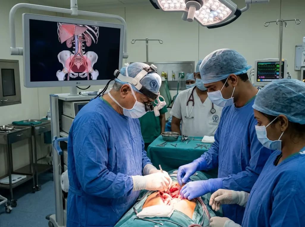
Pelvic oncological procedures frequently present formidable technical hurdles for operative teams across tertiary cancer centers. Performing a complex cancer surgery often requires extensive retroperitoneal dissection close to delicate neurovascular structures. Recently, surgical oncologists at the All India Institute of Medical Sciences (AIIMS) in New Delhi delineated two novel danger territories. Consequently, these landmarks provide clear boundaries that safeguard critical anatomy during radical resection.
Surgeons routinely face life-threatening bleeding when they operate within narrow pelvic planes. Therefore, standardizing anatomical landmarks remains vital to optimize surgical safety and patient survival. Dr. M. D. Ray, a professor of surgical oncology at Dr. BRA-IRCH, AIIMS Delhi, led an extensive clinical initiative to address this issue. His investigative team systematically reviewed 255 patients who underwent radical or modified radical hysterectomies for cervical, endometrial, and ovarian malignancies.
Initially, early cases in the cohort demonstrated substantial intraoperative hemorrhage and inadvertent nerve traction. Therefore, the team thoroughly analyzed specific zones where injuries occurred most frequently. Consequently, researchers formulated an anatomical framework designed to improve surgical precision before dividing tissue planes. Specifically, surgical teams identify the crossing ureters and vascular branches with heightened vigilance. Furthermore, this systematic mapping provides operating teams with an anatomical blueprint for complicated retroperitoneal work. Instead of using blunt dissection, oncologists can anticipate high-risk tissue planes early. Moreover, avoiding blind traction significantly reduces damage to the underlying somatic nerve bundles. As a result, surgical teams systematically minimize unforeseen intraoperative trauma while maintaining clear oncological margins. Ultimately, this structured protocol transforms unpredictable pelvic debulking into a reproducible operative workflow.
The AIIMS team designated the vulnerable territory below the uterine vessels as the Red Alert Zone or Ray's Zone. Geographically, this compact pelvic space harbors the distal ureter, urinary bladder base, sympathetic nerve fibers, and major vascular branches. Because these vital structures lie in immediate proximity, accidental damage can produce severe functional deficits.
Traditionally, aggressive traction or inadvertent lateral spread from energy devices causes covert thermal injuries to the ureter. In addition, disruption of the inferior hypogastric plexus frequently induces chronic neurogenic bladder dysfunction. Postoperative urinary retention and loss of rectal sensation severely diminish quality of life for cancer survivors. Similarly, brisk bleeding from deep paracervical venous plexuses obscures visualization, forcing hasty hemostasis that endangers adjacent conduits.
To counter these hazards, identifying Ray's Zone early establishes a clear perimeter of caution. Surgeons recognize this space as a stop-and-inspect boundary where blind dissection must halt completely. Moreover, defining this specific pelvic area allows trainees to recognize subtle anatomical relationships before entering treacherous planes. By isolating the pelvic ureter and maintaining safe distances from pelvic splanchnic nerves, oncologists achieve radical clearance without sacrificing organ preservation.
Following their initial analysis, the surgical oncology unit implemented targeted technique modifications in 105 consecutive cases. Primarily, the refined protocol mandates meticulous anatomical visualization under optical magnification whenever necessary. Consequently, surgeons identify minute neurovascular networks that standard macroscopic exploration might otherwise miss. Indeed, adequate magnification ensures crystal-clear margins around sensitive pelvic visceral boundaries.
Furthermore, the team replaced blind mass clamping with precise individual vessel skeletonization and ligation. Surgeons now avoid excessive cautery near the ureteric adventitia to prevent vascular devascularization and ischemic fistula formation. Instead, they utilize fine hemoclips or delicate sutures to secure fragile paracervical venous channels individually. Meanwhile, gentle hydrodynamic or sharp micro-dissection separates autonomic nerves cleanly from malignant pelvic fascia.
Importantly, these methodological refinements yielded significant reductions in estimated operative blood loss among treated patients. Patients experienced fewer intraoperative hypotensive episodes, thereby reducing the demand for perioperative allogeneic blood transfusions. In addition, preservation of bladder innervation prevented protracted urinary catheterization after surgery. Patients recovered independent voiding function substantially faster than historical controls. Accordingly, surgical recovery progressed smoothly without recurrent pelvic hematomas. Thus, technical discipline within Ray's Zone directly translated into improved perioperative recovery and lower postoperative morbidity.
Beyond the female pelvis, Dr. Ray characterized an equally hazardous region in upper abdominal oncology known as Ray's Hepatocaval Triangle. This critical operative space resides near the junction of the inferior vena cava and major hepatic veins. During extensive hepatic resections or cytoreductive procedures for advanced ovarian cancer, mobilization of the liver becomes essential. However, the hepatocaval junction presents extreme surgical risks due to thin-walled vessels carrying massive blood volumes.
Uncontrolled traction or blunt finger manipulation in this tight junction can easily avulse short retrohepatic veins. Consequently, catastrophic hemorrhage may ensue within seconds, leading to profound hemodynamic collapse or fatal air embolism. Therefore, delineating Ray's Hepatocaval Triangle provides hepatobiliary surgeons with a clear operative reference point before dividing posterior ligaments.
Specifically, the technique emphasizes deliberate cephalad and lateral traction while maintaining continuous visual control over the retrohepatic cava. Surgeons divide the surrounding peritoneal reflections sharply to expose individual vascular tributaries before applying vascular clamps. Moreover, having a standardized anatomical triangle allows the surgical team to coordinate vascular control maneuvers smoothly with the anesthesia team. Ultimately, this structured approach converts a high-stress, perilous dissection step into a controlled, reproducible procedure.
The establishment of these two anatomical danger zones reinforces a universal surgical maxim: recognize boundaries before entering danger. By conceptualizing Ray's Zone and Ray's Hepatocaval Triangle as anatomical warning zones, clinicians enhance spatial orientation in demanding fields. Indeed, cognitive anatomical roadmaps prevent unintentional surgical deviations. As a result, operative precision improves markedly. Consequently, surgeons transition from reactive crisis management to proactive risk mitigation.
Furthermore, these standardized anatomical concepts hold substantial educational value for surgical oncology residency and fellowship training across India. Novice surgeons often struggle to conceptualize three-dimensional relationships in distorted retroperitoneal or subdiaphragmatic spaces. Therefore, codifying clear anatomical triangles and warning zones provides tangible reference points during complex ablative operations. Surgical mentors can teach trainee oncologists where to deploy magnification, where to curb thermal energy, and where to dissect cautiously.
In addition, preserving autonomic nerves and vascular integrity directly correlates with superior long-term survival metrics. When patients experience fewer surgical complications, they initiate necessary adjuvant chemotherapy or radiotherapy without debilitating delays. Moreover, avoiding chronic pelvic pain, neurogenic bladder, and fecal incontinence allows cancer survivors to regain productive daily lives. Thus, systematic anatomical mapping represents a vital step toward reconciling radical tumor clearance with compassionate functional preservation.
Q1: What anatomical structures define the boundary of Ray's Zone?
Ray's Zone, also designated as the Red Alert Zone, encompasses the retroperitoneal anatomical space situated directly below the uterine vascular pedicles. Crucially, this compact territory contains the distal ureter, the base of the urinary bladder, and branches of the inferior hypogastric nerve plexus. In addition, it houses delicate paracervical venous networks. Identifying this boundary alerts operating teams to exercise extreme care during radical hysterectomy to prevent irreversible visceral injuries.
Q2: Why is Ray's Hepatocaval Triangle considered a high-risk landmark?
Ray's Hepatocaval Triangle designates the delicate junction where major hepatic veins and short retrohepatic branches enter the inferior vena cava. Because these large vascular channels possess extremely thin walls, inadvertent tearing from blunt traction causes massive hemorrhage within seconds. Furthermore, negative thoracic pressure in this region significantly increases the acute risk of fatal venous air embolism. Consequently, surgeons must employ sharp dissection, precise vascular isolation, and direct visual guidance before dividing local ligamentous attachments.
Q3: How does dissecting within Ray's Zone reduce postoperative morbidity?
Controlled dissection within Ray's Zone systematically protects autonomic pelvic nerve fibers that regulate normal urinary bladder voiding, rectal continence, and sexual function. Furthermore, avoiding excessive electrocautery near the pelvic ureter prevents microvascular ischemia, subsequent necrosis, and debilitating ureterovaginal fistulas. Patients treated with these targeted anatomical refinements recover normal physiological voiding much faster. Therefore, they avoid prolonged urethral catheterization, experience fewer pelvic infections, and achieve a significantly higher postoperative quality of life after cancer surgery.
Disclaimer: This content is for informational and educational purposes only. It does not constitute medical advice or replace professional judgment. Refer to the latest local and national guidelines for clinical practice.
References

Read summarized clinical updates, watch expert medical content, and earn CME certifications right from your smartphone.
Open your smartphone camera to scan and install.





Surgical oncologists at AIIMS Delhi have mapped two critical anatomical landmarks: Ray's Zone in the deep pelvis and Ray's Hepatocaval Triangle near the liver. These predefined hazard boundaries guide dissection, dramatically reduce severe intraoperative bleeding, and preserve vital nerves and visceral organs.
Today

Health authorities in the Democratic Republic of Congo confirmed the spread of Ebola to North Ubangi province. With cross-border infections and high case counts, healthcare professionals worldwide must review early clinical recognition, differential diagnosis, infection control measures, and isolation protocols.
Today

A study reveals that the MYLK c.4079G>A, p.(Gly1360Asp) variant is linked to high-penetrance heritable thoracic aortic disease and early-onset aortic dissection, showing a founder effect in Finnish cohorts. Findings emphasize early cascade screening and vigilant surveillance.
Today

A cross-sectional study reveals that reduced upper extremity exercise capacity measured by the 6-minute pegboard and ring test significantly correlates with higher disability in non-specific chronic low back pain, highlighting the necessity of assessing whole-body functional chains in rehabilitation.
Today
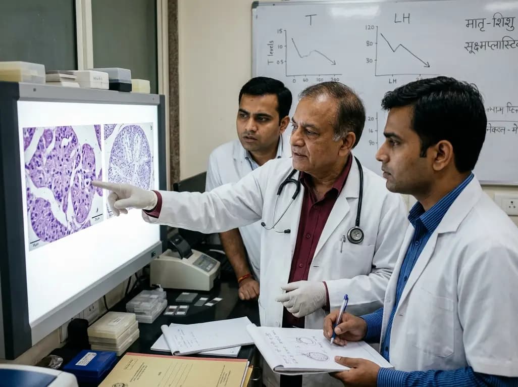
An animal model study reveals that gestational exposure to polystyrene microplastics causes persistent intrauterine growth restriction, testicular hypoplasia, Leydig cell depletion, and an atypical low-testosterone, low-LH endocrine profile in male offspring from birth into adulthood.
Today
