.webp&w=384&q=75)

Loading, please wait...
Loading, please wait...

Quantitative assessment of ventricular mechanics remains a cornerstone of modern diagnostic cardiology. However, accurately measuring transmural myocardial deformation has posed longstanding challenges for non-invasive imaging modalities. Standard two-dimensional speckle-tracking echocardiography often fails to capture through-plane tissue motion, which limits its ability to characterize complex intra-myocardial stress and strain. Although three-dimensional echocardiography theoretically resolves geometric out-of-plane errors, noisy epicardial boundaries and acoustic dropouts frequently degrade tracking accuracy. Consequently, researchers have introduced a physics-informed computational framework that incorporates biomechanical constraints directly into 3D echocardiographic tracking. This breakthrough methodology allows clinicians to reconstruct the complete finite strain tensor across the left ventricular wall with high fidelity.
The human myocardium consists of a complex, multilayered architecture of helical myofibers that contract, twist, and thicken dynamically throughout the cardiac cycle. Subendocardial fibers run longitudinally, whereas mid-wall fibers encircle the ventricle and subepicardial fibers maintain an opposing oblique orientation. Because different pathological states disproportionately affect specific layers, evaluating transmural myocardial deformation provides crucial diagnostic information. For instance, subendocardial ischemia typically diminishes longitudinal shortening early, while transmural infarction disrupts circumferential and radial mechanics entirely.
Traditional two-dimensional strain imaging only evaluates planar projections of this intricate three-dimensional motion. Therefore, subtle regional dysfunctions often remain hidden behind compensatory hyperkinesis of adjacent layers. By resolving deformation across the entire wall thickness, clinicians can detect early subclinical dysfunction in conditions such as hypertensive heart disease, amyloidosis, and non-ischemic cardiomyopathies. Moreover, transmural strain analysis provides fundamental insights into mechanical dyssynchrony and adverse remodeling patterns, enabling more targeted therapeutic strategies.
The newly developed framework integrates image processing with continuum mechanics to overcome traditional speckle-tracking limitations. Initially, automated algorithms segment the endocardial and epicardial boundaries from volumetric 3D echocardiographic datasets. The tracking algorithm then follows acoustic speckle patterns across sequential frames of the cardiac cycle. However, rather than relying solely on image intensity correlations, the model enforces an optimization framework governed by physical laws.
Specifically, the system applies a soft volumetric penalty that permits physiological volume changes at a finite mathematical cost. This biomechanical regularization ensures that myocardial tissue behaves in a physically plausible manner, preserving continuous tissue motion and geometric smoothness. Consequently, the computational solver reconstructs the full three-dimensional finite strain tensor rather than fragmented velocity vectors. The resulting spatially resolved principal strain fields realistically capture the continuous strain distribution across the full myocardial thickness from endocardium to epicardium.
One of the greatest historical obstacles in 3D speckle tracking is the poor acoustic delineation of the epicardium. Because ultrasound attenuation and rib shadowing frequently obscure the outer ventricular boundary, standard automated algorithms generate substantial tracking noise and artificial drift. In contrast, the physics-informed architecture stabilizes regional tracking by coupling epicardial motion directly to endocardial kinematics through constitutive continuum mechanics.
Furthermore, this optimization strategy exhibits remarkable robustness against manual and semi-automated segmentation variability. Even when initial boundary definitions contain minor operator errors, the biomechanical constraints correct unnatural spatial discontinuities. As a result, the reconstructed deformation field maintains physical consistency throughout systole and diastole. Clinicians obtain reproducible global and regional strain indices without encountering the severe artifact-driven variance that previously limited the widespread adoption of 3D echocardiographic strain tools.
To establish clinical credibility, researchers validated this physics-informed echocardiographic framework against cardiac magnetic resonance (CMR) imaging. Cardiovascular magnetic resonance remains the reference standard for non-invasive myocardial characterization due to its superior spatial resolution and tissue contrast. In head-to-head comparisons, global longitudinal strain and global circumferential strain measurements derived from the novel 3D echocardiographic technique demonstrated strong agreement with CMR strain metrics.
Beyond standard global parameters, the framework demonstrated high correlation with CMR when mapping transmural strain gradients across distinct ventricular segments. The model successfully replicated the physiological gradient where endocardial deformation exceeds epicardial strain during normal systolic contraction. Importantly, this concordance validates that echocardiography, when guided by fundamental physical laws, can achieve diagnostic precision comparable to advanced magnetic resonance imaging while remaining faster, more cost-effective, and fully portable at the point of care.
Integrating physics-informed deformation analysis into bedside ultrasound could transform cardiovascular risk stratification and monitoring. Point-of-care echocardiography is widely available across secondary and tertiary medical centers, unlike high-cost cardiac magnetic resonance facilities. Therefore, providing clinicians with high-fidelity, MRI-validated transmural strain data through standard 3D ultrasound scanners bridges a crucial diagnostic accessibility gap.
In coronary artery disease, layer-specific strain assessment facilitates precise differentiation between non-transmural and transmural myocardial infarction without requiring immediate gadolinium administration. Similarly, in patients undergoing chemotherapy, the technique could identify early anthracycline-induced cardiotoxicity at the subendocardial level before left ventricular ejection fraction declines. As research expands to broader clinical cohorts, this physics-informed methodology holds immense potential to enhance valvular assessments, optimize cardiac resynchronization therapy planning, and improve long-term prognostic modeling across diverse patient populations.
Transmural myocardial deformation refers to the complex mechanical contraction, thickening, and shortening that occurs across all layers of the heart muscle wall, including the endocardium, mid-wall, and epicardium. Analyzing these layer-specific mechanical properties helps cardiologists identify subtle structural abnormalities and accurately assess ischemic damage throughout the cardiac cycle.
Conventional echocardiography tracks image pixels solely based on visual contrast, which frequently fails when image quality degrades. In contrast, physics-informed tracking applies biomechanical laws and soft volumetric penalties during optimization. This ensures that calculated tissue deformations remain physically realistic, substantially reducing epicardial tracking errors and smoothing out imaging noise.
Cardiac magnetic resonance serves as the gold standard for non-invasive ventricular quantification due to its high spatial resolution. Demonstrating that physics-informed 3D echocardiography closely matches cardiac magnetic resonance measurements proves that bedside ultrasound can reliably deliver advanced biomechanical insights with accuracy previously attainable only through expensive scanning platforms.
Disclaimer: This content is for informational and educational purposes only and should not be construed as medical advice. Healthcare professionals must exercise their independent clinical judgment when evaluating imaging modalities and patient data. Refer to the latest local and national guidelines for clinical practice.
References
Pradhan SP et al. Physics-Informed Reconstruction of Transmural Myocardial Deformation from 3D Echocardiography: Validation Against Cardiac MRI. Ann Biomed Eng. 2026 Aug 13. doi: 10.1007/s10439-026-04335-y. PMID: 42593608.
Muraru D, Niero A, Rodriguez-Zanella H, Cherata D, Badano L. Three-dimensional speckle-tracking echocardiography: benefits and limitations of integrating myocardial mechanics with three-dimensional imaging. Cardiovasc Diagn Ther. 2018;8(1):101-117. doi:10.21037/cdt.2017.06.11.
Scatteia A, Baritussio A, Bucciarelli-Ducci C. Strain imaging using cardiac magnetic resonance. Heart Fail Rev. 2017;22(4):465-476. doi:10.1007/s10741-017-9621-8.

Read summarized clinical updates, watch expert medical content, and earn CME certifications right from your smartphone.
Open your smartphone camera to scan and install.





A novel physics-informed framework for 3D echocardiography accurately reconstructs transmural myocardial deformation and the full 3D strain tensor across the ventricular wall, showing excellent agreement with cardiac MRI and offering deeper insights into regional cardiac biomechanics.
last month

A secondary analysis of an HIV cure-related trial in South Africa highlights the acceptability and safety of serial excisional lymph node and gut biopsies among young women. Clear communication, empathetic care, and altruistic motives helped mitigate initial apprehension and sustain longitudinal trial engagement.
Today

A nationwide study covering 3.7 million Swedish births reveals that prenatal exposure to TORCH pathogens significantly elevates long-term risks of intellectual disability and autism. Healthcare providers must recognize clinical indicators, promote prenatal prevention, and track early pediatric neurodevelopment.
Today
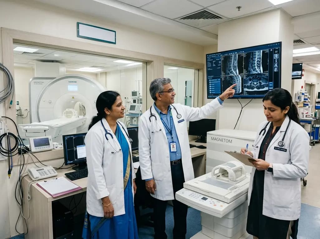
Under India's PLI initiative, domestic production of 57 critical medical devices has commenced, including MRI and CT scanners, cath labs, and heart valves. Global partnerships and local investments are expanding high-end technology, curbing import reliance, and strengthening clinical accessibility nationwide.
Today

A longitudinal study reveals that patients undergoing staged bilateral hip arthroscopy experience progressive contralateral labral deterioration during surgical intervals. However, structural degradation does not compromise 2-year postoperative clinical outcomes or patient-reported satisfaction scores.
Today

New immunological research reveals how PTPN22 forms a delayed inhibitory signaling complex to restrain T cell activation, explaining how the autoimmune-linked R620W variant disrupts feedback control and promotes persistent inflammation in conditions like rheumatoid arthritis and type 1 diabetes.
Today
