.webp&w=384&q=75)

Loading, please wait...
Loading, please wait...
Human social interaction relies heavily on non-verbal communication channels. Among these subtle signals, personal space regulation plays a fundamental protective and communicative role. Personal space describes the immediate physical area surrounding the body where intruders evoke subjective discomfort. For decades, clinicians have observed that individuals with psychotic spectrum disorders often demand greater interpersonal distance. However, the precise neurobiological circuitry driving these defensive boundaries has remained poorly understood. Researchers have recently identified distinct neural pathways that control these interpersonal boundaries. In healthy individuals, frontoparietal networks coordinate sensory inputs to determine physical boundaries smoothly. Conversely, individuals living with schizophrenia frequently experience impaired boundary calibration. This alteration often leads to heightened feelings of intrusion, social withdrawal, and difficulty navigating crowded spaces. Consequently, identifying the exact cortical nodes involved in spatial regulation provides critical biological insights into psychiatric pathology. By bridging neuropsychological assessments with functional neuroimaging, clinicians can better understand why interpersonal discomfort emerges during early and chronic psychosis. This knowledge clarifies the complex link between sensory appraisal, non-verbal communication, and clinical symptom severity in psychiatric disorders.
Dynamic social interactions require rapid neural processing of approaching objects and human faces. Neuroimaging studies demonstrate that a distributed frontoparietal-subcortical network governs spatial appraisal. Within this network, the inferior parietal cortex functions as an essential sensory integration hub. The inferior parietal cortex continuously computes egocentric spatial coordinates and tracks looming threats entering peripersonal space. Simultaneously, subcortical regions such as the amygdala, hippocampus, and striatum process emotional salience and defensive responses. In healthy populations, parietal and frontal cortical areas show heightened activation when stimuli approach the body. Subcortical structures, in contrast, display greater responsiveness during stimulus withdrawal. Recent functional neuroimaging data indicate that individuals with psychotic disorders display altered functional connectivity between the inferior parietal cortex and the ventral or limbic striatum. Furthermore, this approach-biased activation within the inferior parietal cortex correlates directly with physical interpersonal distance requirements. Therefore, disrupted frontoparietal communication distorts distance perception. When frontoparietal modulation falters, subcortical threat networks activate prematurely. As a result, patients perceive distant social contacts as immediate intrusions, prompting compensatory behavioral retreat and persistent social avoidance.
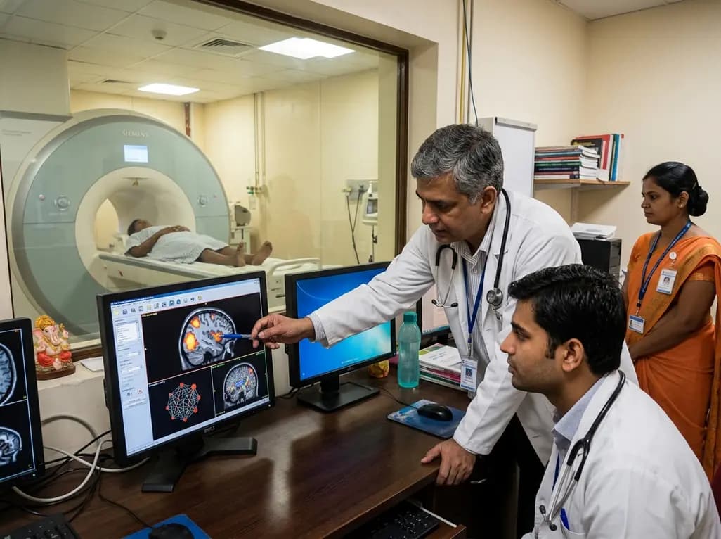
To elucidate these neural pathways, investigators conducted a detailed functional magnetic resonance imaging trial evaluating patients with psychotic disorders and demographically matched healthy controls. During the experimental protocol, participants observed dynamic face stimuli that either moved toward them or retreated into the background. In addition, investigators evaluated resting-state functional connectivity and quantified physical personal space size using the validated Stop Distance Procedure. The behavioral outcomes confirmed that individuals with psychosis maintain significantly larger personal space boundaries than healthy controls. Neuroimaging results revealed that both groups exhibited approach-biased responses in frontoparietal regions alongside withdrawal-biased activity in subcortical structures. Notably, only patients with psychotic disorders showed a robust correlation between personal space dimensions and approach-biased activation within the inferior parietal cortex. Furthermore, the strength of functional connectivity between the inferior parietal cortex and the ventral striatum strongly predicted the degree of interpersonal boundary expansion. These neuroimaging discoveries highlight a specific neurophysiological divergence in how psychotic brains process looming social stimuli. Consequently, frontoparietal-striatal dysconnectivity directly underpins the subjective need for increased physical distance.
Aberrant interpersonal distance regulation is not merely an isolated behavioral curiosity; rather, it reflects profound disruptions in social cognition and motivational processing. In clinical practice, psychiatrists frequently encounter negative symptoms such as social withdrawal, blunted affect, and avolition. Research indicates that personal space expansion and reduced boundary permeability correlate with social anhedonia and negative symptom burdens. When patients feel threatened by normative social distances, they naturally retreat from social environments to avoid distress. Consequently, clinicians must recognize that expanded physical boundaries represent an adaptive physiological response to sensory over-reactivity rather than passive indifference. Moreover, evaluating interpersonal distance during clinical interviews can provide objective behavioral markers of underlying distress or paranoia. By measuring how patients maintain physical space, clinicians can assess the functional integrity of frontoparietal circuits non-invasively. Therefore, incorporating objective behavioral metrics into routine clinical workflows enhances diagnostic precision. Furthermore, understanding the neural basis of spatial discomfort helps clinicians avoid unintentional boundary violations during bedside interactions, fostering greater therapeutic alliance and patient trust.
The identification of specific cortical targets opens transformative avenues for psychiatric rehabilitation and novel therapeutic interventions. Currently, pharmacological treatments for schizophrenia manage positive psychotic symptoms effectively but offer limited benefit for social cognition deficits. Because inferior parietal cortex connectivity correlates directly with measurable interpersonal distance, this circuit represents an objective target for neuromodulation. For instance, non-invasive brain stimulation techniques, such as repetitive transcranial magnetic stimulation or transcranial direct current stimulation, could modulate parietal excitability. Similarly, targeted cognitive remediation therapy and virtual reality training paradigms can systematically retrain spatial perception. By practicing interpersonal interactions in controlled virtual environments, patients can gradually habituate to approaching social stimuli without experiencing overwhelming anxiety. Additionally, biofeedback interventions could help patients regulate autonomic arousal when interpersonal boundaries are challenged. As translational research progresses, combining non-invasive cortical stimulation with behavioral desensitization may restore healthy frontoparietal-striatal communication. Ultimately, these targeted neuromodulatory strategies hold substantial promise for alleviating persistent social withdrawal and improving quality of life in psychotic illness.
Medical practitioners across psychiatry, neurology, and primary care must translate these spatial neurobiology findings into daily clinical encounters. First, clinicians should maintain larger, respectful interpersonal distances during physical and psychiatric evaluations, particularly with acutely symptomatic patients. Standing or sitting too close may inadvertently provoke parietal hyperactivity and subcortical threat responses, worsening agitation or persecutory ideation. Second, healthcare teams should view social withdrawal through a neurobiological lens, recognizing that spatial boundaries reflect altered sensory processing rather than uncooperativeness. Third, multidisciplinary teams should explore behavioral retraining protocols that incorporate gradual spatial habituation for patients experiencing chronic functional impairment. In addition, mental health professionals should monitor changes in personal space requirements as potential surrogate markers of therapeutic progress or impending relapse. Clinicians should also educate family members and caregivers regarding peripersonal space sensitivity to reduce household friction. By aligning clinical communication strategies with underlying neurocircuitry, healthcare providers can cultivate a safer therapeutic environment, optimize patient engagement, and advance holistic psychiatric care across diverse clinical settings.
Individuals with psychotic disorders typically maintain significantly larger personal space boundaries than healthy individuals. Their brains show altered inferior parietal cortex activation and heightened subcortical threat reactivity during social approach. Consequently, normal interpersonal distances feel intrusive, causing distress, reduced spatial permeability, and compensatory behavioral withdrawal during everyday social interactions.
The inferior parietal cortex acts as a central hub for multisensory integration and spatial mapping around the body. It processes egocentric distance and evaluates approaching stimuli. In psychosis, disrupted functional connectivity between the inferior parietal cortex and the ventral striatum directly correlates with expanded personal space requirements.
Yes, measuring personal space boundaries provides an objective, non-invasive behavioral marker of frontoparietal network integrity and social cognitive functioning. Because expanded boundaries correlate with negative symptom severity, paranoia, and social anhedonia, tracking interpersonal distance changes can assist clinicians in assessing treatment response, relapse risk, and social recovery.
Disclaimer: This content is for informational and educational purposes only and should not be considered medical advice. Always consult a qualified healthcare professional regarding any medical condition or before making healthcare decisions. Refer to the latest local and national guidelines for clinical practice.
References

Read summarized clinical updates, watch expert medical content, and earn CME certifications right from your smartphone.
Open your smartphone camera to scan and install.





A recent fMRI study reveals that personal space expansion in psychotic disorders stems from altered inferior parietal cortex function and frontoparietal-striatal dysconnectivity, providing critical biomarkers for negative symptoms and novel neuromodulatory targets.
4 weeks back

Left bundle branch pacing is an emerging physiological resynchronization strategy for heart failure. While observational data and some trials show benefits over biventricular pacing, divergent trial outcomes highlight the importance of confirmed conduction capture and specialized operator technique.
Today

Postoperative adhesions cause severe complications, including bowel obstruction and infertility. This review highlights functional biomaterials, evaluating polysaccharide films and hydrogels while addressing retention, tissue conformability, and translational hurdles in clinical surgery.
Today

Radiofrequency catheter ablation of the cavotricuspid isthmus successfully terminates typical atrial flutter. This article reviews the mechanisms driving coronary sinus electrogram changes, wavefront activation pathways, and clinical criteria to verify bidirectional block during electrophysiology studies.
Today
New biomechanical research reveals that foot-strike angle significantly influences knee energy absorption during cutting tasks in female soccer players. Understanding these kinematic patterns offers clinicians valuable pathways for targeted ACL injury prevention and athletic retraining.
Today

A cross-sectional study demonstrates that increased BMI in adolescents significantly enhances mandibular length and alters salivary adipokines like adiponectin and resistin, providing novel insights into craniofacial growth and orthodontic planning.
Today
