

Loading, please wait...
Loading, please wait...

Pediatric mature B-cell non-Hodgkin lymphomas are aggressive malignancies that typically respond well to front-line immunochemotherapy. However, clinicians occasionally encounter refractory cases that challenge conventional management protocols. A recent clinical report documents a child presenting with pediatric high-grade B-cell lymphoma who harbored an exceptionally rare complex cytogenetic profile. Consequently, the patient exhibited significant chemoresistance and required urgent regimen escalation. This critical case underscores why pediatric oncologists must identify adverse genetic features early to tailor risk-directed interventions effectively.
Historically, mature B-cell lymphomas in children present with recurrent genetic drivers. Burkitt lymphoma frequently exhibits isolated MYC rearrangements, whereas diffuse large B-cell lymphoma displays variable genetic lesions. In contrast, pediatric high-grade B-cell lymphoma with a complex karyotype represents an uncommon, biologically distinct entity. Complex karyotypes typically involve three or more distinct chromosomal abnormalities occurring within the same neoplastic clone.
In adult hematology, complex cytogenetic profiles consistently signify chemotherapy resistance and inferior overall survival. Pediatric protocols, however, have historically cured most patients through pulse-intensive chemotherapy regimens. Despite these historical successes, emerging clinical data indicate that structural chromosomal complexity can subvert standard therapeutic pathways in children. When malignant clones acquire multiple translocations, gains, and deletions, they frequently downregulate apoptotic pathways. Furthermore, these genomic alterations accelerate cell cycle progression and facilitate clonal evolution under therapeutic pressure. Therefore, recognizing structural chromosomal chaos helps hematopathologists and pediatric oncologists anticipate clinical refractoriness before disease progression occurs.
The featured case report details a pediatric patient who presented with a massive tumor burden and systemic symptoms. Initial investigations revealed an advanced-stage mature B-cell malignancy with aggressive histological features. More importantly, comprehensive cytogenetic analyses identified an extraordinarily convoluted karyotypic architecture. The malignant cells demonstrated concurrent chromosomal translocations, inversions, focal deletions, copy number gains, and localized chromosomal losses.
Such extensive structural remodeling creates genetic instability that promotes tumor survival. Clinicians initiated standard pediatric B-cell immunochemotherapy without delay. Nevertheless, interim restaging evaluations demonstrated only a partial radiological response rather than complete metabolic remission. Persistent viable tumor tissue highlighted the intrinsic chemoresistance imparted by this chaotic genomic landscape. In addition, the persistent tumor burden posed substantial risks of rapid clinical deterioration. Consequently, the treating oncology team recognized that continuing conventional standard-intensity therapy would inevitably result in overt treatment failure. This pivotal scenario demonstrates that complex genomic anomalies directly correlate with blunted chemoresponsiveness in the pediatric setting.
Accurate disease classification requires advanced diagnostic technologies that extend beyond routine histopathology and immunohistochemical staining. Although standard tissue biopsy confirms B-cell lineage, it cannot capture the intricate genomic rearrangements that govern clinical behavior. Therefore, clinicians must routinely deploy fluorescence in situ hybridization panels, conventional karyotyping, and next-generation sequencing upon initial presentation.
In this context, cytogenetic testing plays a decisive diagnostic role. For example, fluorescence in situ hybridization can swiftly detect rearrangements in MYC, BCL2, and BCL6 loci. However, targeted probes often fail to detect novel partner translocations, cryptic chromosomal inversions, or structural aneuploidies. Conventional metaphase karyotyping effectively bridges this diagnostic gap by providing a comprehensive overview of the whole genome. Furthermore, high-throughput genomic assays uncover concurrent somatic mutations that amplify transcriptional dysregulation. By integrating cytogenetic analysis with molecular sequencing early in the diagnostic workup, multidisciplinary clinical teams can precisely stratify pediatric patients into true biological risk groups. Ultimately, comprehensive profiling prevents clinicians from underestimating aggressive disease biology during initial therapeutic planning.
When pediatric lymphomas show inadequate clearance following initial therapy cycles, clinical teams must act promptly. In the highlighted case, the patient achieved merely a partial response following standard induction therapy. Consequently, the medical team immediately escalated the therapeutic approach to circumvent chemoresistance and prevent fatal disease progression.
Treatment intensification typically incorporates high-dose cytarabine, etoposide, ifosfamide, and platinum-based rescue regimens. In addition, modern management paradigms incorporate anti-CD20 monoclonal antibodies, such as rituximab, to enhance cytotoxicity. Pediatric clinical trials have proven that adding rituximab to intensive multiagent chemotherapy significantly improves event-free survival in high-risk mature B-cell lymphomas. Moreover, clinicians frequently evaluate refractory patients for cellular therapies or high-dose consolidation followed by autologous stem cell transplantation. Novel targeted agents, including Bruton tyrosine kinase inhibitors and selective BCL2 antagonists, also offer promising salvage options. Thus, timely identification of inadequate early response allows oncologists to pivot rapidly toward intensified consolidation strategies, rescuing patients who would otherwise succumb to refractory disease.
This unusual presentation highlights a critical gap in contemporary pediatric oncology guidelines. Most established risk-stratification models rely predominantly on clinical staging, baseline serum lactate dehydrogenase levels, and central nervous system involvement. However, these clinical criteria do not fully account for rare cytogenetic lesions and complex karyotypic variants. Consequently, some pediatric patients receive standard-risk therapy despite harboring intrinsically high-risk tumor genetics.
To resolve these diagnostic challenges, the international medical community must establish multicenter pediatric registries and collaborative prospective studies. Such cooperative initiatives will systematically document atypical chromosomal rearrangements and characterize their specific molecular drivers. Furthermore, cooperative studies will evaluate whether upfront intensified chemoimmunotherapy or novel targeted therapeutics can eliminate the survival disparities associated with adverse cytogenetics. Clinicians should maintain a low threshold for referring atypical lymphoma cases for comprehensive genomic testing. Ultimately, integrating granular cytogenomic data into baseline risk assessment will enable individualized therapeutic paradigms, transforming outcomes for vulnerable pediatric patients facing aggressive hematologic malignancies.
A complex karyotype significantly worsens the clinical prognosis by fostering genetic instability and drug resistance. Malignant clones carrying multiple structural abnormalities, such as concurrent translocations, deletions, and amplifications, frequently evade standard chemotherapeutic agents. Consequently, affected pediatric patients often exhibit incomplete tumor regression during initial induction. Clinicians must recognize these genomic red flags early to implement intensified consolidation regimens and mitigate the heightened risk of early disease recurrence.
Standard immunohistochemical staining confirms immunophenotype and cell lineage but cannot identify structural chromosomal rearrangements or subtle copy number alterations. Consequently, relying solely on immunohistochemistry risks misclassifying biologically aggressive lymphomas as lower-risk neoplasms. In contrast, comprehensive cytogenetic evaluations, including metaphase karyotyping, fluorescence in situ hybridization, and chromosomal microarray analysis, reveal complex structural rearrangements. These detailed genomic insights empower pediatric oncologists to accurately stratify disease risk and tailor front-line therapeutic intensity.
When pediatric patients exhibit only a partial response to front-line induction, oncologists must escalate therapy immediately to prevent refractory progression. Clinicians typically switch to intensive salvage chemotherapy containing high-dose cytarabine, platinum compounds, and anti-CD20 monoclonal antibodies. Furthermore, pediatric teams evaluate eligible candidates for consolidation with high-dose chemotherapy followed by autologous stem cell transplantation. In refractory cases, novel cellular therapies, bispecific antibodies, or targeted small-molecule inhibitors provide promising alternative pathways to achieve disease remission.
Disclaimer: This content is for informational and educational purposes only and should not be used as a substitute for professional medical judgment. Refer to the latest local and national guidelines for clinical practice.
References

Read summarized clinical updates, watch expert medical content, and earn CME certifications right from your smartphone.
Open your smartphone camera to scan and install.





A pediatric high-grade B-cell lymphoma case reveals a rare complex cytogenetic profile involving translocation, inversion, deletion, gain, and loss. Clinicians must leverage comprehensive karyotyping and molecular profiling to navigate treatment resistance, prompt therapy intensification, and optimize outcomes.
Today
A landmark 12-year longitudinal study highlights significant reductions in opioid prescribing for nonspecific low back pain following clinical guidelines and prescription monitoring systems, emphasizing non-opioid multimodal pain care.
Today

A study in Ultrasound in Obstetrics & Gynecology shows that quantitative vascularization of the anterior fornix/cervix with transvaginal ultrasound reliably identifies placenta accreta spectrum and predicts severe intraoperative hemorrhage, offering vital objective risk stratification.
Today
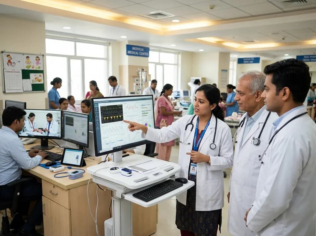
A landmark study details how an enterprise pediatric remote patient management model expanded from a single cardiology pilot to 16 diverse specialty programs by combining centralized digital operations with specialty clinical leadership.
Yesterday
A breakthrough study shows that 3D-planned corrective osteotomy successfully resolves distal radioulnar joint instability in pediatric diaphyseal forearm malunions. The precise osseous realignment eliminates chronic wrist pain and restores joint function without requiring secondary soft-tissue reconstruction.
Today

Protein O-GlcNAcylation couples cellular nutrient status to vital neuronal functions. This review explores how disrupted O-GlcNAc cycling drives both neurodevelopmental delays and neurodegenerative cascades, positioning metabolic post-translational modifications as therapeutic targets in clinical neurology.
Today
