

Loading, please wait...
Loading, please wait...

Placenta accreta spectrum represents one of the most perilous complications in contemporary obstetrics. Across the globe, escalating rates of cesarean delivery have driven a parallel increase in abnormal placental adherence. Consequently, obstetricians encounter complex cases where invasive trophoblasts penetrate deeply into the uterine myometrium. When deliveries occur without prior antenatal recognition, catastrophic hemorrhage frequently ensues, demanding urgent transfusion and emergency hysterectomy. Therefore, accurate prenatal identification remains critical for reducing maternal mortality and morbidity. Standard grayscale ultrasonography assesses morphological signs, such as placental lacunae and disruption of the retroplacental clear zone. However, subjective qualitative interpretation frequently leads to substantial diagnostic variability among imaging specialists. Clinicians often struggle to differentiate true invasiveness from benign superficial vascularity in the lower uterine segment. Because conventional color Doppler depends on visual impressions, clinicians require objective, reproducible assessment tools. Consequently, quantitative metrics that evaluate microvascular density can significantly improve diagnostic accuracy. By transforming subjective imaging into standardized data, clinical teams can anticipate operative challenges and coordinate multidisciplinary care before delivery. Consequently, improving prenatal detection stands as a critical priority for obstetric services.
To eliminate subjective ambiguity, investigators introduced an objective framework evaluating the anterior fornix and cervix region. Sonographers perform standardized transvaginal ultrasound examinations using color Doppler in the midsagittal plane. Notably, the anterior fornix represents an essential anatomical nexus where abnormal neovascularization clusters during invasive placental growth. Clinicians acquire continuous videoclips to record regional vascular patterns across several cardiac cycles. Subsequently, automated computer algorithms analyze these videoclips by measuring the exact proportion of color pixels within the anterior fornix region. The software calculates the average percentage across all frames, generating the final quantitative vascularization value. Importantly, managing obstetricians remained blinded to these quantitative measurements, ensuring that experimental numbers did not alter clinical decisions. By calculating automated pixel density, this innovative approach removes the visual bias inherent in conventional Doppler assessments. Furthermore, transvaginal transducers offer superior resolution compared to transabdominal probes when evaluating cervical architecture. Consequently, clinicians obtain an objective numerical index representing lower uterine segment vascularity. Thus, quantitative vascularization provides a reliable, reproducible measure that reflects the actual biological extent of pelvic neovascularization.
A prospective clinical study evaluated 117 patients presenting with anterior placenta previa and suspected invasive placentation. Histopathological analysis confirmed placenta accreta spectrum in 89 patients, encompassing 11 accreta, 55 increta, and 23 percreta cases. When assessing diagnostic performance, quantitative vascularization demonstrated an outstanding area under the receiver-operating-characteristics curve of 0.821. Furthermore, receiver operating curve analysis identified an optimal cutoff value of 8.5% vascular density. At this 8.5% threshold, quantitative vascularization yielded a sensitivity of 70.5% and a specificity of 89.7%. Most notably, the positive predictive value reached an impressive 93.9%, confirming high diagnostic dependability. Conversely, the negative predictive value was 50.0%, reflecting the recognized difficulty of ruling out focal invasion. Patients exhibiting vascular density at or above 8.5% showed nearly fifteen-fold higher odds of confirmed disease. Therefore, pixel-based analysis provides powerful objective discrimination between straightforward placenta previa and true invasive placentation. Accordingly, maternal-fetal medicine specialists obtain a mathematically validated metric to confirm complex disease before entering the operating room.
Beyond establishing diagnostic accuracy, researchers evaluated the correlation between vascular density and surgical hemorrhage. Among 109 patients with precise blood loss records, 53 women experienced severe hemorrhage exceeding 1500 mL. Crucially, higher quantitative vascularization values correlated strongly with increasing volumes of intraoperative blood loss. In addition, patients with elevated vascular measurements required significantly more transfusions of packed red blood cells and plasma. Neovascular channels invading the anterior cervix create fragile, high-flow vascular networks that rupture during surgical dissection. When preoperative imaging reveals vascular density exceeding the 8.5% threshold, surgical teams can anticipate major blood loss. Consequently, surgical units can mobilize emergency resources well before the patient enters the operating theater. Blood banks can prepare required blood products in advance, while anesthesia teams ready rapid infusers and cell salvage equipment. Furthermore, early surgical risk stratification significantly diminishes the risk of unheralded catastrophic bleeding. Moreover, surgical leaders can assign experienced pelvic surgeons to direct complex dissections. Thus, quantitative vascularization serves as an invaluable prognostic indicator that guides life-saving preparation for massive intraoperative hemorrhage.
In India, rapidly rising cesarean delivery rates have created a significant burden of morbidly adherent placentation. Many tertiary institutions regularly receive complicated cases referred late from peripheral healthcare facilities. However, advanced resources such as massive transfusion protocols and surgical subspecialists are not uniformly available across all centers. Therefore, an objective sonographic metric that predicts both invasion and severe bleeding provides immense practical value. Sonologists can incorporate quantitative vascularization protocols into second- and third-trimester scans for women with anterior placenta previa. Community clinicians identifying vascular scores above 8.5% can arrange timely transfer to high-volume tertiary hospitals. Furthermore, tertiary surgical teams can design tailored operative strategies, including planned cesarean hysterectomy without placental disruption. Surgeons can also plan precise incisions to avoid hypervascular areas and minimize bladder trauma. Additionally, interventional radiologists can schedule prophylactic pelvic arterial embolization when indicated. Ultimately, adopting objective vascular analysis standardizes clinical pathways, limits unexpected surgical hemorrhages, and saves maternal lives across diverse hospital settings.
Conventional color Doppler relies on subjective visual estimation of vascularity, which varies widely among sonographers. In contrast, quantitative vascularization applies post-processing computer software to calculate the exact percentage of color pixels across multiple videoclip frames in the anterior fornix and cervix. Consequently, this method eliminates observer variation, provides reproducible numerical data, and standardizes risk stratification for placenta accreta spectrum across diverse institutions and ultrasound operators.
The prospective study established an optimal quantitative vascularization cutoff value of 8.5% in the anterior fornix and cervix region. Patients exhibiting vascular density at or above this threshold showed nearly fifteen-fold higher odds of confirmed invasive pathology. Furthermore, this 8.5% cutoff demonstrated a sensitivity of 70.5% and an exceptional positive predictive value of 93.9%, providing clinicians with a dependable objective benchmark to identify severe placental invasion preoperatively.
A quantitative vascularization value exceeding 8.5% strongly correlates with severe intraoperative blood loss greater than 1500 mL and higher transfusion volumes. Therefore, identifying elevated vascular density enables surgical teams to activate massive transfusion protocols before surgery begins. Hospital blood banks can crossmatch and thaw required blood components in advance. Consequently, surgical teams prevent critical delays during operative hemorrhage, ensuring rapid hemodynamic resuscitation and significantly improved maternal safety.
Disclaimer: This content is for informational and educational purposes only... Refer to the latest local and national guidelines for clinical practice.
References

Read summarized clinical updates, watch expert medical content, and earn CME certifications right from your smartphone.
Open your smartphone camera to scan and install.





A study in Ultrasound in Obstetrics & Gynecology shows that quantitative vascularization of the anterior fornix/cervix with transvaginal ultrasound reliably identifies placenta accreta spectrum and predicts severe intraoperative hemorrhage, offering vital objective risk stratification.
Today

An ICMR study reveals that nearly 25% of tribal adults in Odisha suffer from renal decline. Driven by untreated hypertension, diabetes, and lifestyle risks, this hidden burden highlights the critical need for point-of-care diagnostics, proactive screening, and targeted non-communicable disease interventions.
Today
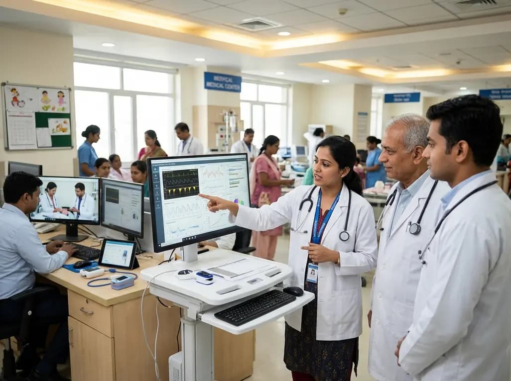
A landmark study details how an enterprise pediatric remote patient management model expanded from a single cardiology pilot to 16 diverse specialty programs by combining centralized digital operations with specialty clinical leadership.
Yesterday

Peripheral nerve injuries present clinical challenges due to regenerative mismatches between sensory and motor neurons. This review explores cutting-edge tissue-engineered nerve grafts designed as central-peripheral signal relays to overcome denervation atrophy and restore functional motor-sensory recovery.
Today
Protein O-GlcNAcylation couples cellular nutrient status to vital neuronal functions. This review explores how disrupted O-GlcNAc cycling drives both neurodevelopmental delays and neurodegenerative cascades, positioning metabolic post-translational modifications as therapeutic targets in clinical neurology.
Today
