

Loading, please wait...
Loading, please wait...

Forearm fractures represent some of the most frequent skeletal injuries treated in pediatric emergency departments. Although natural remodeling corrects many pediatric deformities, significant diaphyseal forearm malunion can still develop when fractures heal with residual displacement. While classical teaching suggests that forearm malunions primarily impair pronation and supination, some individuals retain a normal range of forearm rotation while suffering from disabling distal radioulnar joint instability. Patients frequently present years after the initial injury with progressive ulnar wrist pain, clicking, and reduced grip strength. Fortunately, recent surgical advances demonstrate that three-dimensional planned corrective osteotomy resolves joint instability and restores upper limb function without requiring complex soft-tissue reconstruction.
The distal radioulnar joint functions as a delicate trochoid joint that relies heavily on strict skeletal alignment between the radius and ulna. Consequently, any post-traumatic angular, translational, or rotational malunion of the forearm shafts disrupts this precise kinematic relationship. Diaphyseal forearm malunion alters the spatial orientation of the sigmoid notch relative to the ulnar seat, creating joint incongruence during basic manual tasks. Furthermore, these skeletal deformities drastically alter the normal tension vectors of the interosseous membrane and secondary soft-tissue stabilizers.
Although many clinicians anticipate severe limitations in forearm rotation following a malunion, preserved pronation and supination can sometimes mask underlying structural pathology. In these unique cases, the rotational axis allows functional forearm turning, but the altered osseous architecture forces the ulnar head to subluxate dynamically during loaded activities. As a result, the triangular fibrocartilage complex endures abnormal chronic strain, leading to severe ulnar-sided wrist discomfort and joint instability. Therefore, addressing the underlying osseous deformity provides the only reliable mechanism to restore physiological load transmission across the wrist joint.
Historically, orthopedic surgeons evaluated post-traumatic forearm deformities using standard two-dimensional radiographs. However, conventional plain radiographs often fail to capture complex multiplanar deformities that involve simultaneous sagitta-coronal angulation and axial rotation. Furthermore, clinicians frequently misdiagnose the resulting wrist pain and distal radioulnar joint laxity as isolated ligament tears. Consequently, many surgical teams historically opted for soft-tissue procedures, such as triangular fibrocartilage repairs or ligament tenodeses, to restore joint stability.
Unfortunately, soft-tissue reconstructions consistently demonstrate high failure rates when surgeons perform them in the presence of an uncorrected skeletal deformity. Because abnormal joint kinematics remain unchanged, reconstructed ligaments stretch, attenuate, or rupture under persistent mechanical overload. In addition, conventional freehand corrective osteotomies often yield unpredictable outcomes due to limited intraoperative visibility and inaccurate multiplanar cuts. Therefore, relying solely on two-dimensional radiographic planning or soft-tissue repair exposes patients to recurrent joint instability, continuous pain, and secondary osteoarthritis. Modern clinical paradigms clearly require more comprehensive, three-dimensional skeletal interventions to achieve predictable corrections.
Advanced computer-aided modeling and additive manufacturing now offer unprecedented solutions for complex skeletal reconstructions. Preoperatively, clinicians obtain high-resolution bilateral computed tomography scans of the patient's forearms. Biomedical engineers and surgeons then analyze the multiplanar deformity using dedicated three-dimensional software. By mirroring the patient's healthy contralateral forearm, the surgical team creates a precise virtual template of the native anatomy. Subsequently, the computer algorithm calculates the exact osteotomy location, cut angles, and realignment maneuvers required to restore normal skeletal parameters.
During surgery, orthopedists execute the virtual plan using custom 3D-printed patient-specific drilling and cutting guides. Because these biocompatible nylon guides replicate the unique cortical surface contours, they snap firmly onto the exposed radius or ulna. Surgeons pre-drill screw holes according to the preoperative trajectory before making precise saw cuts through guided slots. Therefore, when the surgeon reduces the bone fragments and fixes them with contoured anatomical plates, the skeleton accurately realigns to the planned position. This advanced technique eliminates intraoperative guesswork, minimizes radiation exposure, and avoids extensive periosteal stripping.
Recent clinical evidence proves the remarkable effectiveness of 3D-guided osseous realignment in patients suffering from diaphyseal forearm malunion with distal radioulnar joint instability. In a cohort of nine patients who sustained childhood forearm fractures at a mean age of 12 years, computer-planned osteotomy restored complete joint stability at twelve months. Most importantly, all nine patients achieved joint stability without needing any concomitant ligament reconstruction or capsular tightening.
The clinical improvements observed across all validated patient-reported metrics were both statistically significant and clinically meaningful. Specifically, the median Patient-Rated Wrist/Hand Evaluation score improved dramatically from 44.5 preoperatively to 5.5 at one year. Furthermore, the Numeric Rating Scale pain score during activity dropped from a severe median of 7 down to 1, while resting pain decreased to 0. Similarly, the Patient-Specific Functional Scale scores improved from 7 to 2. These improvements comfortably surpassed the minimal clinically important differences, illustrating substantial relief from pain, improved functional performance, and restoration of normal upper extremity mechanics.
These clinical findings provide valuable takeaways for reconstructive surgeons, pediatric orthopedists, and hand specialists managing persistent wrist dysfunction. First, surgeons must maintain a high index of suspicion for diaphyseal deformities in any patient presenting with distal radioulnar joint instability, even when pronation and supination remain entirely preserved. Detailed volumetric imaging remains essential to characterize occult multiplanar malalignment.
Second, re-establishing precise osseous anatomy must remain the primary surgical priority. Once three-dimensional bone geometry is restored, native ligamentous tension across the triangular fibrocartilage complex and interosseous membrane normalizes immediately. Therefore, surgeons can safely forgo secondary soft-tissue repairs, avoiding the morbidity of tendon harvesting, prolonged immobilization, and postoperative joint stiffness. In addition, adopting 3D-printed patient-specific instrumentation ensures reliable, reproducible correction of complex deformities that conventional techniques struggle to resolve. As access to surgical planning software expands, 3D-planned osteotomy stands as the definitive standard of care for restoring stability in malunited forearm fractures.
Angular and rotational bony deformities in the forearm shafts alter the delicate kinematic relationship between the radius and ulna. Even when patients retain rotational motion, subtle osseous malalignment changes the tension across the interosseous membrane and incongruently loads the distal radioulnar joint. Consequently, the articular surfaces lose stable contact during loading. Restoring correct skeletal anatomy realigns the joint axis and re-establishes native ligamentous stability without requiring capsular tightening.
Patient-specific 3D surgical guides translate virtual computer-assisted preoperative plans directly to the operating table with high fidelity. Initially, surgeons utilize three-dimensional computed tomography reconstructions of both forearms to model complex multiplanar deformities. Customized drill and cutting jigs snap securely onto the patient's unique bony contours. Therefore, surgeons achieve exact realignment and correction angles intraoperatively, which significantly minimizes surgical error, shortens operative duration, and avoids extensive soft-tissue stripping.
Surgeons can avoid primary soft-tissue reconstruction when distal joint instability stems purely from underlying skeletal misalignment rather than frank ligamentous disruption. If preoperative assessments confirm preserved soft-tissue integrity, precise osseous realignment naturally restores anatomical tension to the triangular fibrocartilage complex. As demonstrated by recent clinical evidence, achieving rigid three-dimensional bony correction stabilizes the distal radioulnar joint immediately, thereby sparing patients the morbidity, prolonged immobilization, and stiffness associated with secondary ligament tenodesis.
Disclaimer: This content is for informational and educational purposes only. It is not intended to provide medical advice or replace the judgment of a healthcare professional. Healthcare professionals must use their clinical judgment when evaluating medical research and applying it to individual clinical situations. Refer to the latest local and national guidelines for clinical practice.
References:

Read summarized clinical updates, watch expert medical content, and earn CME certifications right from your smartphone.
Open your smartphone camera to scan and install.





A breakthrough study shows that 3D-planned corrective osteotomy successfully resolves distal radioulnar joint instability in pediatric diaphyseal forearm malunions. The precise osseous realignment eliminates chronic wrist pain and restores joint function without requiring secondary soft-tissue reconstruction.
Today
A landmark 12-year longitudinal study highlights significant reductions in opioid prescribing for nonspecific low back pain following clinical guidelines and prescription monitoring systems, emphasizing non-opioid multimodal pain care.
Today
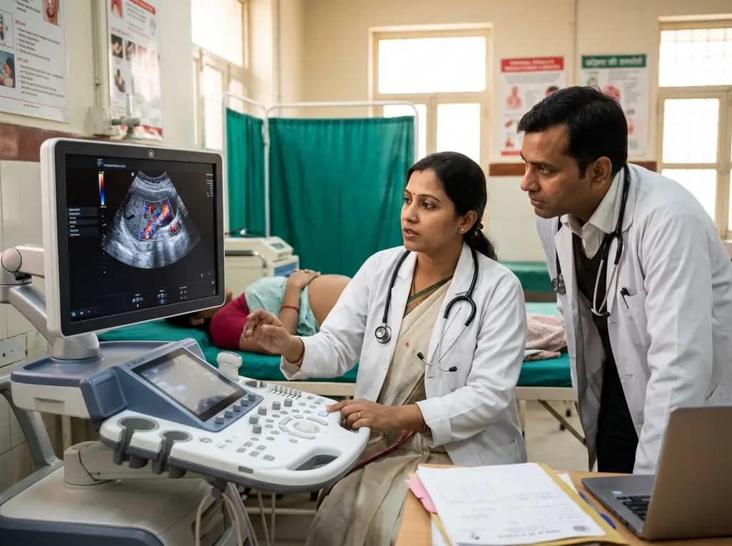
A study in Ultrasound in Obstetrics & Gynecology shows that quantitative vascularization of the anterior fornix/cervix with transvaginal ultrasound reliably identifies placenta accreta spectrum and predicts severe intraoperative hemorrhage, offering vital objective risk stratification.
Today

A landmark study details how an enterprise pediatric remote patient management model expanded from a single cardiology pilot to 16 diverse specialty programs by combining centralized digital operations with specialty clinical leadership.
Yesterday

Protein O-GlcNAcylation couples cellular nutrient status to vital neuronal functions. This review explores how disrupted O-GlcNAc cycling drives both neurodevelopmental delays and neurodegenerative cascades, positioning metabolic post-translational modifications as therapeutic targets in clinical neurology.
Today
