

Loading, please wait...
Loading, please wait...

Diagnosing gastric infections in children presents unique diagnostic challenges for clinicians, especially in resource-limited or high-prevalence settings. Consequently, pediatric H pylori testing requires accurate, practical, and child-friendly modalities that avoid invasive interventions whenever possible. While esophagogastroduodenoscopy with biopsy remains the gold standard reference, invasive endoscopies carry procedural risks, necessitate sedation, and increase emotional stress for young patients and their families. Therefore, noninvasive diagnostic alternatives have attracted considerable clinical interest over recent decades. A comprehensive meta-analysis evaluated the diagnostic performance of noninvasive tests in pediatric cohorts. The investigators systematically analyzed stool antigen assays, urea breath testing, and serum antibody tests to clarify their diagnostic accuracy. Because clinical presentations of infection in children often remain non-specific, clinicians require reliable tools to guide management. Understanding test performance helps pediatricians avoid unnecessary invasive procedures while ensuring appropriate therapeutic decisions.
Helicobacter pylori infection typically begins during early childhood, especially in developing regions where household transmission remains frequent. Although many infected children remain asymptomatic, persistent colonization can lead to chronic active gastritis, peptic ulceration, and refractory iron deficiency anemia. However, adult diagnostic pathways do not automatically translate to pediatric practice because anatomical and immunological responses differ substantially across age groups. Furthermore, children exhibit dynamic antibody responses that complicate standard serological interpretation. In addition, performing routine endoscopies in pediatric outpatient clinics remains impractical due to technical constraints and high costs. Noninvasive assays circumvent these barriers by offering convenient sample collection. Consequently, accurate noninvasive diagnostic modalities facilitate prompt assessment without exposing children to procedural sedation or hospital admissions. Clinicians nevertheless must understand the strengths and diagnostic boundaries of each noninvasive test before selecting an assay for patient care.
The meta-analysis evaluated multiple noninvasive modalities to quantify their diagnostic accuracy, sensitivity, and specificity across pediatric populations. Notably, the stool antigen enzyme-linked immunosorbent assay demonstrated robust diagnostic performance, yielding an estimated sensitivity of 88% and a specificity of 93%. Similarly, the carbon-13 urea breath test demonstrated high diagnostic precision across various age cohorts. In contrast, serum antibody tests exhibited significant discrepancies depending on the measured immunoglobulin class. Serum IgG assays achieved a sensitivity of 89% and a specificity of 97%, whereas serum IgA assays achieved an inadequate sensitivity of only 49%, despite an elevated specificity of 98%. Consequently, IgA serology misses more than half of true pediatric infections. Therefore, current evidence discourages serum antibody assays for routine diagnostic confirmation in children. Instead, stool antigen ELISA and breath testing clearly outperform serology across all standard performance metrics.
Among available modalities, stool antigen ELISA emerges as the most practical and reliable noninvasive option for pediatric practice. Collecting a stool specimen eliminates the fear and discomfort associated with venipuncture or swallowing test substrates. Moreover, younger children and toddlers often fail to cooperate with breath testing protocols because exhalation into specialized bags requires strict coordination. In contrast, stool collection requires no active patient compliance, making it universally feasible from infancy through adolescence. Furthermore, monoclonal stool antigen ELISA assays demonstrate high concordance with endoscopic histology, providing reliable confirmation of active bacterial colonization. However, clinicians must recognize that recent exposure to proton pump inhibitors or antimicrobial agents can induce false-negative results by decreasing bacterial density. Therefore, clinicians must ensure a minimum four-week antibiotic washout and a two-week acid suppression hiatus before performing stool antigen testing.
The carbon-13 urea breath test represents another premier noninvasive diagnostic tool with exceptional diagnostic specificity and sensitivity. The assay relies on bacterial urease activity, which hydrolyzes orally ingested labeled urea into detectable labeled carbon dioxide in exhaled breath. Because carbon-13 is a non-radioactive, stable isotope, the test remains safe for pediatric populations of all ages. However, practical hurdles frequently restrict its utility in routine primary care. For example, the test requires specialized, costly analytical instruments such as isotope-ratio mass spectrometers or infrared analyzers. In addition, infants and preschool children often struggle to maintain an airtight seal on breath collection straws or masks. Furthermore, variations in endogenous carbon dioxide production rates among young children can occasionally skew cut-off values. Thus, while breath testing remains highly accurate in older children, stool antigen ELISA generally offers superior procedural feasibility across diverse clinical environments.
Current international pediatric guidelines recommend against a simple test-and-treat strategy in unselected children with non-specific recurrent abdominal pain. Instead, diagnostic testing should focus on identifying underlying organic disease when symptoms suggest peptic ulceration or unexplained refractory cytopenias. When clinicians establish a legitimate indication for noninvasive assessment, monoclonal stool antigen ELISA represents the most accessible and accurate frontline investigation. Furthermore, this assay provides an excellent method for confirming post-eradication clearance at least four to eight weeks after completing therapy. Clinicians should completely avoid serological tests because circulating antibodies persist for months or years after bacterial clearance, preventing differentiation between active and resolved infections. In high-prevalence settings, standardized testing protocols combined with rigorous pre-test medication screening ensure accurate clinical interpretation and prevent unnecessary antibiotic exposure.
Stool antigen enzyme-linked immunosorbent assay and carbon-13 urea breath tests represent the most reliable noninvasive modalities for children. However, stool antigen testing is generally preferred in routine pediatric practice because it does not require active patient cooperation or expensive breath analysis devices. Consequently, stool antigen ELISA provides the optimal combination of high diagnostic accuracy, cost efficiency, and clinical feasibility for infants, children, and adolescents across primary and secondary healthcare settings.
Serological testing carries substantial limitations in pediatric patients because it cannot differentiate between active infection and past exposure. Circulating antibodies often persist for prolonged periods after successful eradication. Furthermore, serum IgA testing demonstrates an unacceptably low sensitivity of 49%, resulting in frequent false-negative diagnoses. Therefore, international pediatric guidelines advise against blood antibody tests and recommend direct antigen or breath detection methods to confirm active Helicobacter pylori colonization.
Clinicians must verify that the pediatric patient has avoided specific medications that suppress bacterial load before performing active antigen detection. Specifically, patients must discontinue all systemic antibiotic therapy and bismuth compounds for at least four weeks prior to testing. Additionally, clinicians should pause proton pump inhibitor medications for a minimum of two weeks. Adhering strictly to these timing precautions prevents false-negative results in both stool antigen assays and breath tests.
Disclaimer: This content is for informational and educational purposes only... Refer to the latest local and national guidelines for clinical practice.
References
Montoya-Arango MA et al. Meta-analysis of diagnostic validation studies on noninvasive methods for detecting Helicobacter pylori in the pediatric population. Rev Gastroenterol Mex (Engl Ed). 2026 Oct 24. doi: undefined. PMID: 42926158.
Joint ESPGHAN/NASPGHAN Guidelines for the Management of Helicobacter pylori in Children and Adolescents (Update 2016). J Pediatr Gastroenterol Nutr. 2017;64(6):991-1003.
Best LM, Takwoingi Y, Siddique S, et al. Non-invasive diagnostic tests for Helicobacter pylori infection. Cochrane Database Syst Rev. 2018;3(3):CD012080.

Read summarized clinical updates, watch expert medical content, and earn CME certifications right from your smartphone.
Open your smartphone camera to scan and install.





A comprehensive meta-analysis evaluates noninvasive pediatric H pylori testing methods, highlighting the diagnostic performance and clinical utility of stool antigen ELISA and urea breath tests over serology.
Today

This methodological viewpoint establishes ten rules to constrain noise and enhance precision across image scoring, database handling, and data analysis in rheumatology research.
Today

A study in JAMIA Open shows lightweight open-source reasoning LLMs achieve up to 95% sensitivity and accelerate evidence synthesis screening by nearly twentyfold, presenting a cost-effective, scalable AI solution for biomedical research and systematic reviews.
Yesterday
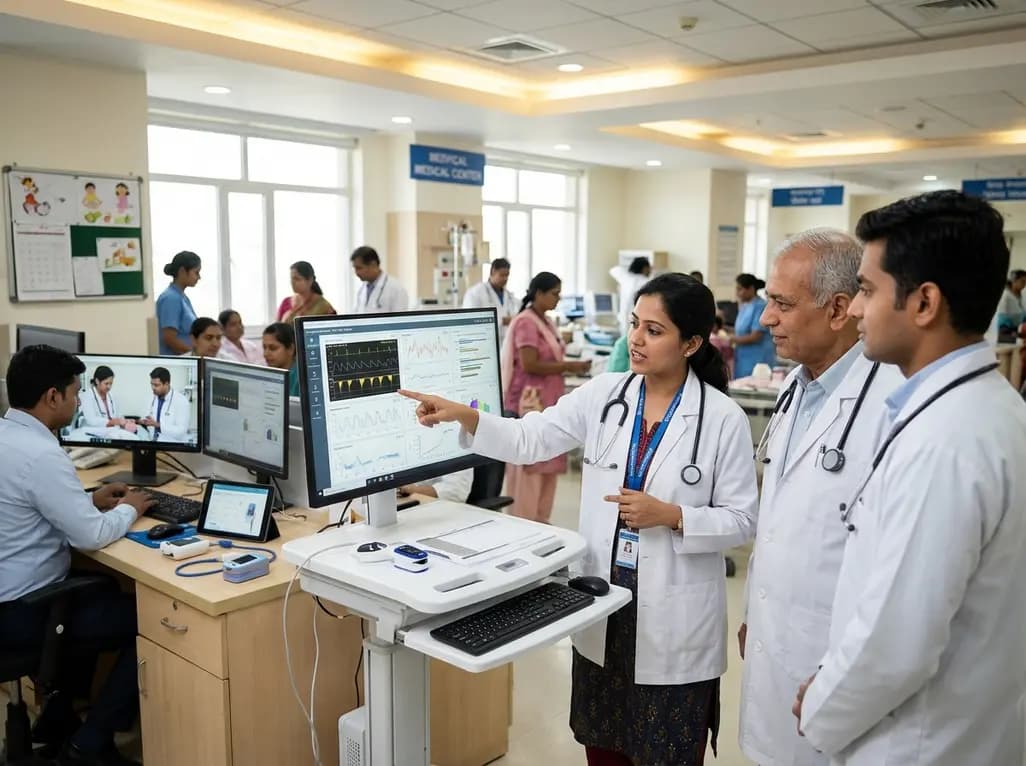
A landmark study details how an enterprise pediatric remote patient management model expanded from a single cardiology pilot to 16 diverse specialty programs by combining centralized digital operations with specialty clinical leadership.
Yesterday

A comparative review of tension band wiring and intramedullary screw fixation in thumb metacarpophalangeal arthrodesis, evaluating union rates, hardware complications, and clinical outcomes.
Yesterday

A nationwide observational study from Norway investigates whether SGLT2 inhibitor therapy improves 5-year metabolic parameters in kidney transplant recipients with post-transplantation diabetes mellitus (PTDM), finding comparable glycemic, renal, and blood pressure profiles between user and non-user cohorts.
Today
