.webp&w=384&q=75)

Loading, please wait...
Loading, please wait...
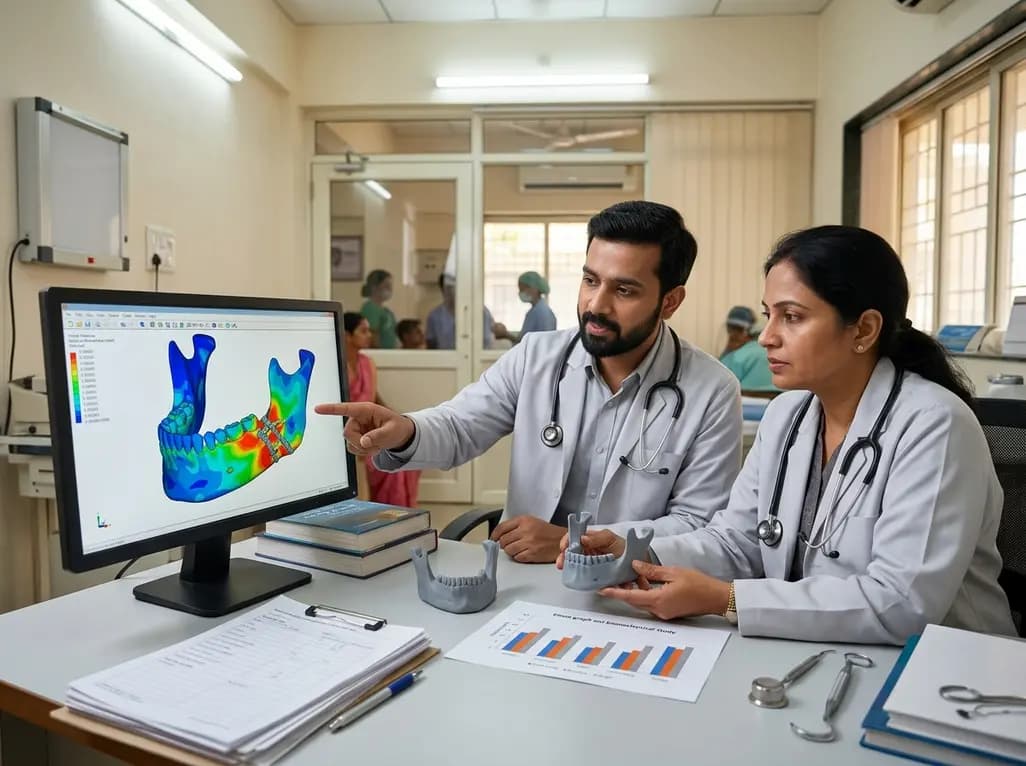
Mandibular angle fractures represent common and clinically complex injuries encountered in oral and maxillofacial surgery. Achieving stable mandibular angle fracture fixation remains challenging due to the complex functional loads exerted by masticatory muscles in this region. Traditionally, surgeons rely on conventional two miniplate fixation along established lines of osteosynthesis to restore anatomical stability. Although standard miniplates deliver acceptable clinical results, localized stress concentration within titanium hardware frequently raises concerns regarding potential mechanical failure or screw loosening. Recently, advances in computer-aided design and manufacturing have introduced patient-specific three-dimensional titanium plates tailored to individual patient anatomy. These custom implants aim to optimize stress distribution and improve plate adaptation. However, comparing their biomechanical behavior against traditional hardware requires rigorous computational evaluation under physiological loading conditions.
The human mandible undergoes significant mechanical strain during daily functions like chewing, swallowing, and clenching. When a fracture occurs at the mandibular angle, opposing muscle forces create high tensile stresses along the superior border and compressive forces along the inferior border. Effective fixation must withstand these dynamic multidirectional loads to prevent fracture displacement. Conventional two miniplate systems address these stresses by placing one plate at the superior tension zone and another at the inferior border. However, intraoperative manual bending of stock miniplates can introduce microscopic structural defects, increasing susceptibility to hardware fatigue. Additionally, incomplete plate adaptation to irregular cortical surfaces can cause localized stress concentrations around screw holes. Consequently, high mechanical stress may compromise long-term fixation stability or induce localized bone resorption. Evaluating novel patient-specific hardware designed to fit anatomical contours without manual manipulation offers a promising approach to overcome these persistent clinical limitations.
To evaluate these fixation techniques, researchers performed a three-dimensional finite element analysis based on computed tomography data from a dentate male patient. Advanced software tools, including Mimics and Geomagic Design X, enabled accurate reconstruction of cortical and cancellous bone architecture. Fixation hardware was then assembled using SolidWorks software to create two distinct computational models. Model A incorporated a conventional two miniplate system, whereas Model B featured a patient-specific three-dimensional titanium plate contoured along the mandibular tension zone. Both assembled models were imported into ANSYS software to simulate unilateral clenching under identical physiological muscle loading and boundary conditions. Investigators systematically evaluated von Mises stress in the titanium plates, maximum principal stress within cortical bone, and total interfragmentary displacement. This controlled simulation provided objective biomechanical data comparing custom and standard osteosynthesis configurations.
The simulation results demonstrated notable differences in mechanical performance between the two fixation approaches. The patient-specific three-dimensional plate model exhibited a lower maximum von Mises stress within the titanium plate at 326.19 MPa, compared to 417.48 MPa in the conventional two miniplate system. This marked reduction in hardware stress indicates that custom plates distribute functional loads more evenly, significantly lowering the risk of implant fracture or fatigue failure. Conversely, maximum principal stress in cortical bone was higher in the patient-specific model at 121.03 MPa, compared with 87.759 MPa in conventional miniplate fixation. While higher cortical bone stress requires careful consideration, controlled mechanical transfer from implant to bone helps prevent stress shielding. Consequently, the custom plate design promotes favorable biomechanical interaction while shielding the titanium hardware from excessive localized forces.
In addition to stress patterns, fracture line displacement plays a vital role in determining clinical healing outcomes. In this study, total displacement reached 157.32 micrometers in the patient-specific plate model, compared to 131.47 micrometers in the conventional miniplate model. Although the custom plate permitted slightly greater micro-motion, both values remained well within clinically acceptable limits for bone union. Standard literature indicates that interfragmentary movements under 200 micrometers support primary bone healing without predisposing the fracture to nonunion or construct destabilization. Therefore, the patient-specific plate provides sufficient stiffness while accommodating micro-displacement that stimulates physiological callus formation. Furthermore, custom plates reduce surgical complexity by eliminating manual plate bending during operation. This anatomical pre-contouring enhances precise adaptation to cortical bone, potentially reducing operative times and improving overall surgical predictability for facial trauma patients.
These finite element findings offer valuable insights for oral and maxillofacial surgeons considering patient-specific hardware for complex trauma cases. Custom three-dimensional titanium plates deliver superior hardware stress distribution while maintaining biomechanically sound fracture stability. Their pre-contoured geometry simplifies intraoperative placement, minimizes soft tissue disruption, and eliminates bending-induced material weakness. However, clinicians must consider potential limitations, including manufacturing lead times and increased healthcare costs compared to stock miniplate inventory. In urgent clinical scenarios requiring immediate reduction, conventional miniplates remain a highly effective and versatile primary treatment option. Nevertheless, as digital workflows, virtual surgical planning, and rapid additive manufacturing become increasingly integrated into hospital systems, custom three-dimensional fixation will become progressively accessible. Adopting these advanced technologies thoughtfully will allow surgeons to tailor fixation strategies to specific biomechanical demands, ultimately enhancing patient recovery and clinical success.
The primary advantage of patient-specific three-dimensional titanium plates is their superior stress distribution across the fixation hardware. By conforming precisely to individual anatomical contours, custom plates reduce peak von Mises stress compared to conventional miniplates. Consequently, this lower stress concentration minimizes the risk of implant fatigue failure, screw loosening, and hardware breakage, providing reliable structural stability during functional jaw movement and early postoperative healing.
No, the slight increase in displacement observed with patient-specific plates does not adversely affect fracture healing. Although the custom plate demonstrated a displacement of 157.32 micrometers compared to 131.47 micrometers for conventional miniplates, both values fall well within clinically accepted physiological thresholds. Micro-motion under 200 micrometers actually promotes secondary bone healing by stimulating callus formation without causing interfragmentary instability or nonunion risks.
Finite element analysis provides a powerful non-invasive computational method to evaluate biomechanical responses in complex maxillofacial structures. It allows researchers to simulate realistic muscular forces and precisely measure stress patterns across bone and fixation hardware. Consequently, finite element analysis helps clinicians compare competing osteosynthesis techniques, optimize implant geometry, and predict mechanical performance before clinical application, ultimately improving patient outcomes in facial trauma repair.
Disclaimer: This content is for informational and educational purposes only. It does not constitute medical advice or substitute for professional clinical judgment. Refer to the latest local and national guidelines for clinical practice.
References
1. Abd Elfattah AM et al. Finite element analysis of patient specific three-dimensional titanium plate versus conventional two miniplate fixation for mandibular angle fractures. BMC Oral Health. 2026 Aug 08. doi: undefined. PMID: 42568091.
2. Arora L, Bhardwaj S, Hashmi GS, Anwar SF, Rahman SA. Finite Element Analysis (FEA) of Perpendicular Plating versus Conventional Plating in Mandibular Symphysis Fracture. J Maxillofac Oral Surg. 2020;19(1):143-148.
3. Liokatis P, Tzortzinis G, Gerasimidis S, Smolka W. Finite Element Analysis of Different Titanium Miniplates: Evaluation of Three-Dimensional Designs Applied on Condylar Neck Fractures. J Stomatol Oral Maxillofac Surg. 2021;123(2):184-190.

Read summarized clinical updates, watch expert medical content, and earn CME certifications right from your smartphone.
Open your smartphone camera to scan and install.





A finite element analysis evaluated patient-specific 3D titanium plates versus conventional two miniplate fixation for mandibular angle fractures. Custom plates showed lower peak hardware stress (326.19 MPa vs 417.48 MPa) and clinically acceptable displacement, highlighting key biomechanical benefits.
last month
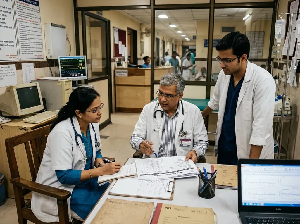
A quaternary centre study of 143 patients undergoing TIPS revealed high 90-day rates of hepatic encephalopathy (43.8%) and mortality (18.6%). Type 2 diabetes independently doubled encephalopathy risk, while advancing age and higher baseline MELD scores strongly predicted short-term mortality.
Today

Meghalaya recorded a historic 55% reduction in maternal deaths, dropping from 244 to 107 over five years. Through the MOTHER app, dedicated transit homes, and expanded infrastructure like the 150-bed Tura Maternity Hospital with its first public NICU, the state demonstrates an effective public health model.
Today

Explore modern perioperative management protocols for clinical xenotransplantation. Learn how costimulation blockade, anti-inflammatory perfusion, and coagulopathy rescue overcome interspecies hurdles.
Today
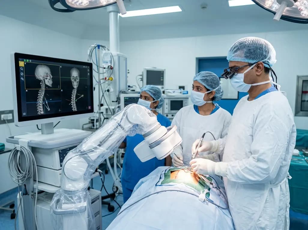
North India's first Centre of Excellence for Brain and Spine at Amrita Hospital Faridabad integrates Brainlab's Loop-X mobile robot. This landmark facility combines cone-beam computed tomography with surgical navigation to elevate precision, optimize operative workflows, and enhance specialized clinician training.
Today

At Homa CME 2026 at Hyderabad's T-Hub, healthcare leaders emphasized moving beyond routine metrics through digital calculators and precision stratification. Discover why characterizing individual insulin secretion, resistance patterns, and vascular risks transforms chronic metabolic care in India.
Today
