.webp&w=384&q=75)

Loading, please wait...
Loading, please wait...

Oncology has actively searched for targeted, non-invasive therapeutic options that selectively destroy malignant tissues while preserving healthy structures. Consequently, researchers at the Indian Institute of Science have made a groundbreaking discovery regarding oral cancer ultrasound therapy. Their study reveals that low-frequency ultrasound waves can selectively eliminate malignant cells while leaving healthy cells unharmed. Indeed, this remarkable finding holds profound clinical significance for India, where oral malignancies represent a major public health challenge. Specifically, because conventional treatments often damage surrounding tissues, this novel biomechanical approach could transform local oncological management.
The core of this clinical breakthrough lies in exploiting the specific physical differences between healthy and malignant cells. To understand this mechanism, we must analyze how cells respond to external mechanical stimulation. Healthy cells typically possess robust internal cytoskeletal structures that help them withstand external physical forces. In contrast, researchers discovered that oral cancer cells exhibit a significant mechanical vulnerability. Specifically, this vulnerability occurs due to reduced levels of Tropomyosin 2.1, a key cytoskeletal protein. Usually, this protein helps normal epithelial cells sense, adapt to, and resist mechanical stress in their microenvironment. However, lacking adequate Tropomyosin 2.1, oral cancer cells cannot cope with moderate mechanical forces. Consequently, when researchers exposed patient-derived tumor samples to low-frequency ultrasound, the cancer cells suffered catastrophic structural damage. Meanwhile, healthy oral epithelial cells remained largely unaffected by the acoustic energy. Therefore, this finding highlights a biological weakness that clinicians can selectively exploit without resorting to cytotoxic drugs. Ultimately, targeting physical rather than chemical pathways represents a massive shift in local oncology protocols. Indeed, it opens new pathways for localized, side-effect-free tumor ablation therapies in the near future.
Another significant hurdle in clinical oncology is the presence of a dense, protective barrier surrounding tumor masses. This barrier is composed of a complex extracellular matrix that restricts therapeutic drug penetration. Furthermore, it actively prevents host immune cells from reaching and destroying the tumor core. Interestingly, researchers observed that the mechanical forces of the ultrasound successfully disrupted this dense biological barrier. Consequently, the acoustic waves effectively loosened the tightly packed matrix and increased its permeability. This disruption could potentially allow administered chemotherapeutic drugs to penetrate deeper into the tumor. Moreover, the study demonstrated that ultrasound exposure significantly reduced the overall migration and invasion capabilities of the cancer cells. Therefore, this therapy does not merely kill localized cells. Instead, it concurrently weakens the entire defensive structure of the tumor. As a result, combining ultrasound with traditional pharmacological agents might significantly boost overall treatment efficacy. Ultimately, this approach could dismantle the tumor’s defenses, allowing host defenses to reclaim control. Consequently, clinical outcomes could improve dramatically.
Developing effective treatments requires testing methodologies that accurately reflect actual patient pathology. Consequently, the researchers chose to work directly with patient-derived oral tumor samples instead of conventional, laboratory-grown cell lines. These primary clinical samples were obtained in collaboration with clinicians from MS Ramaiah Medical College and Hospitals. Furthermore, this clinical relevance ensures that the experimental findings are highly applicable to real-world scenarios. During the testing process, the team observed a remarkably consistent therapeutic response across multiple patient samples. Regardless of individual tumor stages, the malignant cells consistently exhibited profound vulnerability to the oral cancer ultrasound protocol. Conversely, the healthy control cells maintained their viability throughout the treatment sessions. Therefore, this technology shows promise as a standardized, predictable local therapy. In the future, clinicians might utilize customized acoustic frequencies to safely target diverse patient profiles. Ultimately, this consistency minimizes the risk of treatment resistance, which often plagues molecularly targeted therapies. Indeed, these outcomes offer strong validation for translating this research into clinical protocols.
When compared to existing treatment options, acoustic stimulation offers several unique clinical advantages. Traditional interventions like radical surgery often cause permanent cosmetic and functional deficits in the oral cavity. Similarly, chemotherapy and external beam radiation frequently induce severe systemic toxicities and local tissue necrosis. In contrast, low-frequency acoustic therapy utilizes gentle, non-thermal mechanical forces to achieve therapeutic outcomes. Therefore, this method completely avoids the risks of thermal tissue damage or systemic drug accumulation. Furthermore, ultrasound equipment is already highly accessible and widely integrated into standard medical infrastructure globally. Consequently, introducing this specialized modality into oncology clinics would require minimal capital expenditure. Additionally, because the procedure is entirely non-invasive, it can be performed repeatedly on an outpatient basis. Indeed, this approach could significantly lower overall healthcare costs while simultaneously improving the patient's quality of life. Moreover, it eliminates the psychological anxiety often associated with invasive surgeries and hospitalizations. Ultimately, it represents an extremely patient-centric alternative.
Although these patient-derived lab results are highly encouraging, several translational steps remain before widespread clinical implementation. Currently, the research team plans to transition their testing into advanced, living preclinical models. These animal models will help clarify how the complex host circulatory and immune systems interact with local acoustic stimulation. Furthermore, researchers want to study whether combining ultrasound with established chemotherapeutic regimens can enhance local drug delivery. Consequently, this combination therapy could minimize systemic side effects while maximizing local cytotoxicity. Additionally, investigators believe this mechanical targeting strategy can be extended to other easily accessible malignancies. For instance, breast and skin cancers represent excellent candidates for targeted low-frequency acoustic treatment due to their anatomical locations. Therefore, successful validation in these fields could expand the scope of acoustic oncology. Ultimately, this research paves the way for a safer, non-pharmacological pillar in multi-modal cancer care. Indeed, clinicians could soon have access to a highly precise tool that complements traditional therapeutic paradigms.
Q1: Why are oral cancer cells more vulnerable to low-frequency ultrasound compared to healthy cells?
Specifically, this selective vulnerability occurs because oral cancer cells have significantly lower levels of a crucial protein called Tropomyosin 2.1. In healthy oral epithelial cells, this protein acts as a mechanosensor, allowing the cellular cytoskeleton to withstand external mechanical forces. However, lacking adequate levels of Tropomyosin 2.1, malignant cells cannot tolerate the moderate physical stress generated by the ultrasound waves. Consequently, they experience mechanical failure and undergo selective cell death, whereas healthy cells remain unaffected.
Q2: What role does the extracellular matrix play in oral cancer, and how does ultrasound address this?
Indeed, the tumor microenvironment is characterized by a dense, protective extracellular matrix that chokes the tissue and limits drug delivery. This capsule-like barrier effectively shields malignant cells from chemotherapy and host immune responses. However, the study demonstrated that low-frequency ultrasound waves successfully disrupt this dense matrix. Consequently, the mechanical forces loosen the protective barrier and increase tumor permeability. Ultimately, this disruption allows therapeutics to penetrate deeper into the tumor core, significantly boosting overall treatment efficacy.
Q3: How was the clinical relevance of this study ensured during the laboratory testing phase?
To guarantee high clinical relevance, the researchers collaborated with clinicians from MS Ramaiah Medical College and Hospitals. Instead of using conventional, lab-grown cell lines, they tested the ultrasound on primary tumor samples obtained directly from patients. Consequently, this approach allowed them to capture the vast heterogeneous nature of oral malignancies seen across the Indian population. Furthermore, the consistent therapeutic response observed across multiple patient stages highlights the clinical potential and predictability of this treatment.
Disclaimer: This content is for informational and educational purposes only. It does not constitute medical advice or replace professional judgment. Refer to the latest local and national guidelines for clinical practice.
References

Read summarized clinical updates, watch expert medical content, and earn CME certifications right from your smartphone.
Open your smartphone camera to scan and install.





A pioneering study by the Indian Institute of Science (IISc) reveals that low-frequency ultrasound can selectively destroy oral cancer cells. By exploiting the mechanical vulnerability of malignant cells lacking Tropomyosin 2.1, this non-invasive approach destroys tumors while leaving healthy tissues unharmed.
3 months ago

Following regulatory scrutiny from FSSAI regarding promotional representations and lean muscle preservation claims, Dr. Reddy's Laboratories and Nestlé Health Science are currently reviewing the branding and marketing strategy for their joint nutritional supplement Celevida GLP+.
Today

A comprehensive scoping review synthesises evidence linking prenatal maternal mental health to long-term offspring neurodevelopment, evaluating neuroendocrine, inflammatory, and epigenetic pathways while highlighting clinical screening and future research priorities.
Today

The Directorate General of Health Services directs Indian states to halt the hazardous reuse and reprocessing of single-use medical devices. Discover regulatory implications under the Drugs and Cosmetics Act, critical infection risks, legal liabilities, and operational changes required across clinical specialties.
Today
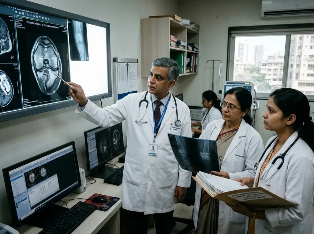
A 25-year multi-institutional cohort reveals high recurrence rates in pediatric giant cell tumor of bone, particularly in patients with open physes and bone marrow edema on preoperative MRI. Early surveillance and careful balance between growth plate preservation and tumor clearance are essential.
Today

India is advancing preventive healthcare by integrating genomics, artificial intelligence, and quantum technologies. With the SANGAM programme sequencing one million Indian genomes, clinicians can unlock personalized therapies, detect cardiometabolic risks early, and curb the burden of diabetes and heart disease.
Today
