.webp&w=384&q=75)

Loading, please wait...
Loading, please wait...
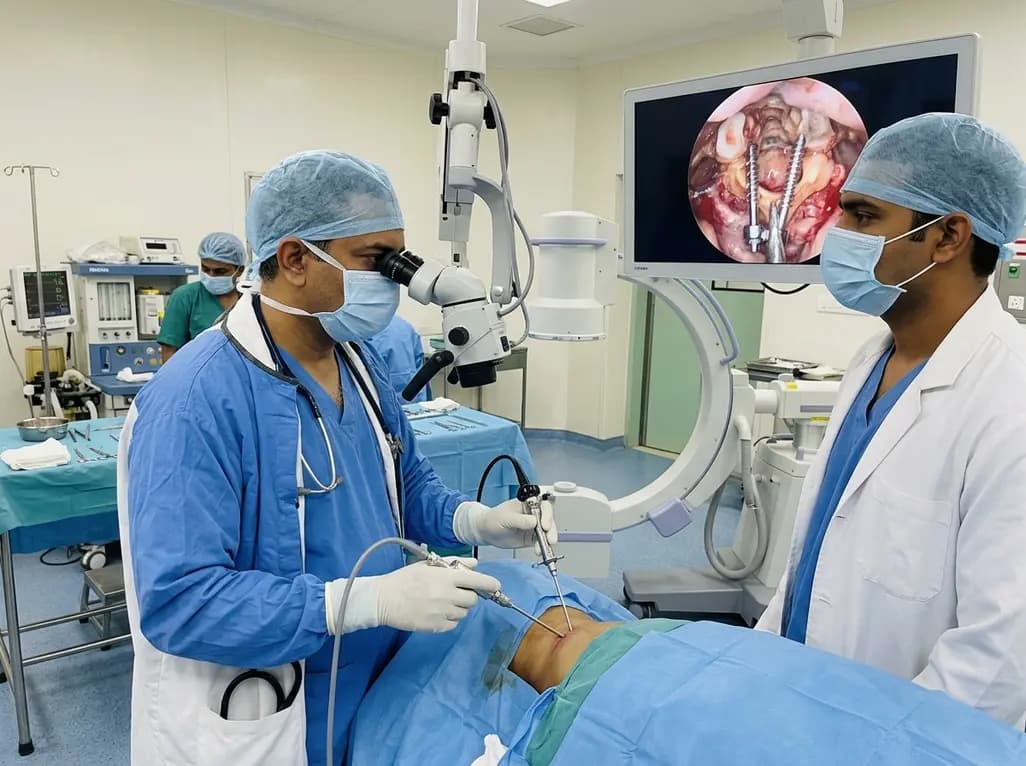
Endoscopic posterior cervical fusion represents a transformative milestone in minimally invasive spine surgery. Historically, surgical stabilization of the upper cervical spine required extensive posterior suboccipital and subaxial muscle dissection. Consequently, open approaches frequently caused severe postoperative axial neck pain, substantial blood loss, and protracted rehabilitation periods. While spinal endoscopy has reshaped lumbar interventions, applying endoscopic techniques to the atlantoaxial joint remained challenging. Specifically, simultaneous neural decompression, joint preparation, bone grafting, and rigid internal instrumentation through an endoscope presented formidable obstacles.
However, recent pioneering work now demonstrates the technical feasibility and safety of this integrated approach. By utilizing an arthroscopic-assisted or uniportal non-coaxial endoscopic spine surgery platform, clinicians can address complex upper cervical instability with unmatched direct visualization. Furthermore, this method preserves the critical posterior cervical muscular tension band. As a result, patients experience significantly decreased muscular detachment, reduced operative trauma, and accelerated functional recovery. This innovative procedure marks a crucial shift toward full endoscopic reconstruction at the craniovertebral junction.
The atlantoaxial junction possesses unique biomechanical flexibility alongside unforgiving neurovascular anatomy. Specifically, the C1-C2 articulation accounts for approximately fifty percent of normal cervical spine rotation. Because of this high mobility, pathological disruption from trauma, inflammatory rheumatoid arthritis, or congenital anomalies causes debilitating instability. Traditional open posterior stabilization relies on constructs like C1-C2 transarticular screws or Harms-Goel fixations. Although these classic techniques provide rigid stability, they require wide surgical exposure that jeopardizes vital structures.
Moreover, the proximity of the vertebral arteries, the C2 spinal nerve root, and the upper cervical cord heightens surgical risk. During exposure, accidental injury to the complex venous plexus surrounding the C2 nerve produces relentless intraoperative bleeding. Similarly, aberrant courses of the vertebral artery, such as a high-riding transverse foramen, drastically elevate the risk of vascular catastrophe. Therefore, any endoscopic adaptation must deliver precise anatomical orientation, visual clarity, and steady instrument handling. Continuous fluid irrigation steadily clears small hemorrhages, providing crisp visualization of bony landmarks throughout dissection.
Executing an endoscopic posterior stabilization requires meticulous preoperative planning and specialized instrumentation. First, surgeons position the patient prone under general anesthesia with continuous multimodal neurophysiological monitoring. Next, fluoroscopic guidance establishes accurate trajectory corridors before clinicians place a dedicated tubular portal. Using an arthroscopic-assisted or uniportal endoscopic system, the operator directs motorized burrs and radiofrequency wands through targeted corridors. This dual-instrument configuration separates the viewing axis from the working corridor, granting dynamic triangulation and unobstructed freedom of movement.
Subsequently, the surgical team carries out neural decompression under continuous high-definition endoscopic magnification. The surgeon carefully mobilizes the C2 nerve root and coagulates the surrounding epidural venous plexus. Next, specialized curettes thoroughly denude the cartilaginous surfaces of the C1-C2 lateral mass joint. The operator then packs autologous or allogeneic bone graft into the prepared articular space. Finally, surgeons place rigid screws under direct visualization, selecting C1 pedicle screws, arch screws, or C2 laminar screws. Clinicians then seat the connecting rods and tighten locking caps to secure rigid stabilization.
Recent clinical cohorts provide robust evidence supporting the safety and efficacy of this procedure. In the pioneering clinical series, surgeons successfully treated ten consecutive patients without encountering conversion to an open operation. Over the study course, the team placed forty posterior cervical screws. Postoperative computed tomography confirmed exceptional hardware accuracy; only two left C1 pedicle screws exhibited slight medial deviation without causing neurological impingement. Importantly, clinicians documented zero vertebral artery injuries, neurological deficits, or infections throughout follow-up.
Additionally, patient-reported outcome measures revealed dramatic, statistically significant reductions in neck pain and disability scores. Patients resumed independent ambulation rapidly, requiring fewer narcotic analgesics compared to historical open cohorts. Furthermore, radiographic studies confirmed solid osseous fusion across the atlantoaxial joint in every patient. One patient experienced an isolated, low-flow cerebrospinal fluid leak that resolved conservatively. Consequently, this study demonstrates that endoscopic fixation achieves rigid stabilization and high fusion rates without compromising clinical safety.
Achieving successful outcomes in endoscopic posterior cervical stabilization requires adherence to rigorous technical principles. First, surgeons must obtain high-resolution preoperative CT angiography to evaluate vertebral artery course anomalies and osseous landmarks. Identifying a persistent intersegmental artery or a high-riding vertebral artery prevents disastrous vessel laceration. Second, maintaining optimal hydraulic pressure throughout the procedure remains essential. Controlled hydrostatic pressure gently suppresses epidural venous oozing while preserving visual clarity, but excessive fluid pressure can elevate intracranial pressure.
Furthermore, surgeons must avoid aggressive traction on the C2 ganglion during lateral mass decortication. Bipolar radiofrequency ablation must remain strictly subperiosteal to prevent permanent occipital neuralgia or neuropathic pain. In addition, surgeons must prepare the lateral mass joint thoroughly to facilitate robust arthrodesis. Leaving cartilage on the articular facings invites pseudarthrosis and hardware loosening. Finally, integrating intraoperative navigation or advanced fluoroscopy ensures optimal screw entry points. These combined measures safeguard vital neurological structures while promoting rapid, durable bony arthrodesis.
The adoption of advanced endoscopic spine interventions has expanded rapidly across leading tertiary medical centers in India. However, performing endoscopic fusion at the craniovertebral junction requires specialized fellowship training and substantial endoscopic experience. Surgeons should ideally master subaxial endoscopic procedures and open craniovertebral reconstructions before attempting endoscopic C1-C2 fusion. In Indian clinical settings, trauma cases, rheumatoid subluxation, and congenital os odontoideum represent common indications that benefit from this approach.
Moreover, reducing soft-tissue injury substantially curtails postoperative wound complications, which remains vital in elderly or immunocompromised individuals. Smaller incisions also decrease postoperative surgical site infection rates and promote quicker return to functional independence. As medical technology advances, incorporating intraoperative three-dimensional navigation and specialized endoscopic drill systems will further flatten the learning curve. Therefore, while open fixation remains a reliable standard, endoscopic cervical fusion offers an innovative alternative for experienced spine teams.
Surgeons primarily consider endoscopic posterior cervical fusion for patients with atlantoaxial instability, traumatic odontoid fractures, congenital os odontoideum, and inflammatory subluxation secondary to rheumatoid arthritis. Additionally, patients who have failed conservative treatment and present with intractable neck pain or compressive cervical myelopathy represent prime surgical candidates. However, clinicians must carefully review vascular anatomy and ensure the pathology does not require extensive irreducible ventral decompression before choosing an endoscopic posterior stabilization corridor.
Traditional open Goel-Harms fixation requires extensive stripping of paraspinal musculature from the occiput and upper cervical vertebrae. Consequently, open approaches frequently result in notable blood loss, muscular atrophy, and chronic postoperative axial neck pain. In contrast, endoscopic posterior cervical fusion preserves the posterior ligamentous complex and paraspinal muscles. Furthermore, the high-magnification endoscopic camera enhances lighting around critical neurovascular anatomy, reducing intraoperative tissue trauma while achieving comparable fusion rates and rigid biomechanical stability.
Surgeons mitigate vertebral artery risks through comprehensive preoperative three-dimensional CT angiography and careful trajectory planning. Identifying high-riding vertebral arteries or anomalies such as a persistent intersegmental artery enables tailored screw selection. During surgery, continuous endoscopic visualization and steady fluid irrigation ensure complete clarity around the C1-C2 lateral mass. Moreover, surgeons maintain a strictly subperiosteal plane, apply careful bipolar coagulation, and utilize intraoperative fluoroscopy to prevent lateral cortical wall perforation during screw placement.
Disclaimer: This content is for informational and educational purposes only and is not intended to replace professional medical assessment, advice, diagnosis, or treatment. Refer to the latest local and national guidelines for clinical practice.
References
Yan YG et al. Endoscopic Posterior Cervical Fusion for the Treatment of Atlantoaxial Pathologies. Oper Neurosurg. 2026 Sep 09. doi: 10.1227/ons.0000000000002194. PMID: 42714433.
Chen Q, Brahimaj BC, Khanna R, Kerolus MG, Tan LA, Fessler RG. Posterior atlantoaxial fusion: a comprehensive review of surgical techniques and relevant vascular anomalies. J Spine Surg. 2020;6(1):164-170.
Li Z, Guan L, Hai Y, Liu Y, Zhang X. Endoscopic-Assisted Posterior Atlantoaxial Fusion: Technical Innovation and Clinical Experience. Orthop Surg. 2024;16(5):1150-1160.

Read summarized clinical updates, watch expert medical content, and earn CME certifications right from your smartphone.
Open your smartphone camera to scan and install.





Endoscopic posterior cervical fusion combines minimally invasive decompression, joint preparation, and rigid screw-rod fixation for atlantoaxial pathologies. Early clinical findings demonstrate solid bony union, excellent symptom relief, and minimal soft-tissue morbidity without significant vascular compromise.
last month

Review how tailored exercise programs benefit frail older patients with heart failure. Explore pathophysiological mechanisms, individualized training prescriptions, and strategies to overcome adherence barriers in clinical practice.
Today

Leucine Rich Bio has introduced South Asia's first comprehensive vaginal microbiome test, BugSpeaks. By profiling microbial dysbiosis, pathogens, and antimicrobial resistance, this next-generation diagnostic platform provides clinicians with deeper insights into recurrent infections, fertility, and reproductive health.
Today

In high-risk pulmonary embolism complicated by severe anemia and active uterine bleeding, systemic thrombolysis is contraindicated. Large-bore aspiration thrombectomy provides rapid hemodynamic rescue without fibrinolysis, supported by inferior vena cava filter placement and multidisciplinary critical care.
Today

This comparative study evaluates posterior labral tears with and without glenohumeral instability, highlighting distinct demographic patterns, MRI underestimation in stable shoulders, and successful arthroscopic repair outcomes.
Today

A clinical study demonstrates that early intervention with tocilizumab, either as monotherapy or bridged with corticosteroids, achieves significant proptosis reduction and resolves active inflammation in treatment-naive thyroid eye disease.
Today
