.webp&w=384&q=75)

Loading, please wait...
Loading, please wait...
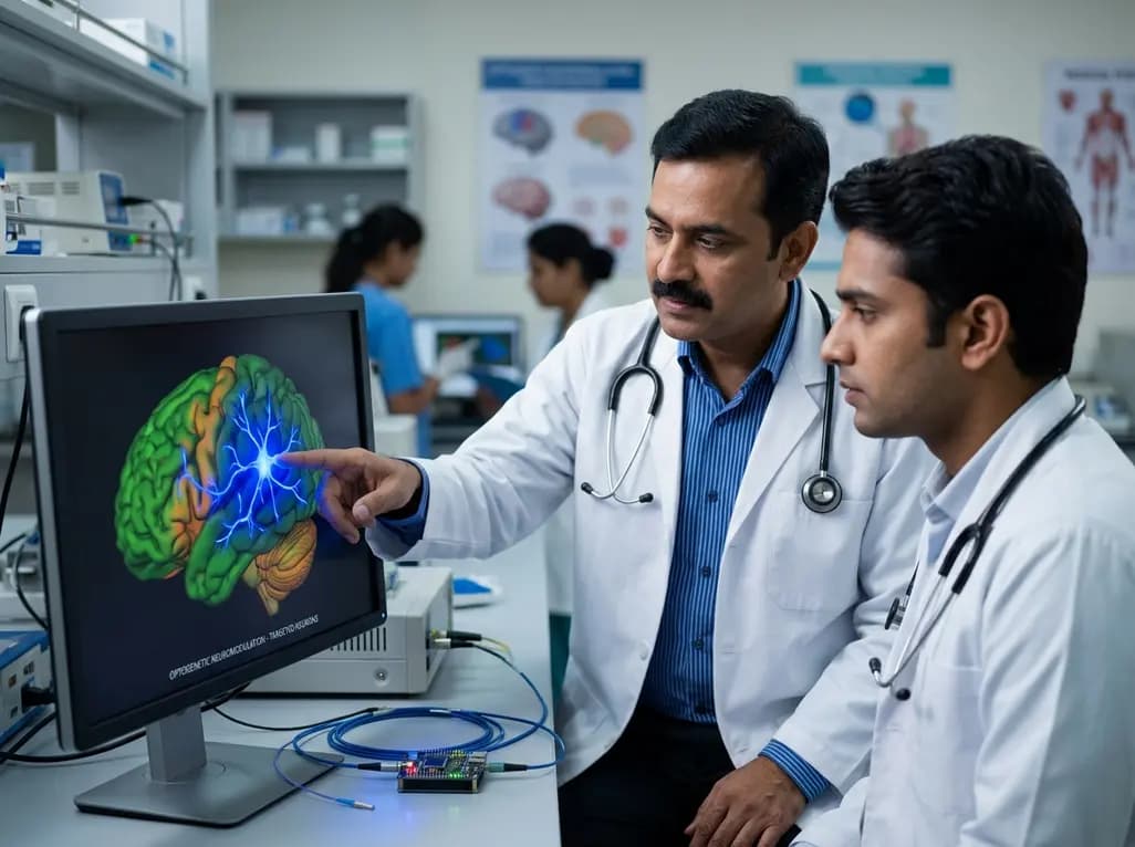
Refractory epilepsy poses one of the most stubborn therapeutic dilemmas in clinical neurology. Nearly one-third of individuals with epilepsy fail to attain seizure freedom despite multi-drug regimens. Consequently, clinicians urgently seek alternative solutions that can target epileptogenic circuits with absolute precision. The emerging application of optogenetics for epilepsy represents a transformative therapeutic avenue. By utilizing light-sensitive proteins to modulate specific neuronal networks, investigators are moving closer to switching off hyperexcitable seizure generators on demand.
The 2026 Nobel Prize in Physiology or Medicine honored Peter Hegemann, Georg Nagel, and Karl Deisseroth for uncovering light-gated ion channels. Their groundbreaking work established optogenetics, an innovative discipline uniting molecular genetics with optical physics. In this system, researchers harvest genes encoding microbial opsins, such as channelrhodopsin and halorhodopsin, from unicellular algae. Subsequently, scientists package these genetic sequences into benign viral delivery vectors. Clinicians can then introduce these vectors into targeted populations of cerebral neurons. When exposed to specific wavelengths of visible light, opsins undergo rapid conformational changes. Channelrhodopsin facilitates cation influx upon blue light illumination, thereby depolarizing and exciting the host neuron. Conversely, halorhodopsin and archaerhodopsin drive chloride influx or proton efflux under yellow-green light, immediately hyperpolarizing the cell membrane. This optical mechanism allows researchers to switch neuronal activity on or off within milliseconds. Therefore, optogenetics surpasses conventional pharmacological agents that diffuse broadly across non-target neural pathways. Dr. Nitin Kumar Sethi, chairman of the PSRI Institute of Neurosciences, highlights that optogenetics offers unparalleled cell-type specificity and real-time control. As a result, this platform provides neuroscientists with an unprecedented tool to interrogate intricate human neural circuits without unintended off-target electrophysiological disruption.
Standard neuromodulation strategies, such as vagus nerve stimulation and responsive neurostimulation, deliver electrical pulses across mixed brain tissue. Although these interventions reduce seizure burden in selected patients, electrical stimulation inevitably activates nearby bypass pathways and inhibitory interneurons indiscriminately. In contrast, optogenetics for epilepsy enables precise interrogation and suppression of isolated epileptogenic generators. Dr. Manjari Tripathi, head of neurology at the All India Institute of Medical Sciences (AIIMS), emphasizes that optogenetics allows clinicians to control hyperactive neurons with unmatched precision. During ictogenesis, abnormal synchronized bursting rapidly recruits surrounding cortical and subcortical networks. However, an implanted optical closed-loop system can detect early electrographic seizure onset and immediately trigger an inhibitory light pulse. The light instantaneously halts bursting pyramidal cells, extinguishing the seizure before clinical manifestations emerge. Furthermore, optogenetic inhibition acts locally without inducing widespread sedative consequences or systemic drug toxicity. Preclinical trials in animal models confirm that transient optical silencing of epileptic foci completely aborts behavioral seizures. Meanwhile, the surrounding physiological networks continue their normal functional tasks without cognitive deterioration. Consequently, optogenetic neuromodulation could soon transform surgical and interventional management for focal cortical dysplasia and mesial temporal sclerosis.
Currently, medical practitioners manage intractable focal epilepsy through extensive resective surgery, stereotactic radiofrequency ablation, or permanent tissue transection. Although resective surgery achieves favorable outcomes in well-defined temporal lobe cases, it carries substantial risks of postoperative cognitive decline, memory impairment, or visual field defects. Moreover, patients with seizure foci located within eloquent cortex often remain ineligible for surgical resection because tissue removal would cause devastating neurological deficits. Optogenetic technology presents a non-destructive alternative that preserves vital cerebral parenchyma. Instead of permanently excising critical functional areas, neurosurgeons could introduce inhibitory opsins directly into the epileptogenic focus. When aberrant high-frequency discharges begin, an optical interface silences the aberrant focus while leaving adjacent language or motor circuits undamaged. In addition, this approach provides substantial benefits over lifelong multidrug therapy. Antiseizure medications frequently induce systemic adverse events, including hepatotoxicity, severe cognitive slowing, bone density loss, and dangerous dermatological reactions. In contrast, localized optical interventions confine therapeutic action strictly to pathological nodes. Thus, the clinical application of light-mediated silencing could significantly alter the prognosis for individuals who have exhausted standard antiepileptic drug combinations.
The translation of optogenetics from animal models to human clinical reality is advancing rapidly through translational laboratory studies. Recently, collaborative teams at academic medical centers, including the University of California, San Francisco and the University of California, Los Angeles, achieved notable experimental milestones. Researchers applied adeno-associated viral vectors carrying engineered opsins to resected human neocortical and hippocampal brain slices obtained during epilepsy surgery. Upon exposure to targeted light pulses, these human neurons demonstrated robust suppression of spontaneous epileptiform discharges. Importantly, sustained opsin expression did not produce measurable cellular toxicity or alter normal membrane resistance. Furthermore, clinical trials in ophthalmology have already demonstrated the feasibility of human opsin gene therapy. Clinicians successfully introduced light-sensitive proteins into retinal ganglion cells to partially restore visual perception in patients suffering from end-stage retinitis pigmentosa. This monumental ocular milestone confirms that viral delivery of microbial opsins does not provoke unacceptable systemic immune rejection. Consequently, neurologists view the eye as an essential proof-of-concept proving ground. Building on these ocular accomplishments, neuroscientists are preparing protocols to validate similar gene transfer approaches for intractable focal neocortical epilepsies.
Despite remarkable preclinical progress, widespread clinical deployment of intracranial optogenetics faces significant bioengineering obstacles. First, delivering visible light deep into the human brain requires safe, biocompatible hardware. Photons scatter rapidly in dense neural tissue, meaning blue and yellow light penetrate only a few millimeters. Therefore, investigators are engineering flexible micro-LED arrays and wireless optical probes that minimize mechanical trauma during long-term implantation. Additionally, researchers are developing red-shifted opsins because near-infrared wavelengths penetrate deeper through cerebral layers with negligible photothermal tissue heating. Second, vector-mediated gene delivery demands rigorous long-term safety evaluation. Clinicians must ensure that adeno-associated virus serotypes remain stable for decades without triggering neuroinflammatory cascades or losing transcriptional efficacy. Third, neurosurgeons require dependable closed-loop electroencephalographic detectors that recognize pre-ictal biomarkers accurately. If an algorithm fails to detect seizure initialization promptly, optical intervention cannot abort the subsequent generalization. Dr. Tripathi emphasizes that resolving these technical challenges will require sustained multidisciplinary collaboration among neurosurgeons, optical engineers, and geneticists. Nevertheless, solving these biocompatibility and photonic hurdles will establish optogenetics as a reliable, revolutionary therapy for previously untreatable neurological conditions.
Q1: What is the primary difference between optogenetics and conventional electrical brain stimulation?
Conventional electrical brain stimulation delivers electrical currents non-selectively across local tissue, exciting all neighboring neurons and axonal tracts indiscriminately. In contrast, optogenetics utilizes cell-type-specific promoters to direct opsin expression exclusively into defined neuronal subtypes. Consequently, optogenetics allows clinicians to modulate only the seizure-generating pyramidal cells or protective inhibitory interneurons. This extraordinary specificity prevents off-target cognitive disruption, suppresses collateral network excitation, and avoids the tissue-damaging current spread frequently observed with standard deep brain stimulation electrodes.
Q2: How does a closed-loop optogenetic system detect and abort a seizure in real time?
A closed-loop optogenetic system pairs continuous local field potential recording electrodes with an implantable optical stimulator. Sophisticated machine-learning algorithms monitor real-time neurophysiological data to detect subtle pre-ictal oscillations indicative of impending seizure onset. Once the system identifies an abnormal discharge pattern, it instantly activates a miniature light source. This rapid optical pulse triggers inhibitory opsins within milliseconds, hyperpolarizing hyperactive focal neurons and terminating the emergent epileptiform cascade before overt clinical convulsions can manifest.
Q3: Why has optogenetics succeeded in human ophthalmology before direct intracranial neurological applications?
The human eye represents an ideal translational environment because the cornea and lens naturally transmit light directly to the retina without invasive cranial surgery. Furthermore, the ocular chamber maintains immune privilege, which dramatically minimizes the risk of inflammatory rejection against foreign microbial opsin proteins. In contrast, intracranial applications require invasive craniotomy, complex intracranial optical hardware implants, and extensive viral transduction across dense brain tissue, necessitating significantly more exhaustive preclinical safety and biocompatibility validations.
Disclaimer: This content is for informational and educational purposes only. It does not constitute medical advice or replace professional judgment. Refer to the latest local and national guidelines for clinical practice.
References

Read summarized clinical updates, watch expert medical content, and earn CME certifications right from your smartphone.
Open your smartphone camera to scan and install.





The 2026 Nobel Prize highlights optogenetics, an innovative optical neuromodulation approach that activates or silences targeted neuronal populations. Clinicians and researchers are now investigating how light-sensitive proteins can abort refractory focal seizures without damaging healthy surrounding neural tissue.
Today

Hospitalization for severe viral lower respiratory tract disease causes lasting physical, psychological, and productivity burdens for patients and their caregivers. Real-world multinational data reveal major gaps in discharge planning, emphasizing the urgent need for structured transitional pulmonary care.
Today

New evidence from Karolinska Institutet shows women vaccinated against HPV prior to conception have significantly lower rates of preterm birth and major perinatal complications. Clinicians in India can leverage these findings to strengthen adolescent immunisation and reproductive health advocacy.
Today

Cinematic rendering utilizes advanced global illumination to transform coronary CT angiography datasets. This imaging technique provides lifelike depth perception, helping cardiologists and surgeons precisely evaluate anomalous coronary artery origins, high-risk interarterial courses, and post-surgical anatomy.
Today
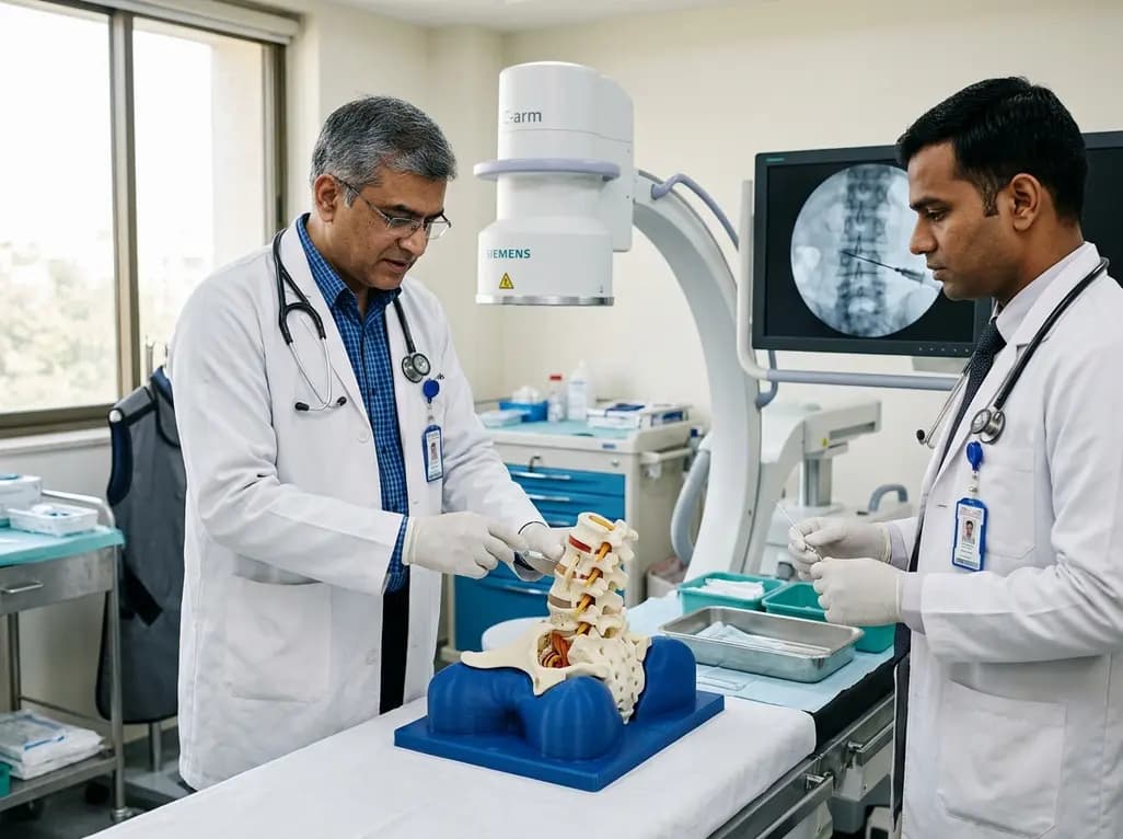
A novel 3D-printed lumbar spine simulator with tunable radiopacity provides realistic tactile and radiographic feedback for fluoroscopy-guided intervertebral disc puncture training, offering a low-cost, ethical alternative to cadaver models.
Today

A breakthrough study reveals that loss of SELENOT in hypothalamic POMC neurons triggers premature cellular senescence, ATF6α stress activation, and ER calcium depletion, linking neuroendocrine decay to metabolic disease.
Today
