.webp&w=384&q=75)

Loading, please wait...
Loading, please wait...

Cinematic rendering represents a major technological leap forward in post-processing algorithms for cardiovascular computed tomography. Unlike traditional volume rendering techniques that apply direct, localized ray-casting illumination models, this advanced algorithm utilizes Monte Carlo path tracing. Consequently, the technique simulates billions of physical light rays interacting naturally with volumetric tissue data, accurately reproducing absorption, scattering, ambient occlusion, and soft shadows. This sophisticated physics-based illumination creates photorealistic volumetric reconstructions that dramatically enhance human depth perception and surface relief. In addition, the method preserves subtle variations in soft-tissue density across complex anatomic boundaries. Cardiac radiologists can adjust window levels, color gradients, and lighting transfer functions to highlight specific cardiovascular tissue planes with remarkable clarity. Furthermore, cinematic rendering transforms dense volumetric datasets into intuitive anatomical models that simplify intricate spatial geometry. Cardiovascular specialists can therefore track tortuous vessels with greater confidence than conventional greyscale imaging typically permits. The resulting volumetric clarity effectively bridges the gap between diagnostic cross-sectional imaging and the operative field. Thus, clinicians gain an invaluable tool for conceptualizing complex anatomy before initiating invasive interventions.
Congenital anomalies of coronary artery origin occur in approximately one percent of the general population worldwide. Although many anatomical variants remain clinically silent and benign throughout life, specific configurations carry substantial risks of severe morbidity and mortality. In particular, anomalous origins from the opposite sinus of Valsalva represent hazardous anatomical variations with profound clinical consequences. Young competitive athletes face especially elevated risks of sudden cardiac arrest when unheralded coronary anomalies exist during intense physical exertion. Therefore, early and precise anatomical delineation plays an indispensable role in contemporary clinical risk stratification. Traditional catheter-based coronary angiography often struggles to clarify acute takeoff angles and three-dimensional pathways because it relies on two-dimensional projections. Coronary computed tomography angiography has consequently emerged as the reference standard imaging modality for noninvasive vascular evaluation. It reliably visualizes the ostial origin, proximal course, and adjacent cardiovascular structures with isotropic resolution. However, complex spatial relationships can still challenge clinicians when viewing standard multiplanar reformations alone. Cinematic rendering addresses this limitation by rendering anomalous vascular geometries with unmatched depth and clarity. Consequently, medical teams can promptly recognize malignant patterns and formulate tailored therapeutic management strategies.
The interarterial trajectory represents the most perilous anatomical course for an anomalous coronary artery. In this dangerous pattern, the aberrant coronary vessel traverses the narrow space between the ascending aorta and the main pulmonary trunk. During strenuous physical exercise, transient increases in cardiac output cause marked systolic expansion of both great vessels. Consequently, the stiff adjacent arterial walls mechanically compress the interarterial segment, provoking acute myocardial ischemia and life-threatening ventricular arrhythmias. Furthermore, high-risk morphological features frequently accompany this interarterial course, including acute-angle takeoffs, ostial kinking, and slit-like orifices. Intramural coursing within the aortic wall tunic further amplifies luminal collapse during cyclic aortic systolic expansion. Cinematic rendering provides exceptional anatomical visualization when delineating these critical geometric factors. The photorealistic lighting accentuates the external spatial relationships between the coronary trunk and the great vessels. In addition, realistic shadow casting highlights subtle tissue compression and geometric narrowing along the interarterial pathway. Clinicians can continuously track the anomalous vessel from its aortic origin down to its distal myocardial distribution. Therefore, cinematic rendering substantially improves clinical confidence during high-risk surgical decision-making and patient risk stratification.
Surgical unroofing serves as the primary operative strategy for repairing anomalous coronary arteries that feature intramural segments. During this corrective procedure, cardiac surgeons carefully excise the inner vascular wall shared between the aorta and the coronary artery. This surgical maneuver relocates the functional ostium directly into the appropriate aortic sinus, successfully eliminating dynamic systolic compression. However, meticulous postoperative imaging assessment remains crucial to verify adequate luminal decompression and procedural success. Coronary CT angiography provides essential cross-sectional surveillance following surgical intervention. Nevertheless, subtle postoperative tissue remodeling, local edema, and surgical clip artifacts can obscure fine anatomic detail on traditional planar views. Cinematic rendering significantly enhances post-surgical evaluation by clearly depicting the reconstructed neo-ostium in true photorealistic depth. Moreover, the algorithm illuminates the expanded lumen while accurately outlining residual adjacent cardiovascular structures. Radiologists and cardiac surgeons can easily examine the repaired vascular segment for potential complications such as persistent stenosis or flap creation. Consequently, cinematic rendering delivers comprehensive anatomical verification that reassures surgical teams and optimizes long-term patient recovery.
Standard multiplanar reformations and curved planar reconstructions remain the foundational cornerstones of coronary CT angiography interpretation. These established planar techniques provide precise quantitative measurements of luminal diameter, stenosis severity, and vessel wall thickness. However, planar slices intrinsically remove global spatial context, requiring interpreting radiologists to mentally reconstruct three-dimensional anatomy. Cinematic rendering effectively complements these standard diagnostic reconstructions by restoring intuitive volumetric perspectives and depth cues. The technique excels at illustrating complex spatial relationships during multidisciplinary heart team discussions. Furthermore, cardiothoracic surgeons benefit immensely from realistic depictions that closely match their operative field of view in the operating theatre. Clear photorealistic models also enhance communication with patients and families during pre-procedural informed consent discussions. Clinicians must recognize that cinematic rendering currently serves as a qualitative adjunctive tool rather than a standalone replacement for axial analysis. Future prospective studies will likely establish formal quantitative protocols and validate reader diagnostic performance across larger patient cohorts. Nevertheless, cinematic rendering already enriches clinical practice by making complex coronary anomalies immediately understandable to all medical providers.
Standard volume rendering relies on simplified ray-casting models that simulate direct surface lighting without natural ambient scattering. In contrast, cinematic rendering implements Monte Carlo path tracing to simulate complex physical light interactions, including absorption, diffusion, and soft shadows. Consequently, this advanced method generates photorealistic volumetric context with realistic depth perception. Clinicians therefore achieve clearer differentiation of overlapping cardiac structures and complex spatial relationships between coronary vessels and great arteries.
An anomalous coronary artery coursing between the ascending aorta and pulmonary trunk poses significant clinical dangers. During vigorous physical exertion, expansion of these great vessels can mechanically compress the anomalous vessel. Furthermore, acute ostial angulation, slit-like orifices, and intramural aortic pathways exacerbate dynamic luminal collapse. These anatomical factors severely restrict myocardial perfusion. Consequently, patients face heightened risks of exertional angina, syncope, myocardial infarction, and sudden cardiac death, particularly in young competitive athletes.
Surgical unroofing relieves intramural coronary segments by excising the common tissue wall shared with the aorta. Cinematic rendering excels in post-surgical assessment by clearly depicting the newly created, widened neo-ostium and residual vascular contours. Moreover, the enhanced soft-tissue contrast allows radiologists to verify complete decompression of the intramural tract. Thus, surgical teams can reliably confirm surgical success, assess anatomic remodeling, and detect complications such as persistent narrowing or flap formation.
Disclaimer: This content is for informational and educational purposes only... Refer to the latest local and national guidelines for clinical practice.
References

Read summarized clinical updates, watch expert medical content, and earn CME certifications right from your smartphone.
Open your smartphone camera to scan and install.





Cinematic rendering utilizes advanced global illumination to transform coronary CT angiography datasets. This imaging technique provides lifelike depth perception, helping cardiologists and surgeons precisely evaluate anomalous coronary artery origins, high-risk interarterial courses, and post-surgical anatomy.
Today
Hospitalization for severe viral lower respiratory tract disease causes lasting physical, psychological, and productivity burdens for patients and their caregivers. Real-world multinational data reveal major gaps in discharge planning, emphasizing the urgent need for structured transitional pulmonary care.
Today
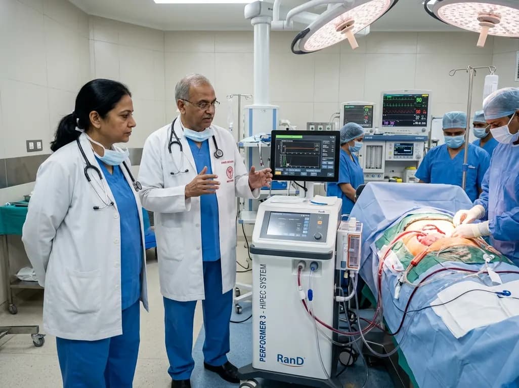
AIIMS Bathinda has inaugurated a specialized peritoneal oncology service featuring the RanD Performer 3 HIPEC platform. The facility offers cytoreductive surgery alongside hyperthermic intraperitoneal chemotherapy, transforming regional management for advanced ovarian, colorectal, and appendiceal malignancies.
Today
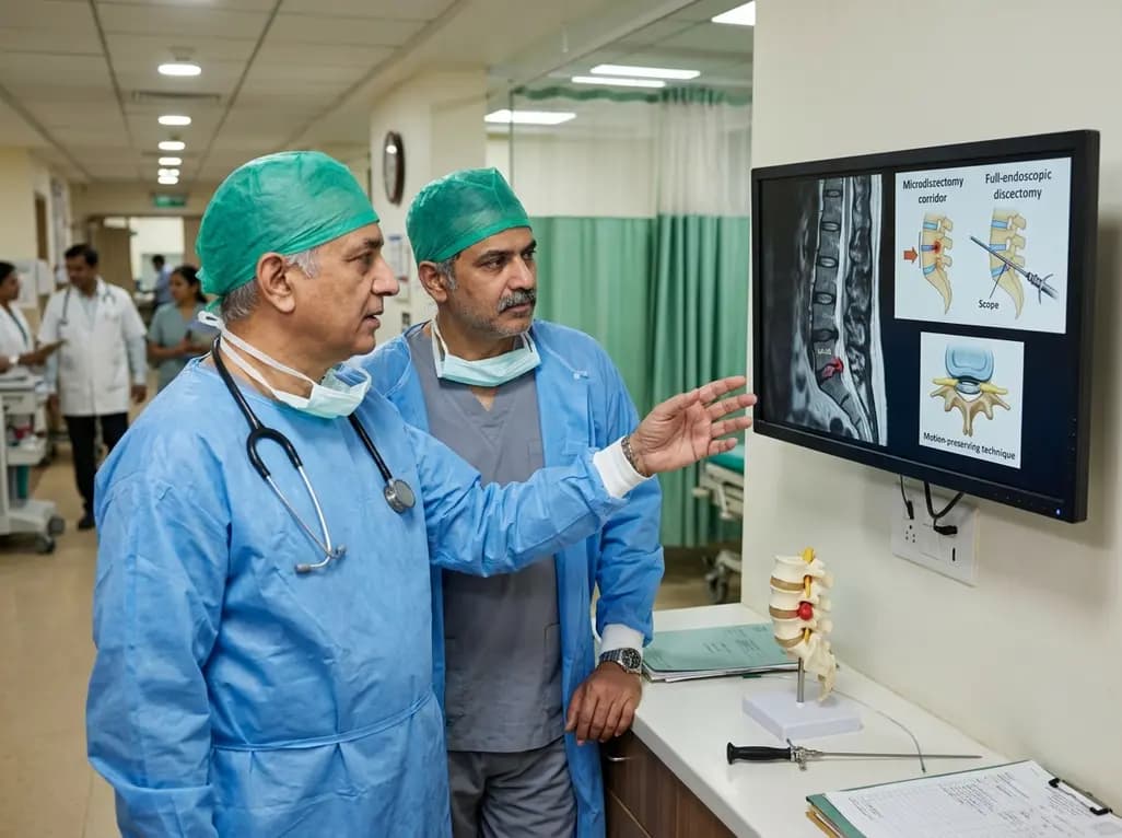
This clinical overview examines recent advancements in lumbar disc herniation surgery, comparing microscopic and full-endoscopic discectomy while highlighting biportal techniques and emerging motion-preserving innovations for spine specialists.
Today

A breakthrough study reveals that loss of SELENOT in hypothalamic POMC neurons triggers premature cellular senescence, ATF6α stress activation, and ER calcium depletion, linking neuroendocrine decay to metabolic disease.
Today
