.webp&w=384&q=75)

Loading, please wait...
Loading, please wait...
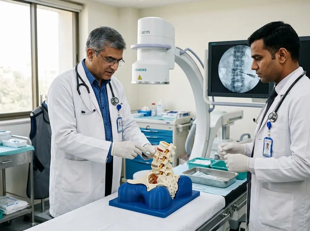
Interventional spine procedures demand exceptional needle precision, three-dimensional spatial orientation, and seamless fluoroscopic navigation. Traditionally, residency and fellowship programs have relied on cadaveric specimens to teach complex percutaneous spinal techniques. However, cadavers present substantial logistical hurdles for academic departments. They involve high procurement expenses, rigorous biohazard storage protocols, ethical constraints, and finite anatomical durability. Animal models offer compliant tissue, but their structural spinal anatomy diverges markedly from human morphology. Consequently, trainees often encounter their initial patient cases with limited tactile experience under real-time C-arm guidance. To resolve this educational challenge, researchers designed an innovative lumbar spine simulator. An ideal training model must combine anatomical fidelity, realistic haptic feedback, and authentic radiographic transparency. By integrating additive manufacturing with tailored casting polymers, modern bioengineers now produce durable physical phantoms. These accessible systems enable trainees to practice delicate intervertebral disc access repeatedly without compromising patient safety or exhausting departmental budgets. Furthermore, structured phantom simulation instills procedural discipline and calms resident anxiety before live operating theatre exposure.
Developing a dependable lumbar spine simulator begins with precise digital anatomical reconstruction. Investigators generated detailed lumbar vertebrae and pelvic bony structures from open-source stereolithography files. Next, they fabricated master prototypes using desktop fused deposition modeling. Because raw additive prints often exhibit surface layer striations, technicians polished the polymer surfaces to achieve authentic anatomical contours. They subsequently constructed flexible silicone molds around these polished templates to enable rapid, reproducible batch casting. For the structural bone matrix, the team utilized a durable two-component polyurethane casting resin. This polyurethane matrix replicates the rigid structural density of human cortical and cancellous bone. Furthermore, the mold-based workflow permits rapid fabrication of multiple identical vertebral sets. Educators can replace damaged vertebral units or depleted discs seamlessly during intensive procedural workshops. As a result, teaching hospitals can maintain high-throughput simulation curricula without investing in proprietary, cost-prohibitive commercial manikins. Ultimately, this modular approach empowers academic programs to scale their interventional training sustainably.
Achieving realistic radiographic transparency remains the primary engineering hurdle in spinal phantom development. Untreated printing polymers appear completely radiolucent under standard X-ray beams. Conversely, excessive mineral loading produces intense radiographic shadows that obscure needle visibility. Therefore, investigators systematically evaluated diverse radiopaque additives, including barium sulfate, iohexol, and calcium sulfate. While barium sulfate demonstrated high radiopacity, it frequently triggered image saturation and obscuring blooming artifacts around key bone landmarks. Iohexol yielded variable chemical stability within the curing polyurethane polymer. Ultimately, a composite incorporating five weight percent calcium sulfate achieved the ideal balance. Under C-arm fluoroscopy, this formulation clearly demarcated vertebral endplates, pedicles, and spinous processes. Simultaneously, the material permitted unhindered visualization of the advancing metallic needle tip. Thus, trainees can master the fluoroscopic parallax technique and accurately align the C-arm beam along the target vertebral axis. In addition, this tunable opacity mimics true osseous attenuation across varying tube voltages.
Procedural success during intervertebral disc access depends heavily on tactile discrimination across layered soft tissues. To emulate human tissue compliance, the engineers assembled the vertebrae using pliable silicone interfaces to create puncturable intervertebral disc spaces. They replicated paraspinal musculature and subcutaneous tissue using an optimized gelatin-glycerin foam. In addition, pigmented silicone served as an external cutaneous barrier. When clinicians introduce a spinal needle, they experience lifelike resistance changes as the needle transverses simulated skin, subcutaneous fat, and paraspinal muscle. Crucially, the needle encounters a distinctive give and firmer resistance upon engaging the silicone disc annulus. Fluoroscopic confirmation proves that the needle traverses the correct trajectory without premature deflection. Moreover, design optimization yielded a compact, lightweight housing that educators can easily transport between training suites. This portable architecture ensures convenient integration into busy clinical teaching schedules. As a result, fellows develop nuanced haptic reflexes alongside real-time radiological confirmation.
Integrating synthetic procedural models into training programs yields substantial educational dividends for modern healthcare systems. Spinal interventions carry inherent risks of nerve injury, vascular trauma, and discitis. By practicing on high-fidelity phantoms, novices overcome the steep learning curve in a safe, zero-risk setting. Trainees cultivate critical needle manipulation habits, manual dexterity, and biplanar fluoroscopic correlation. Furthermore, repeating needle passes on the simulator refines procedural efficiency and significantly reduces fluoroscopy screening time. Consequently, both the interventionalist and prospective patients receive lower cumulative radiation doses during future clinical encounters. In developing healthcare ecosystems like India, where expensive commercial simulation suites remain scarce, cost-effective open-source designs democratize access to advanced interventional training. Academic departments can produce these modular platforms locally using standard desktop 3D printers and common workshop polymers. Ultimately, this accessible training modality bridges the crucial gap between didactic anatomical theory and independent clinical execution, fostering safer patient care.
Researchers selected calcium sulfate because it produces balanced radiographic contrast under C-arm fluoroscopy. While dense additives like barium sulfate or iohexol can cause excessive blooming artifacts, five weight percent calcium sulfate accurately mirrors native vertebral attenuation. Consequently, trainees can discern critical bony anatomical landmarks clearly. In addition, this formulation preserves fluoroscopic needle tip visualization, ensuring realistic practice during complex interlaminar and transforaminal puncture simulations.
Traditional spinal intervention training relies heavily on expensive cadaveric specimens or proprietary commercial manikins. In contrast, this model utilizes open-source stereolithography files, fused deposition modeling, and inexpensive polyurethane casting resins. Therefore, academic medical institutions can fabricate durable, anatomically accurate training stations at minimal expense. Furthermore, departments can rapidly replace punctured intervertebral discs without buying entirely new assemblies, significantly lowering long-term maintenance costs for repetitive procedural education.
Trainees can utilize this phantom to rehearse various fluoroscopy-guided spinal interventions. Specifically, the model supports discography, percutaneous intervertebral disc puncture, and lumbar transforaminal epidural injections. Moreover, clinicians can refine essential C-arm positioning skills, such as obtaining true anteroposterior, lateral, and oblique views. Because the phantom provides realistic tactile resistance across simulated tissue layers, fellows build vital muscle memory before performing interventions on actual human patients.
Disclaimer: This content is for informational and educational purposes only and should not be considered medical advice. Always consult a qualified healthcare professional regarding any medical condition or treatment. While we strive to provide accurate, up-to-date information, medicine is an evolving field, and clinical practices may vary. Readers must independently verify all content. The opinions expressed are those of the authors and do not necessarily reflect the official policy or position of any medical institution or regulatory body. Refer to the latest local and national guidelines for clinical practice.
References

Read summarized clinical updates, watch expert medical content, and earn CME certifications right from your smartphone.
Open your smartphone camera to scan and install.





A novel 3D-printed lumbar spine simulator with tunable radiopacity provides realistic tactile and radiographic feedback for fluoroscopy-guided intervertebral disc puncture training, offering a low-cost, ethical alternative to cadaver models.
Today
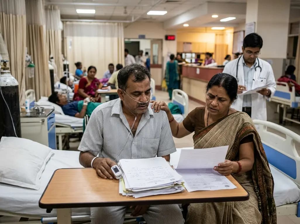
Hospitalization for severe viral lower respiratory tract disease causes lasting physical, psychological, and productivity burdens for patients and their caregivers. Real-world multinational data reveal major gaps in discharge planning, emphasizing the urgent need for structured transitional pulmonary care.
Today
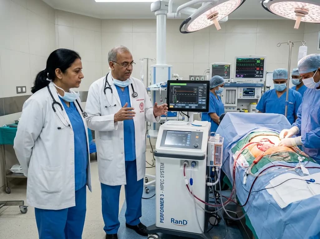
AIIMS Bathinda has inaugurated a specialized peritoneal oncology service featuring the RanD Performer 3 HIPEC platform. The facility offers cytoreductive surgery alongside hyperthermic intraperitoneal chemotherapy, transforming regional management for advanced ovarian, colorectal, and appendiceal malignancies.
Today
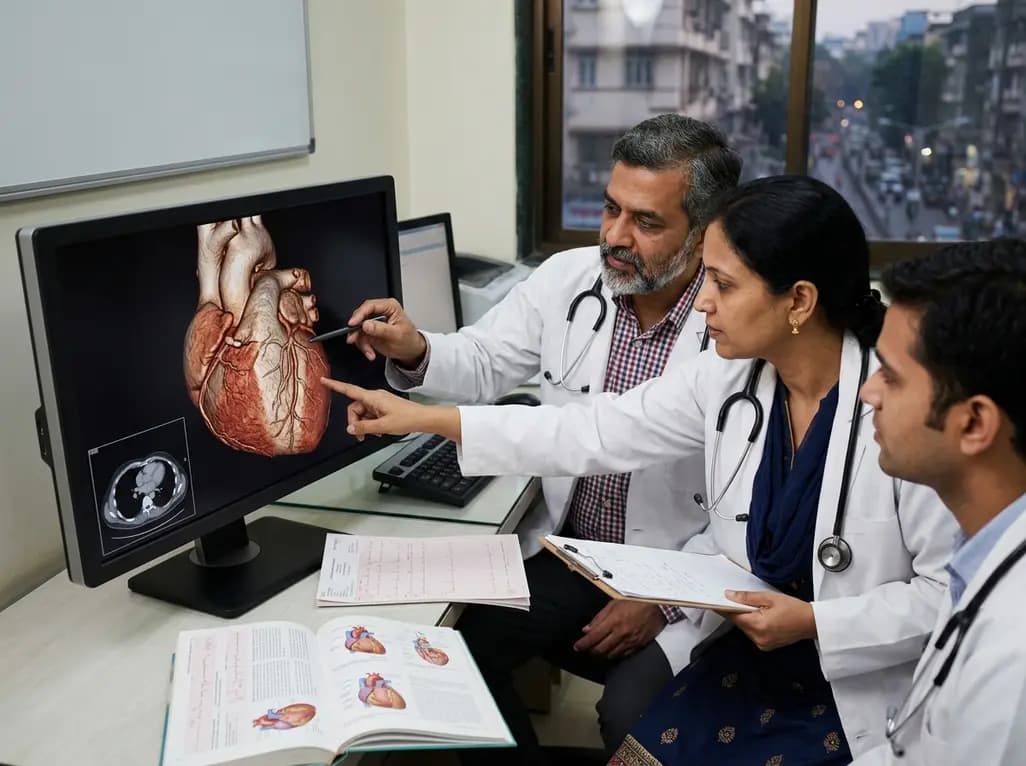
Cinematic rendering utilizes advanced global illumination to transform coronary CT angiography datasets. This imaging technique provides lifelike depth perception, helping cardiologists and surgeons precisely evaluate anomalous coronary artery origins, high-risk interarterial courses, and post-surgical anatomy.
Today

A breakthrough study reveals that loss of SELENOT in hypothalamic POMC neurons triggers premature cellular senescence, ATF6α stress activation, and ER calcium depletion, linking neuroendocrine decay to metabolic disease.
Today
