

Loading, please wait...
Loading, please wait...

Early hepatocellular carcinoma detection remains an urgent priority in clinical hepatology and surgical oncology worldwide. Primary liver malignancy often advances without evident clinical symptoms, which delays critical therapeutic intervention for high-risk patients. Recent advances in molecular diagnostics offer powerful optical tools to overcome this challenge. Specifically, multi-responsive fluorescent sensors now provide exceptional spatial resolution and chemical specificity for identifying early cellular transformation.
Traditional imaging modalities such as ultrasonography, contrast-enhanced computed tomography, and magnetic resonance imaging face significant limitations in resolving microscopic hepatic lesions. In contrast, fluorescent probes provide real-time visualization of dynamic cellular processes at the subcellular level. Researchers recently introduced an innovative tri-lock fluorescent probe named CMA, designed specifically to target mitochondrial abnormalities. Hepatocellular carcinoma cells demonstrate distinctive biochemical alterations, including enzymatic upregulation and biophysical microenvironmental changes. Consequently, relying on a single biomarker often produces false-positive readings or insufficient diagnostic specificity. To overcome these diagnostic hurdles, the CMA probe simultaneously monitors two distinct physiological parameters: mitochondrial carboxylesterase activity and local viscosity. Furthermore, the alkaline mitochondrial microenvironment acts as a third regulatory stimulus. When these three conditions coincide within malignant hepatocytes, the probe generates an extraordinary 654-fold fluorescence enhancement. Therefore, this multi-responsive platform establishes a highly sensitive threshold that clearly separates early malignant cells from surrounding non-tumorous liver tissue. As a result, optical bioimaging enters a new era of biochemical precision.
Carboxylesterases represent a superfamily of serine hydrolases localized predominantly in the endoplasmic reticulum and mitochondrial matrix of hepatocytes. These essential enzymes catalyze the hydrolysis of diverse ester-bearing compounds, regulating lipid homeostasis and processing exogenous pharmaceutical agents. In malignant hepatic cells, carboxylesterase expression increases dramatically to accommodate accelerated metabolic demands and fuel rapid tumor proliferation. Moreover, elevated carboxylesterase levels directly contribute to multidrug resistance by inactivating ester-containing chemotherapeutic drugs before they reach nuclear targets. Consequently, tracking carboxylesterase upregulation provides vital clinical insights into tumor phenotype, metabolic reprogramming, and therapeutic vulnerability. The CMA probe integrates a tailored benzoate ester moiety that functions as an enzymatic lock. When mitochondrial carboxylesterase hydrolyzes this protective ester group, it triggers an intramolecular charge transfer process that shifts probe absorption into the red spectrum. This enzymatic cleavage confirms the presence of hyperactive esterase activity in malignant hepatocytes. Accordingly, selective enzymatic cleavage prevents non-specific background fluorescence in healthy tissue, ensuring reliable diagnostic discrimination during microscopic analysis.
Beyond enzymatic alterations, the physical microenvironment within cellular organelles undergoes substantial reorganization during malignant progression. Viscosity serves as a fundamental physical property that dictates the rate of biomolecular diffusion, enzymatic collision, and intracellular signal propagation. In normal hepatocytes, mitochondria maintain fluid membranes and a strictly regulated internal matrix to facilitate rapid respiratory chain electron transfer. Conversely, cancer cells experience profound metabolic stress, organelle swelling, and abnormal accumulation of macromolecular protein aggregates. These structural modifications markedly elevate mitochondrial matrix viscosity. In addition, exposure to cytotoxic drugs and reactive oxygen species rapidly alters membrane fluidity, intensifying mitochondrial viscosity further. The CMA probe incorporates an indolium-coumarin conjugated rotor system that sensitively responds to these mechanical changes. Under standard low-viscosity conditions, the rotor dissipates absorbed photonic energy through non-radiative intramolecular rotation. However, elevated viscosity physically restricts this free molecular twisting, suppressing twisted intramolecular charge transfer pathways. Consequently, the probe emits intense radiative fluorescence, converting physical microviscosity into a measurable optical signal.
The remarkable performance of the CMA probe relies on an elegant tri-lock activation mechanism that prevents premature optical signal generation. First, the benzoate ester functional group acts as an enzymatic cage, masking the core fluorophore until mitochondrial carboxylesterase performs specific ester bond cleavage. Second, the molecular rotor mechanism formed by the conjugated indolium-coumarin backbone requires high environmental viscosity to halt rotational energy dissipation. Third, the physiological alkaline pH of the mitochondrial matrix promotes deprotonation of the newly liberated phenolic hydroxyl group. Furthermore, this deprotonation enhances the push-pull electronic architecture, thereby optimizing internal charge transfer. Thus, full optical activation occurs only when the probe encounters hyperactive esterase, high viscosity, and mitochondrial alkalinity at the exact same location. In experimental evaluations, this three-tier convergence produced an unprecedented 654-fold amplification in emission intensity. In contrast, single-stimulus triggers produced negligible background fluorescence. Cellular confocal imaging successfully confirmed that CMA preferentially accumulates in mitochondria and distinctly illuminates human hepatocellular carcinoma cells while remaining dark in healthy hepatocytes. Hence, this design achieves exceptional signal-to-noise contrast.
The development of multi-responsive fluorescent sensors provides meaningful practical avenues for modern oncology and clinical hepatology. Early detection of hepatocellular carcinoma remains a pressing clinical dilemma, especially among patients suffering from underlying cirrhosis, chronic viral hepatitis, or metabolic dysfunction-associated steatotic liver disease. Because conventional surveillance with ultrasound and serum alpha-fetoprotein exhibits limited sensitivity for sub-centimeter tumors, novel chemical tools can bridge critical diagnostic gaps. In future clinical workflows, probes like CMA could facilitate precise optical biopsy guidance during real-time laparoscopy, fluorescence-guided liver resection, and intraoperative margin evaluation. Surgeons could accurately identify microscopic tumor borders, sparing healthy functional parenchyma and reducing local recurrence rates. In addition, because the CMA probe sensitively records drug-induced viscosity fluctuations, it offers immense utility in preclinical pharmacological screening. Clinicians and pharmaceutical researchers could rapidly assess drug efficacy, monitor hepatotoxicity, and identify emerging chemoresistance patterns in patient-derived organoids. Therefore, integrating biophysical and biochemical organelle sensing heralds transformative improvements in liver cancer management.
The CMA probe significantly improves early cancer identification by tracking two distinct biophysical alterations simultaneously. Specifically, it senses both mitochondrial carboxylesterase upregulation and increased microviscosity within malignant hepatocytes. Conventional diagnostic modalities often overlook these subtle metabolic shifts during early tumor evolution. Consequently, by triggering a robust 654-fold fluorescent signal enhancement, this tri-lock molecular tool allows clinicians and pathologists to differentiate nascent malignant hepatic tissue from healthy liver parenchyma with remarkable diagnostic precision.
Mitochondrial viscosity directly influences molecular diffusion, signal transduction, and enzymatic interactions within cellular compartments. During hepatic malignant transformation, abnormal metabolic stress, dense protein accumulation, and structural cytoskeletal remodeling significantly increase mitochondrial microviscosity. Furthermore, elevated viscosity impairs normal organelle dynamics and promotes resistance to standard chemotherapeutic drugs. Therefore, monitoring mitochondrial viscosity provides physicians with crucial biological insight into tumor aggressiveness, cell membrane stiffness, and immediate pharmacological therapeutic response during cancer intervention.
Carboxylesterases play an essential physiological role in metabolizing various endogenous lipids and hydrolyzing xenobiotic pharmaceutical compounds. In hepatocellular tumors, malignant cells frequently overexpress mitochondrial carboxylesterases to accelerate drug deactivation, which facilitates substantial therapeutic evasion. In addition, this elevated enzymatic activity accelerates organelle detoxification pathways, preventing drug-induced apoptosis. Thus, measuring mitochondrial carboxylesterase levels enables oncologists to anticipate prospective chemotherapeutic resistance, stratify individual patient risks, and adjust personalized anticancer pharmacotherapy protocols effectively.
Disclaimer: This content is for informational and educational purposes only... Refer to the latest local and national guidelines for clinical practice.
References
Yue J et al. A multi-responsive fluorescent probe for simultaneous detection of mitochondrial esterase and viscosity. Anal Methods. 2026 Oct 26. doi: 10.1039/d6ay01913g. PMID: 42929085.
Yin J, Peng M, Lin W. Visualization of Mitochondrial Viscosity in Inflammation, Fatty Liver, and Cancer Living Mice by a Robust Fluorescent Probe. Anal Chem. 2019 Jul 2;91(13):8415-8421. doi: 10.1021/acs.analchem.9b01293. PMID: 31179692.
European Association for the Study of the Liver. EASL Clinical Practice Guidelines: Management of hepatocellular carcinoma. J Hepatol. 2018 Jul;69(1):182-236. doi: 10.1016/j.jhep.2018.03.019.

Read summarized clinical updates, watch expert medical content, and earn CME certifications right from your smartphone.
Open your smartphone camera to scan and install.





Discover how CMA, a tri-lock multi-responsive fluorescent probe, simultaneously detects mitochondrial esterase and microviscosity. This novel optical sensing platform produces a 654-fold emission enhancement, offering breakthrough diagnostic potential for early hepatocellular carcinoma detection.
Today
A cross-sectional study evaluates physician knowledge and counseling practices regarding e-cigarettes. Discover how these clinical insights align with India's prohibition framework under PECA 2019 and evidence-based tobacco cessation strategies.
Today

A KGMU study reveals over half of women with breast abnormalities fail to undergo diagnostic confirmation, leading to advanced-stage detection. Discover the socio-cultural barriers, clinical risks of incomplete resection, and community-level solutions needed to bridge the critical screening-to-diagnosis gap in India.
Today
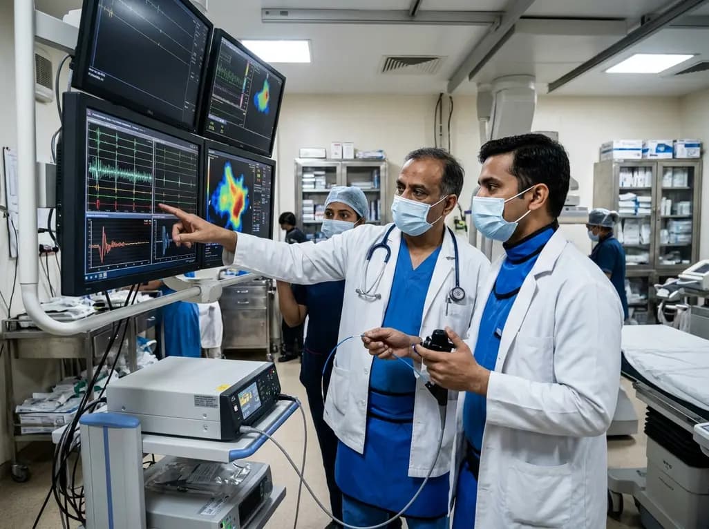
A comparative non-contact study reveals that high-power radiofrequency delivery with the QDOT MICRO catheter induces an artifactual contact force rise. Electrophysiologists must understand this power-dependent thermal and electrical phenomenon to avoid inadvertent catheter withdrawal and ineffective ablation.
Today
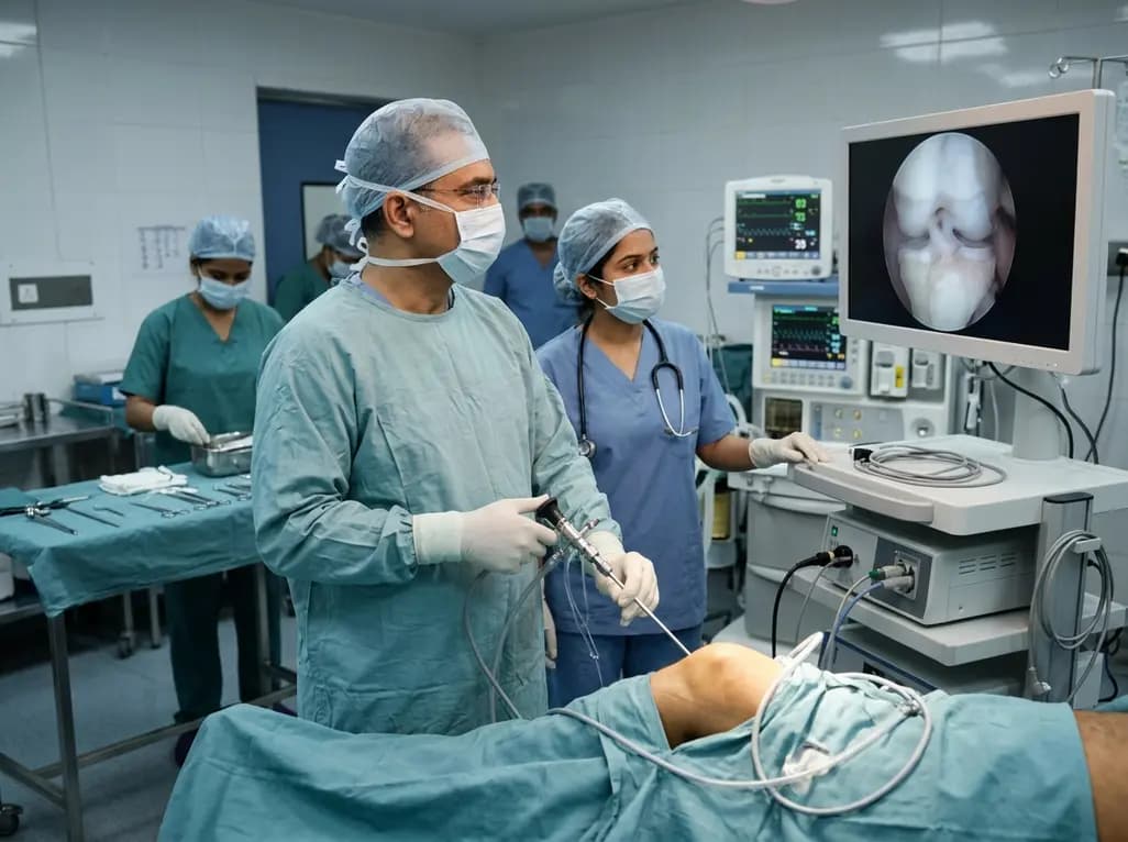
Healthium Medtech has transitioned its arthroscopy vertical into SIRONIX Orthopedics, aiming to capture key segments of the global joint care market. The strategic shift introduces dedicated portfolios in sports medicine, joint regeneration, surgical reconstruction, and digital operating technologies.
Today
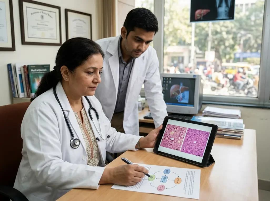
Preclinical research demonstrates that BMI1 overexpression alleviates metabolic syndrome-related liver injury. By triggering ubiquitin-mediated METTL3 degradation, BMI1 prevents AHNAK m6A methylation and suppresses fibrogenic TGF-β/SMAD signaling, significantly improving hepatic steatosis and insulin resistance.
Today
