.webp&w=384&q=75)

Loading, please wait...
Loading, please wait...
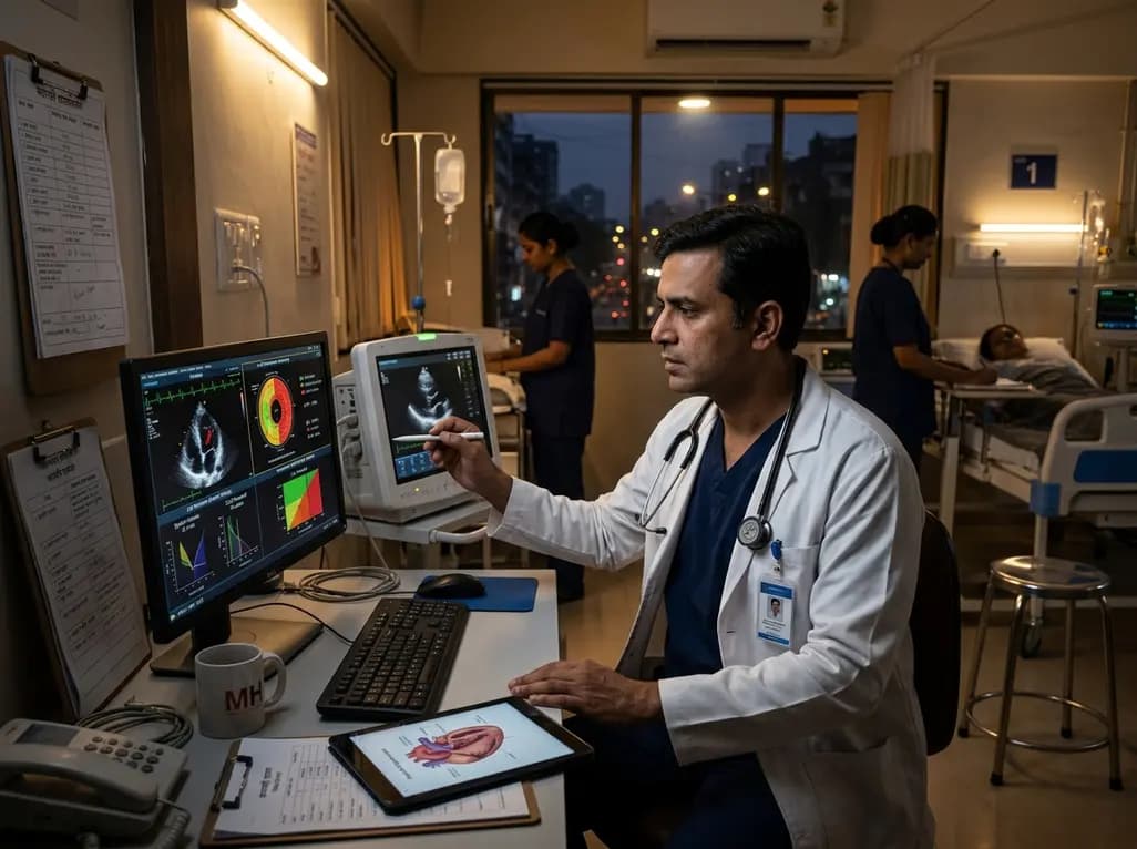
Modern lifestyles have drastically altered natural circadian cycles through widespread artificial lighting. Recent investigations demonstrate that even low levels of night-time light exposure trigger deleterious changes in myocardial structure and function. While clinicians traditionally focus on conventional cardiovascular hazards, environmental photic disruption represents a potent risk factor. Consequently, chronic nighttime illumination destabilizes autonomic regulation and impairs restorative nocturnal physiology, initiating cardiac remodeling before overt clinical symptoms appear.
A groundbreaking cohort investigation evaluated the cardiac consequences of nocturnal illumination across 11,017 participants in the UK Biobank. Investigators objectively quantified baseline sleep photic exposure using wrist-worn accelerometers equipped with sensitive light sensors over seven days. Subsequently, participants underwent comprehensive cardiovascular magnetic resonance imaging after three years of follow-up. The resulting imaging revealed clear morphological discrepancies between cohorts. Specifically, participants exposed to night-time illumination exceeding three lux displayed distinct left ventricular concentric hypertrophy. Adjusted regression analyses demonstrated a 2.4 percent increase in left ventricular mass indexed to height. Furthermore, subjects exhibited a 1.5 percent increase in mean wall thickness and a 1.9 percent lower contraction fraction. These structural alterations narrowed ventricular cavity volume while restricting overall cardiac flexibility. In addition, the researchers observed compromised global strain values across circumferential, radial, and longitudinal axes. Therefore, objective imaging proves that minimal nighttime illumination induces measurable subclinical cardiac decline in previously healthy adults. Importantly, three lux represents an exceedingly low illuminance threshold. For instance, a glowing mobile display on a bedside table easily exceeds this level. Hallway lighting seeping beneath a cracked bedroom door also produces equivalent ambient exposure. Consequently, millions of urban sleepers routinely encounter light intensities sufficient to stimulate harmful cardiac adaptation.
The biological mechanisms connecting dim nighttime illumination to structural myocardial changes involve complex neuroendocrine pathways. Specialized intrinsically photosensitive retinal ganglion cells detect low-intensity light through closed eyelids. These sensitive photoreceptors transmit photic impulses directly to the suprachiasmatic nucleus, the central circadian pacemaker in the brain. Consequently, this inappropriate nocturnal stimulus sharply suppresses pineal melatonin production. Melatonin serves as a crucial endogenous antioxidant and anti-inflammatory mediator throughout the vascular tree. In addition, circadian disruption impairs peripheral clock genes residing inside cardiomyocytes, disturbing regular mitochondrial energetic metabolism. Furthermore, photic stimulation prevents normal nocturnal parasympathetic activation, triggering sustained sympathetic nervous system dominance during sleep. Patients lose the normal nocturnal blood pressure dipping pattern that permits cardiovascular recuperation. As a result, persistent systemic vascular resistance elevates continuous mechanical afterload against the left ventricle. Over time, chronically elevated wall tension compels myocardial cells to synthesize sarcomeric proteins in parallel. Therefore, the left ventricular walls thicken concentrically to distribute excess mechanical strain. This maladaptive biological remodeling progressively impairs diastolic compliance, elevating the eventual risk of clinical heart failure. Indeed, animal models show that persistent light accelerates myocardial fibrosis and activates pro-inflammatory cytokine pathways within cardiac tissues.
Adverse remodeling driven by light pollution does not restrict itself to the left ventricle. On the contrary, advanced cardiovascular magnetic resonance revealed substantial structural modifications across multiple cardiac chambers. Specifically, the researchers observed significant morphological alterations within both the left atrium and the right ventricle. Exposed subjects showed a 2.3 percent higher left atrial volume index and a 1.1 percent lower ejection fraction. Similarly, right ventricular measurements revealed a 0.9 percent greater end-diastolic volume and a 1.6 percent elevation in end-systolic volume. These widespread chamber alterations indicate systemic functional impairment rather than isolated focal remodeling. Moreover, extensive epidemiological tracking over eight to ten years confirmed that these subtle imaging phenotypes herald severe downstream clinical disease. High nocturnal light exposure was associated with a 29 percent higher risk of incident heart failure. Additionally, exposed cohorts experienced a 24 percent higher risk of myocardial infarction and a 33 percent increase in stroke risk. Participants also exhibited a 15 percent greater incidence of atrial fibrillation and a 26 percent elevation in cardiovascular mortality. Hence, subclinical chamber deformation directly predicts catastrophic clinical endpoints.
The cardiovascular implications of nocturnal light exposure carry exceptional relevance for medical practice across modern India. Unprecedented urbanization has illuminated Indian metropolitan centers with dense street lighting, luminous commercial hoardings, and continuous traffic glare. Concurrently, Indian populations face a heightened genetic susceptibility to coronary artery disease, early-onset heart failure, and metabolic syndrome. Many urban residences feature compact, multigenerational living arrangements where televisions, hallway lights, and digital electronics remain continuously active overnight. Moreover, pervasive smartphone usage prior to sleep exposes individuals to potent short-wavelength blue light right in bed. Consequently, physicians frequently encounter patients with resistant nocturnal hypertension characterized by absent physiological blood pressure dipping. Clinicians regularly prescribe statins, antihypertensive agents, and lifestyle diets during busy outpatient consultations. However, few healthcare providers routinely assess bedroom lighting hygiene or environmental photic disruption during clinical reviews. Given that artificial light pollution represents a completely modifiable environmental pollutant, physicians possess a valuable preventative opportunity. Proactively addressing sleep hygiene and nocturnal darkness can reinforce pharmacological therapies and reduce premature cardiovascular events across vulnerable Indian communities. Thus, simple environmental counseling empowers clinicians to mitigate cardiometabolic risks without adding costly pharmacotherapy.
Unlike genetic susceptibility or advancing age, nocturnal photic exposure represents an easily modifiable cardiovascular risk factor. Clinicians should incorporate targeted environmental guidance into routine preventive consultations. First, physicians must encourage patients to eliminate all illuminated electronic devices from the immediate sleep environment. Charging docks, standby light-emitting diodes, and television sets should remain completely turned off or covered. Furthermore, patients should banish smartphones and tablets from their bedside tables to prevent late-night retinal stimulation. Second, individuals residing near urban streetlamps should install heavy blackout curtains or light-blocking window shades. When home modifications remain financially or structurally unfeasible, wearing a contoured eye mask provides an exceptionally cost-effective barrier. Third, individuals requiring nocturnal illumination for bathroom visits should install motion-activated dim red or amber lights. Red light wavelengths exert minimal influence on melanopsin photopigments compared to alerting blue spectra. In addition, healthcare providers should emphasize consistent sleep schedules to strengthen natural circadian rhythmicity. By implementing these inexpensive non-pharmacological interventions, patients can effectively suppress nighttime sympathetic arousal and preserve cardiac structure. Ultimately, preserving biological darkness serves as accessible, non-invasive preventive medicine for the heart.
Q1: What threshold of nocturnal light exposure produces measurable cardiac changes?
Researchers detected significant adverse remodeling at illuminance levels exceeding just 3 lux. In practical terms, this minimal intensity corresponds to a dim smartphone display or distant ambient illumination from a hallway bulb. Consequently, even faint ambient light disrupts normal biological dark periods during sleep. Thus, ambient bedroom illumination does not need to appear glaringly bright to impose sustained physical strain on myocardial tissue over extended timeframes.
Q2: Why does dim light exposure during sleep cause left ventricular hypertrophy?
Light stimulates intrinsically photosensitive retinal ganglion cells even through closed human eyelids. Consequently, this continuous sensory input suppresses pineal melatonin production and prompts excessive nocturnal sympathetic nerve firing. Furthermore, the autonomic nervous system sustains elevated vascular resistance and prevents standard nocturnal blood pressure dipping. Therefore, chronic hemodynamic afterload forces left ventricular cardiomyocytes to remodel concentrically, gradually thickening the chamber walls and restricting filling capacity over several consecutive years.
Q3: How can physicians counsel patients to reduce nocturnal light risks effectively?
Clinicians should actively advise patients to treat bedroom darkness as an essential cardiovascular health pillar. Specifically, individuals should eliminate illuminated electronics, remove glowing chargers, and relocate phones away from nightstands. Moreover, families living in dense urban settings ought to install blackout window shades or wear contoured eye masks. These simple non-pharmacological interventions rapidly restore circadian entrainment, lower autonomic night-time stress, and provide accessible preventative protection against progressive cardiac remodeling.
Disclaimer: This content is for informational and educational purposes only. It does not constitute medical advice or replace professional judgment. Refer to the latest local and national guidelines for clinical practice.
References

Read summarized clinical updates, watch expert medical content, and earn CME certifications right from your smartphone.
Open your smartphone camera to scan and install.





Emerging evidence shows that even dim night-time light exposure alters cardiac geometry and weakens contractility. In a landmark cohort study, nocturnal light above 3 lux triggered concentric left ventricular hypertrophy and subclinical dysfunction, highlighting nocturnal illumination as a modifiable cardiac risk factor.
Today

A new scoping review protocol systematically maps global evidence on how climatic shifts, ambient heat, and pollution impact human fertility. Discover key physiological mechanisms, clinical preconception strategies, and critical public health alignments under Sustainable Development Goals 3 and 13.
Today

Pharmacogenomics and artificial intelligence are revolutionizing cardiology by tailoring therapies to individual genetic profiles and clinical data, reducing adverse drug events, and improving cardiovascular patient outcomes.
Today
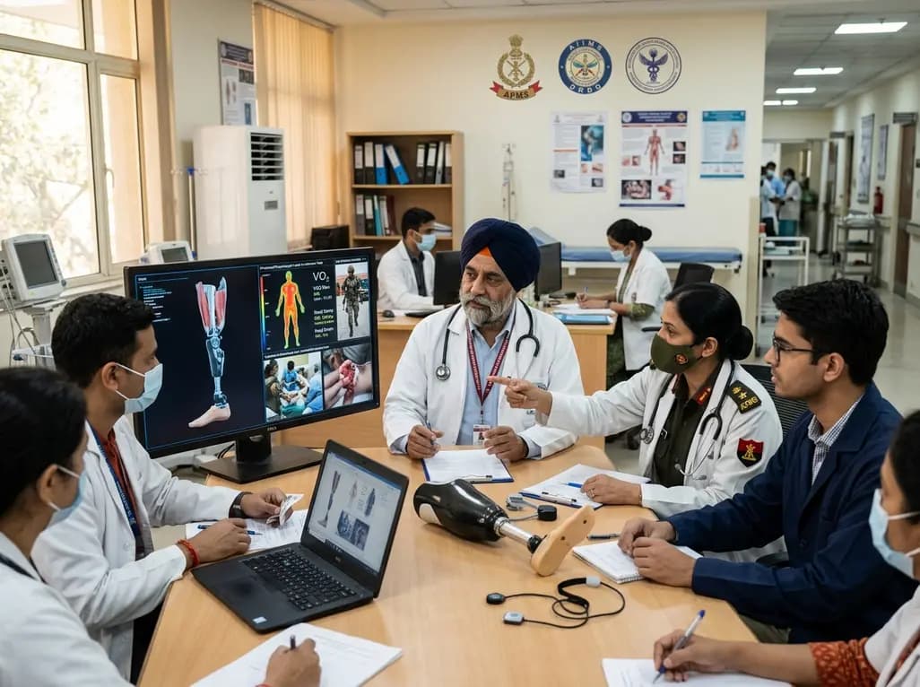
India's premier scientific bodies have joined the Armed Forces Medical Services to solve unique physiological and operational challenges faced by soldiers. The national collaboration covers combat casualty care, artificial intelligence diagnostics, bionic prosthetics, and extreme-environment physiological resilience.
Today

A retrospective cohort study evaluates the short- and long-term impacts of SARS-CoV-2 infection on patients with Graves' disease receiving antithyroid drug therapy, highlighting thyroid status destabilization, clinical symptom exacerbations, and post-viral sequelae.
Today
