

Loading, please wait...
Loading, please wait...

Motor dysfunction remains one of the most debilitating outcomes of stroke, traumatic spinal cord injury, and cerebral palsy. Fortunately, modern bioengineering offers transformative pathways to restore lost mobility through brain-computer interfaces. These systems translate cortical motor intentions into functional actions by controlling robotic exoskeletons and neuroprostheses. However, achieving sustained clinical efficacy requires stable communication between neural tissue and electronic hardware. Consequently, investigators are designing advanced neural interface materials to overcome signal degradation and tissue scarring. In addition, integrating intelligent computational decoding into these platforms accelerates functional restoration. Clinicians must now understand how material science and computational neuroscience converge to reshape neurorehabilitation.
Traditional rigid microelectrodes frequently trigger adverse biological reactions in brain tissue. Specifically, the mechanical mismatch between stiff silicon and delicate neural parenchyma generates chronic shear stress. Consequently, microglia and astrocytes activate around the insertion tract within days. This inflammatory cascade promotes robust glial scar formation around recording sites. Over time, the insulating capsule increases electrical impedance and dramatically diminishes signal amplitude. Therefore, modern material science prioritizes flexible and biomimetic substrates to resolve this chronic foreign body response. Conductive polymers, such as PEDOT:PSS, provide high charge-injection capacity and lower interfacial impedance. Furthermore, researchers engineer stretchable elastomers like polydimethylsiloxane to match the elastic modulus of native brain tissue. In addition, biocompatible hydrogel coatings mimic the extracellular matrix and release anti-inflammatory therapeutics locally. These bioinspired coatings suppress neuroinflammation and promote long-term neuronal survival near recording sites. Thus, advanced neural interface materials preserve cellular proximity and sustain high signal-to-noise ratios over extended implantation periods. Ultimately, these biocompatible innovations ensure that neural signals remain robust across months of intensive clinical neurorehabilitation. As a result, patients avoid repeat revision surgeries, while clinicians secure unbroken data streams for motor learning. Such reliable structural integration establishes the biological foundation for dependable therapeutic interventions.
Clinical neurotechnologies capture electrophysiological signals through distinct recording modalities that offer differing trade-offs. Noninvasive electroencephalography records electrical potentials from the scalp surface without surgical risk. Because scalp electrodes avoid tissue penetration, clinicians readily deploy them in outpatient therapy settings. However, volume conduction through the skull and cerebrospinal fluid attenuates high-frequency signals. Consequently, scalp sensors provide lower spatial resolution and suffer from motion artifacts during active movement. To mitigate these drawbacks, engineers design advanced dry electrodes and conductive textile sensors that improve skin contact. Conversely, invasive intracortical microelectrodes penetrate directly into the motor cortex to capture action potentials from individual neurons. This approach delivers exceptional spatial precision and decodes complex kinematic trajectories with high fidelity. Nevertheless, intracortical arrays require craniotomy and face persistent biological rejection risks. Semi-invasive electrocorticography bridges this clinical gap by placing electrode grids directly over the dura or subdural cortex. Electrocorticography records high-gamma power with superior temporal accuracy compared to scalp sensors while avoiding parenchymal tissue damage. Moreover, endovascular stent-electrode arrays now access cortical vessels without open neurosurgery. Hence, selecting the appropriate recording modality depends on the clinical risk profile and the motor resolution required for each patient. Additionally, multidisciplinary teams tailor sensor configurations to align with individual recovery targets and anatomical integrity.
Decoded neural commands require rapid and precise computational processing to execute realistic movements. However, raw neural recordings exhibit substantial nonstationarity due to micro-motion, fluctuating attentional states, and baseline biological drift. Therefore, modern brain interfaces employ advanced artificial intelligence algorithms to maintain decoding accuracy across sessions. Deep learning architectures, including convolutional neural networks and recurrent models, capture complex spatiotemporal patterns within multichannel brain signals. Furthermore, graph neural networks map functional connectivity across disparate motor regions, extracting relational features that reflect motor intention. In addition, transfer learning and self-supervised representations allow models to generalize across multiple training days without lengthy recalibration. This algorithmic capability directly mitigates neural distribution shifts that previously caused decoding failure during rehabilitation sessions. Efficient edge-computing chips also execute these deep networks with minimal latency, ensuring immediate feedback delivery. Consequently, when a stroke patient imagines wrist extension, the algorithm detects the intention within milliseconds. Such real-time responsiveness reinforces Hebbian plasticity by synchronizing cortical intention with peripheral sensory feedback. Thus, artificial intelligence transforms noisy, erratic electrophysiological data into stable and intuitive motor commands. These computational advances bridge the gap between static lab demonstrations and responsive, clinic-ready neuroprosthetic devices. As a result, patients experience smooth, immediate control that maintains engagement throughout exhausting therapy protocols.
Translating brain interfaces into practical neurorehabilitation addresses severe motor deficits caused by central nervous system damage. In post-stroke care, motor imagery-based interfaces activate ipsilesional and contralesional motor networks during attempted movement. When the system detects sensorimotor rhythm desynchronization, it triggers an external robotic exoskeleton or neuromuscular electrical stimulation. Consequently, this paired associative stimulation closes the sensorimotor loop and stimulates latent corticospinal tracts. Meta-analyses demonstrate clinically meaningful improvements on the Fugl-Meyer Assessment for stroke survivors who complete interface-driven training. Similarly, individuals with complete or incomplete spinal cord injury benefit from brain-controlled robotic gait trainers. By bypassing disrupted spinal pathways, the interface decodes supraspinal locomotion intentions and drives robotic lower-limb orthoses. In addition, chronic stimulation protocols promote spinal cord plasticity and elicit voluntary electromyographic recovery below the injury level. Pediatric patients with cerebral palsy also demonstrate promising functional gains through gamified, noninvasive neurofeedback protocols. These engaging environments encourage active participation, helping young patients refine coordination and motor planning skills. Furthermore, combining assistive robotics with direct neural control accelerates functional independence in activities of daily living. As clinical trials expand, these neurotechnologies are transitioning from investigational tools into standard neurorehabilitation workflows. Ultimately, clinicians gain objective biomarkers to measure neuroplastic progress alongside observable physical recovery.
Despite impressive scientific breakthroughs, widespread adoption of neural interfaces faces several practical and technical hurdles. First, the phenomenon of interface illiteracy affects approximately fifteen to thirty percent of prospective users. These individuals struggle to generate reliable sensorimotor rhythm modulation despite sustained practice and coaching. Therefore, researchers must establish individualized priming techniques, such as sensory vibrotactile cueing, to enhance cortical responsiveness. Second, clinical usability demands streamlined hardware that therapy teams can operate without specialized biomedical engineers. Lengthy setup times and conductive gel application frequently reduce the time available for active patient rehabilitation. Consequently, industry partners are manufacturing user-friendly wireless headsets and self-calibrating software packages. Third, regulatory approval pathways require rigorous proof of long-term safety, especially for chronically implanted materials. Sterilization protocols, hermetic packaging, and electrochemical stability under continuous stimulation remain demanding engineering benchmarks. In addition, reimbursement frameworks in healthcare systems must evolve to support high-cost robotic and neurotechnological equipment. Clinicians, biomedical engineers, and regulatory agencies must collaborate closely to establish standardized outcome measures and safety protocols. By systematically addressing these material, computational, and practical obstacles, clinical centers can unlock the full therapeutic potential of next-generation neurorehabilitation systems. Thus, sustained multidisciplinary innovation will expand restorative treatments to millions living with profound neurological impairments worldwide.
Rigid silicon probes induce micromotion against soft parenchymal tissue, causing continuous mechanical microtrauma, neuroinflammation, and thick glial encapsulation. In contrast, flexible neural interface materials, including conductive polymers and elastomeric substrates, closely match the mechanical compliance of native brain tissue. This mechanical biomimicry drastically diminishes shear strain and foreign body reactions. Consequently, the electrode remains close to active neurons, reducing electrical impedance and sustaining high-fidelity signal acquisition over months of chronic implantation.
Neural recordings inherently fluctuate due to patient fatigue, physiological shifts, and minor sensor movements. Artificial intelligence architectures, such as recurrent and graph neural networks, resolve these dynamic distribution shifts by extracting robust spatiotemporal representations. Additionally, deep learning algorithms decode intent within milliseconds, facilitating immediate closed-loop feedback through robotic orthoses or electrical stimulators. This rapid synchronization between cortical motor imagination and peripheral movement reinforces Hebbian synaptic plasticity, significantly accelerating functional recovery during neurorehabilitation.
Stroke lesions frequently disrupt corticospinal pathways, preventing intentional commands from executing limb movement. When stroke survivors practice motor imagery, brain-computer interfaces detect sensorimotor rhythm desynchronization across surviving cortical networks. The system immediately activates assistive devices, such as robotic exoskeletons or neuromuscular electrical stimulation, executing the intended limb movement. Consequently, this contingent feedback closes the sensorimotor loop, reorganizing peri-infarct neural networks and promoting functional motor recovery far beyond spontaneous healing capacity.
Disclaimer: This content is for informational and educational purposes only and should not be considered as medical advice or used for diagnostic or treatment purposes. Refer to the latest local and national guidelines for clinical practice.
References

Read summarized clinical updates, watch expert medical content, and earn CME certifications right from your smartphone.
Open your smartphone camera to scan and install.





Advanced neural interface materials and artificial intelligence are transforming brain-computer interfaces. By enhancing biocompatibility and real-time signal decoding, these systems establish reliable closed-loop neurorehabilitation for patients recovering from stroke, spinal cord injury, and cerebral palsy.
Today
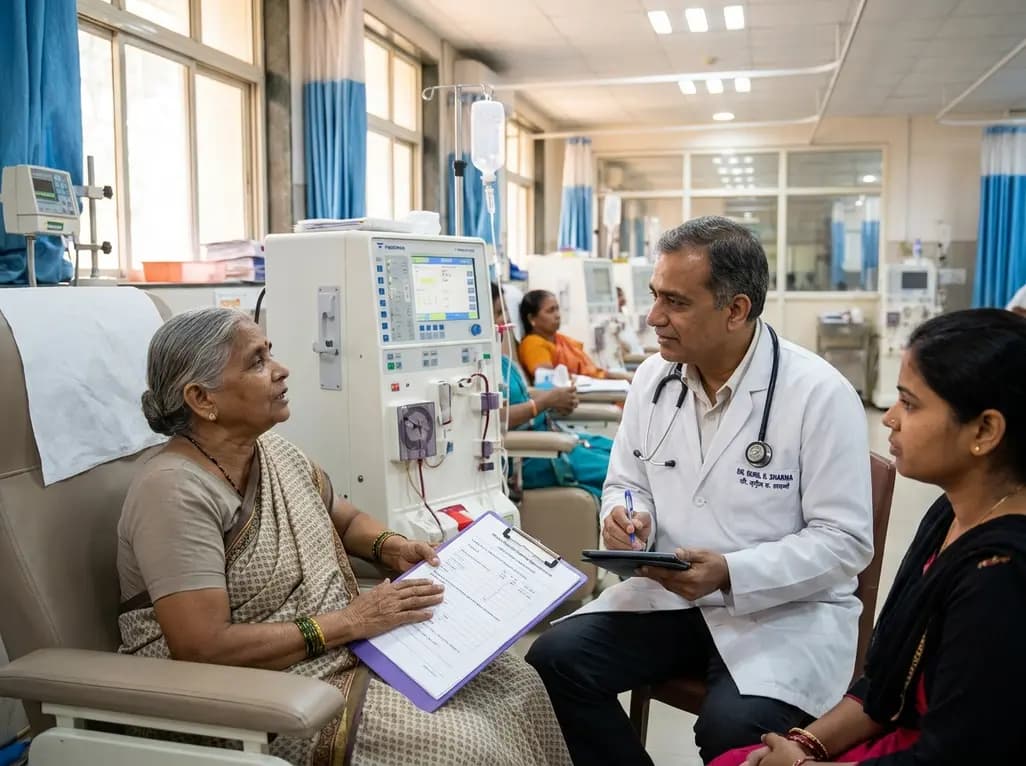
A prospective multicenter cohort study demonstrates that baseline health-related quality of life, particularly physical functioning, independently predicts all-cause maintenance hemodialysis mortality. Assessing patient-reported outcomes provides vital prognostic value beyond standard biochemical markers.
Today
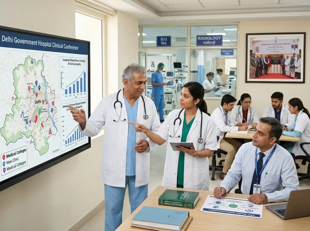
Delhi is undergoing a major healthcare overhaul with enhanced Ayushman Bharat insurance up to ₹10 lakh, increased MBBS and PG medical training seats, expanded primary network mandirs, new tertiary neuro-ICUs, and public-private reproductive technology initiatives to build a resilient, preventative healthcare model.
Today
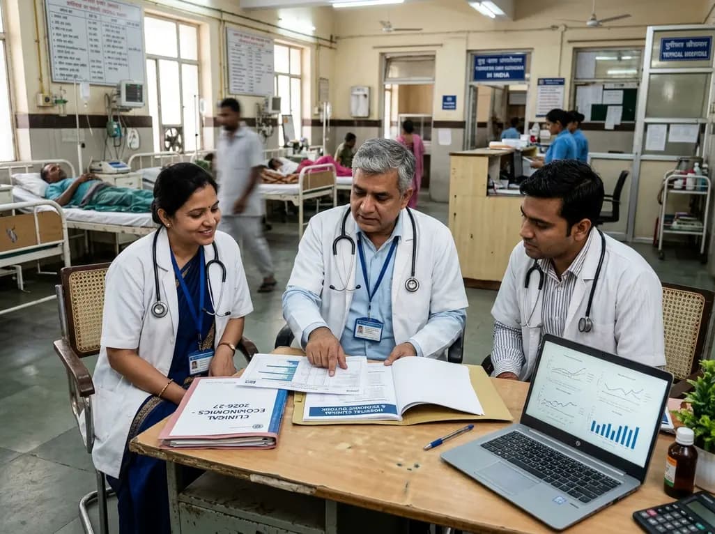
Aon forecasts India's healthcare inflation will ease to 10.1% in 2027 from 11.5% in 2026. Despite moderation, healthcare expenses continue to outpace general inflation, propelled by non-communicable diseases, advanced medical therapeutics, and expanding utilization patterns across diverse patient populations.
Today
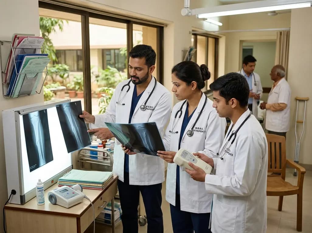
Non-union fractures occur in 2-5% of cases, causing significant disability and cost. Conservative modalities such as LIPUS, PEMF, and focused ESWT offer non-surgical healing options for defects up to 5-10 mm when adequate mechanical stability exists without infection.
Today

Explore how novel oxygen-releasing PVA hydrogels prevent early post-transplant hypoxic damage in pancreatic beta cells, sustaining viability and insulin secretory function.
Today
