.webp&w=384&q=75)

Loading, please wait...
Loading, please wait...
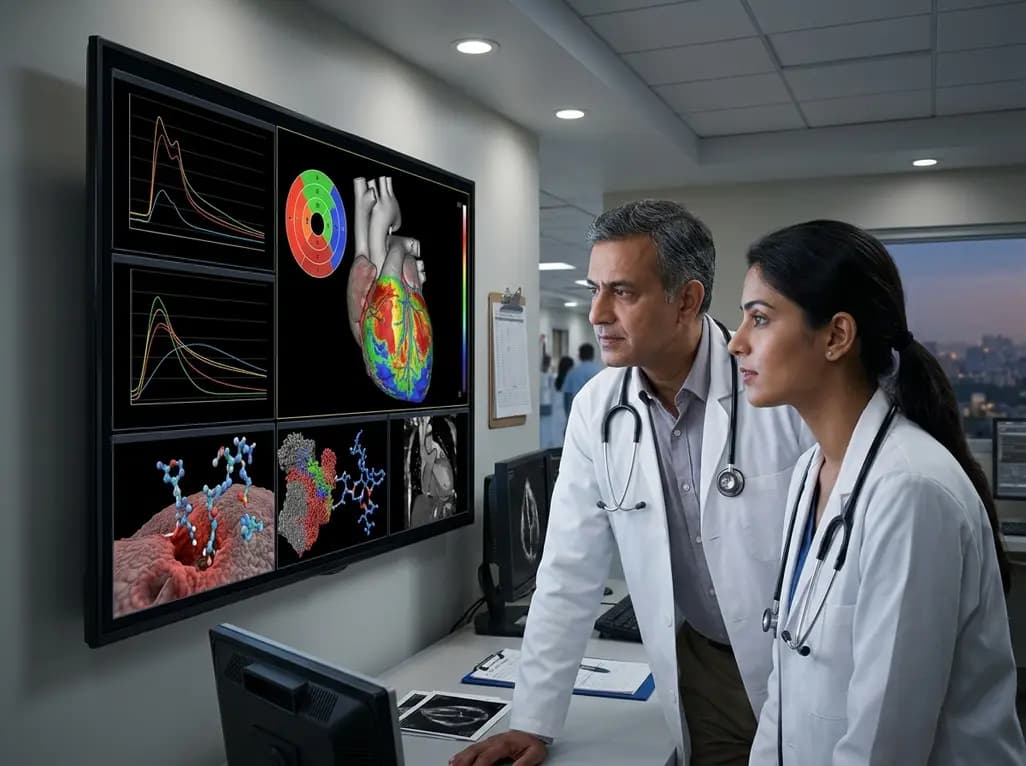
Ischemic heart disease remains a leading cause of morbidity and mortality across India, despite major advances in emergency revascularization techniques. While timely restoration of blood flow is essential for salvaging threatened myocardium, the process of reperfusion itself can paradoxically trigger further damage. This phenomenon, known as myocardial ischemia reperfusion injury (MIRI), represents a critical therapeutic hurdle in acute cardiac care. Modern clinicians recognize that successful percutaneous coronary intervention (PCI) is not the end of the journey, as I/R injury continues to evolve over minutes, days, and even weeks. This complex injury network contributes significantly to the final infarct size, often accounting for up to 50% of the total myocardial damage. Consequently, identifying high-risk patients who suffer from severe MIRI is vital for improving long-term outcomes and preventing adverse ventricular remodeling. Researchers are now shifting focus from simple diagnosis to deep phenotyping, using molecular and imaging signatures to understand the unique biological trajectory of each patient. By refining our understanding of these pathways, we can better tailor post-reperfusion management and secondary prevention strategies.
Myocardial ischemia reperfusion injury is not a single event but rather a multi-phase process characterized by distinct biological transitions. The initial phase involves the sudden burst of reactive oxygen species and mitochondrial dysfunction upon the return of oxygenated blood. Furthermore, calcium overload and the opening of the mitochondrial permeability transition pore lead to immediate cardiomyocyte death. As the process matures, microvascular obstruction—often termed the "no-reflow" phenomenon—compromises tissue perfusion despite the patency of the epicardial artery. In addition, an intense inflammatory response follows, where neutrophils and macrophages infiltrate the damaged tissue to clear debris, inadvertently causing further collateral damage. Over the subsequent weeks, this injury leads to intramyocardial hemorrhage and structural changes that define adverse ventricular remodeling. Therefore, understanding these temporal windows is essential for selecting the appropriate biomarker at the right time. For instance, markers of oxidative stress may be most relevant in the hyperacute phase, whereas proteins related to extracellular matrix turnover provide insights into long-term remodeling and the risk of heart failure development. Clinicians must view these phases as a continuum requiring dedicated monitoring.
High-sensitivity cardiac troponins (hs-cTn) currently serve as the diagnostic anchor for identifying myocardial necrosis in acute settings. They offer unmatched sensitivity for detecting cell death; however, they provide relatively limited insight into the upstream pathogenetic mechanisms of MIRI. While peak troponin levels correlate roughly with total infarct size, they do not distinguish between the initial ischemic insult and the secondary injury caused by reperfusion. Moreover, troponin kinetics can be influenced by renal function and chronic comorbidities, potentially confounding the assessment of acute I/R severity. This gap in clinical knowledge necessitates the search for more specific tools that can phenotype the "quality" of the injury rather than just the "quantity" of cell death. For example, markers that specifically reflect endothelial dysfunction or microvascular integrity could provide earlier warnings of impending complications like no-reflow. In the Indian context, where patients often present late to the emergency department, the limitations of troponins become even more apparent. We require biomarkers that can provide prognostic value beyond mere diagnosis, helping clinicians allocate intensive care resources more effectively to those at the highest risk of post-infarct heart failure.
The search for novel MIRI biomarkers has led to the exploration of diverse molecular targets beyond traditional proteins. Emerging candidates include tissue-derived markers, non-coding RNAs, and cell-free DNA, which offer high specificity for cardiac injury processes. Notably, extracellular vesicles (EVs) have gained attention as messengers of intercellular communication during the inflammatory phase of reperfusion. Additionally, the integration of multi-omics platforms—encompassing genomics, proteomics, and metabolomics—allows for a holistic view of the patient’s metabolic state. For instance, metabolomic signatures can reveal shifts in fatty acid oxidation and amino acid imbalances that occur during the ischemic transition. These high-dimensional data points enable a level of precision medicine previously thought impossible in acute cardiology. In practice, a multi-marker approach might involve combining a traditional necrosis marker with a specific inflammatory mediator and a microRNA associated with angiogenesis. Such a panel could refine risk stratification by identifying patients likely to benefit from adjunct cardioprotective therapies. However, the translation of these markers into routine clinical use requires standardized assays and rigorous validation in large, multi-ethnic cohorts to ensure their incremental value over existing diagnostic standards.
Complementing molecular markers, advanced imaging techniques like cardiovascular magnetic resonance (CMR) and positron emission tomography (PET) provide essential tissue characterization. CMR is widely considered the gold standard for assessing myocardial viability and quantifying the area at risk. Specifically, late gadolinium enhancement (LGE) allows for the precise measurement of infarct size, while T2-weighted imaging can detect myocardial edema and intramyocardial hemorrhage. Furthermore, PET imaging provides quantitative assessment of myocardial blood flow and metabolic activity, which is particularly useful for identifying microvascular dysfunction. These imaging biomarkers offer spatial resolution that blood-based markers cannot provide, allowing clinicians to see exactly where the injury is most severe. Integrating these findings with molecular data creates a powerful prognostic framework. For example, a patient with high levels of inflammatory biomarkers and significant microvascular obstruction on CMR would be at the highest risk for adverse remodeling. Despite the higher costs associated with these technologies, their role in selective post-reperfusion phenotyping is becoming increasingly realistic. In specialized cardiac centers, these tools are already helping to guide complex decision-making regarding the intensity of follow-up and the use of expensive neurohormonal blockade therapies.
The successful translation of MIRI biomarkers into the clinical workflow depends on several critical factors, especially within the resource-diverse healthcare environment of India. First, there is an urgent need for standardized assays that can be performed rapidly at the point of care. Second, researchers must demonstrate that biomarker-guided decisions actually lead to improved patient outcomes or better resource allocation. Consequently, prospective clinical trials are necessary to move these markers from the "research only" category into evidence-based guidelines. In addition, the cost-effectiveness of multi-marker panels and advanced imaging must be established to justify their use in routine practice. Expert opinion suggests that the primary value of these novel tools will likely be prognostic rather than diagnostic, helping to refine the intensity of post-discharge monitoring. As we move toward 2026 and beyond, the focus will remain on harmonizing sampling protocols and integrating artificial intelligence to interpret complex multi-omic data. Ultimately, the goal is to develop a personalized approach to myocardial salvage, where every patient receives a tailored management plan based on their unique molecular and imaging profile of ischemia-reperfusion injury.
While a heart attack or myocardial infarction involves the initial blockage of a coronary artery, myocardial ischemia reperfusion injury specifically refers to the damage that occurs when blood flow is restored. This paradoxical injury is caused by oxidative stress, inflammation, and mitochondrial dysfunction that happens during the re-oxygenation phase. It can significantly increase the total amount of damaged heart tissue even after the artery is successfully opened by doctors.
Traditional troponins are excellent at identifying that heart muscle cells have died, but they do not explain the underlying cause or specific mechanism of that death. They cannot differentiate between the initial damage from the blockage and the secondary damage from the reperfusion process. Therefore, additional biomarkers are needed to identify specific issues like microvascular obstruction or intense inflammation, which require different management strategies than simple necrosis.
Cardiovascular magnetic resonance (CMR) acts as a high-resolution tool for tissue characterization, allowing clinicians to visualize the physical consequences of reperfusion injury. It can identify edema, intramyocardial hemorrhage, and the "no-reflow" phenomenon within the heart wall. This information is vital for predicting which patients are likely to develop heart failure or heart rhythm problems later, allowing for more aggressive early intervention and closer long-term monitoring.
Disclaimer: This content is for informational and educational purposes only and does not constitute medical advice. It is not intended to be a substitute for professional medical advice, diagnosis, or treatment. Always seek the advice of your physician or other qualified health provider with any questions you may have regarding a medical condition. Refer to the latest local and national guidelines for clinical practice.
References

Read summarized clinical updates, watch expert medical content, and earn CME certifications right from your smartphone.
Open your smartphone camera to scan and install.





Myocardial ischemia/reperfusion injury (MIRI) remains a significant challenge in cardiology. This review explores emerging molecular and imaging biomarkers beyond traditional troponins to enhance prognostic stratification and post-reperfusion phenotyping in patients with acute myocardial infarction.
3 months ago

Following Nigeria's first recorded Ebola case linked to regional travel, the Africa CDC has convened an emergency ministerial summit. This alert underscores critical clinical priorities for healthcare professionals, including differentiating early viral infection from malaria, implementing barrier nursing, and triage.
Today

Leucine Rich Bio has introduced South Asia's first comprehensive vaginal microbiome test, BugSpeaks. By profiling microbial dysbiosis, pathogens, and antimicrobial resistance, this next-generation diagnostic platform provides clinicians with deeper insights into recurrent infections, fertility, and reproductive health.
Today

Review how tailored exercise programs benefit frail older patients with heart failure. Explore pathophysiological mechanisms, individualized training prescriptions, and strategies to overcome adherence barriers in clinical practice.
Today

This comparative study evaluates posterior labral tears with and without glenohumeral instability, highlighting distinct demographic patterns, MRI underestimation in stable shoulders, and successful arthroscopic repair outcomes.
Today

A clinical study demonstrates that early intervention with tocilizumab, either as monotherapy or bridged with corticosteroids, achieves significant proptosis reduction and resolves active inflammation in treatment-naive thyroid eye disease.
Today
