.webp&w=384&q=75)

Loading, please wait...
Loading, please wait...
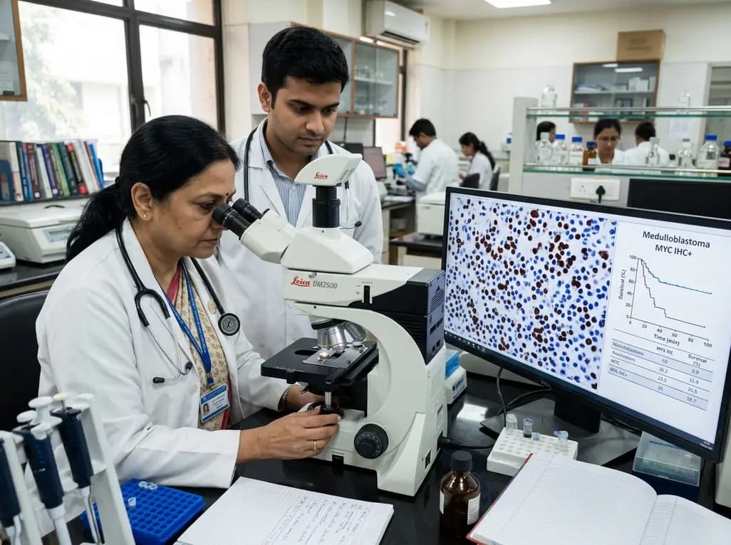
Medulloblastoma represents the most common malignant central nervous system tumor in children. Although modern multimodal management improves survival rates, oncologists continue to encounter therapy-resistant cases that fail standard craniospinal irradiation and chemotherapy. Historically, clinical risk stratification has relied on clinical staging, extent of surgical resection, and histopathology. The 2021 World Health Organization classification highlights distinct molecular subgroups, yet implementing advanced genomic profiling remains challenging in everyday practice. Fortunately, high-resolution proteomic profiling has identified MYC immunohistochemistry as a robust, practical biomarker. This tissue-based assessment reliably predicts treatment failure and guides risk-adapted management in pediatric neuro-oncology.
Current international consensus recognizes four core molecular medulloblastoma entities: WNT-activated, SHH-activated, Group 3, and Group 4. Furthermore, genomic and epigenomic studies divide these categories into twelve nuanced subtypes with divergent prognostic trajectories. However, translating these complex classifications into daily clinical workflows presents formidable logistical obstacles. Most pathology laboratories in low- and middle-income regions lack DNA methylation arrays or whole-genome sequencing platforms. Consequently, oncologists frequently rely on imperfect surrogate markers or conventional clinicopathological staging alone.
This diagnostic gap frequently delays optimal therapeutic decision-making. Standard treatment protocols deliver intensive craniospinal radiotherapy and high-dose alkylating chemotherapy to eradicate residual disease. Nevertheless, aggressive cytotoxic regimens induce profound neurocognitive impairments, endocrine deficiencies, and secondary malignancies in developing children. Conversely, undertreating aggressive biological variants leads to early relapse and dismal salvage rates. Therefore, neuro-oncologists urgently need cost-effective, clinically tractable assays that reliably distinguish indolent tumors from lethal phenotypes. Identifying protein-level drivers from routinely preserved tissue specimens bridges this critical translational divide.
To resolve biological heterogeneity directly from archival tissue, researchers conducted data-independent acquisition mass spectrometry on 56 formalin-fixed paraffin-embedded medulloblastoma specimens. This deep proteomic mapping uncovered a prominent protein signature driven by the MYC proto-oncogene, specifically enriched within aggressive, treatment-refractory Group 3 tumors. Importantly, this molecular pattern correlated directly with therapeutic failure under standard regimens.
To confirm these findings, the investigative team evaluated MYC immunohistochemistry across two large, independent validation cohorts comprising 362 Group 3 and Group 4 medulloblastoma cases. Pathologists performed standardized immunohistochemical staining using standard automated platforms available in routine diagnostic laboratories. After excluding WNT-driven neoplasms, which inherently carry favorable prognoses, positive nuclear staining for MYC emerged as an independent predictor of treatment resistance and overall mortality. Patients harboring MYC-positive tumors experienced an adjusted hazard ratio of 23.6 for therapy resistance and 3.23 for overall mortality. Consequently, this simple antibody assay demonstrated remarkable statistical power in identifying high-risk individuals before treatment initiation.
Historically, neuro-oncologists evaluated MYC gene status strictly through fluorescence in situ hybridization or genomic copy-number analysis. Clinicians assumed that high MYC oncogenic activity required focal DNA amplification. Remarkably, the proteomic investigation demonstrated that only approximately 50 percent of MYC immunohistochemistry-positive tumors harbored genomic MYC amplification. Thus, conventional cytogenetic testing missed half of all patients with high, clinically impactful MYC oncoprotein expression.
Multiple biological mechanisms explain this substantial discrepancy. Specifically, post-transcriptional stabilization, aberrant translational enhancement, and disrupted protein degradation pathways drive robust MYC protein accumulation without underlying gene amplification. Epigenetic alterations and upstream pathway activations can also elevate protein synthesis independent of copy number gains. Because functional proteins directly execute cellular transformation and therapy evasion, quantifying the actual protein product reflects true tumor behavior far better than genomic copy enumeration. Cross-validated survival models incorporating MYC immunohistochemistry substantially outperformed existing stratification models. In fact, adding protein-level assessment reclassified approximately 20 percent of patients into a more accurate, very high-risk category.
The biological implications of elevated MYC protein expression translate directly into dramatic differences in clinical outcomes. In pediatric cohorts, patients with MYC-overexpressing tumors exhibited significantly accelerated disease progression, early leptomeningeal dissemination, and poor response to initial craniospinal irradiation. Furthermore, standard alkylating chemotherapy combinations failed to achieve durable remissions in this cohort. These observations demonstrate that MYC-driven tumor cells possess intrinsic radiobiological and chemoresistant properties that blunt standard cytotoxic insults.
Accordingly, identifying MYC protein positivity should prompt multidisciplinary teams to reconsider conventional therapeutic approaches. Standard-risk regimens prove inadequate for these patients, resulting in unnecessary toxicity without achieving disease control. Instead, these children represent prime candidates for novel therapeutic combinations, such as targeted BET bromodomain inhibitors, CDK9 inhibitors, or intensified upfront induction protocols. Moreover, accurately segregating this very high-risk population prevents inappropriate de-escalation of therapy, protecting vulnerable children from preventable recurrences. Incorporating this biomarker into prospective clinical trial designs will clarify whether intensified interventions or targeted biological agents can overcome treatment resistance.
The most compelling aspect of MYC immunohistochemistry lies in its immediate clinical applicability across diverse global healthcare environments. High-throughput mass spectrometry and comprehensive DNA methylation profiling provide exquisite research insights, but their operational costs and technical demands restrict them to elite research institutions. In contrast, immunohistochemistry represents a foundational technique readily accessible in nearly all tertiary hospital pathology laboratories, including those in low- and middle-income countries.
Additionally, using standard formalin-fixed paraffin-embedded tissue blocks eliminates the need for fresh-frozen tissue preservation, which is rarely feasible in routine clinical workflows. Pathologists can perform and interpret MYC staining within standard turnaround times, delivering crucial prognostic data before oncologists initiate post-surgical adjuvant therapy. In regions where molecular diagnostics are cost-prohibitive, this assay provides a pragmatic gateway to biology-driven risk stratification. Medical oncologists, neuropathologists, and pediatric teams should collaborate to standardize scoring thresholds and staining protocols across diagnostic networks. Ultimately, adopting this accessible biomarker democratizes precision neuro-oncology, ensuring that every child receives treatment matched to tumor biology.
MYC immunohistochemistry directly measures functional nuclear oncoprotein expression on tissue sections, whereas genetic tests evaluate DNA copy-number gains. Remarkably, approximately half of all MYC protein-positive medulloblastomas lack gene amplification. Immunohistochemistry therefore identifies aggressive, high-risk cases driven by post-transcriptional mechanisms that conventional cytogenetic testing fails to detect.
Testing primarily benefits patients diagnosed with non-WNT medulloblastoma, particularly Group 3 and Group 4 tumors. Because WNT-activated neoplasms inherently have an excellent prognosis, excluding them allows MYC immunohistochemistry to function as an independent, highly accurate predictor of therapy resistance, disease relapse, and overall mortality across remaining cohorts.
Yes, standard pathology laboratories can easily implement MYC immunohistochemistry on routine formalin-fixed paraffin-embedded biopsy sections using automated immunostainers. Unlike costly genomic sequencing or methylation arrays, this assay provides rapid, cost-effective prognostic insights, making modern biology-driven risk stratification accessible even in resource-constrained tertiary care centers worldwide.
Disclaimer: This content is for informational and educational purposes only and should not be considered medical advice or relied upon for diagnosis or treatment. Guidelines, regulations, and recommendations are subject to change. Physicians and other qualified medical professionals should exercise their independent judgment when making diagnostic or clinical decisions. While we strive to provide accurate and up-to-date information, we make no representations or warranties, express or implied, regarding completeness, accuracy, or reliability. Refer to the latest local and national guidelines for clinical practice.
References

Read summarized clinical updates, watch expert medical content, and earn CME certifications right from your smartphone.
Open your smartphone camera to scan and install.





A landmark study reveals that high-resolution proteomics identifies MYC immunohistochemistry as an independent predictor of therapy resistance and death in medulloblastoma, outperforming genetic amplification alone and enabling rapid risk stratification in resource-limited clinical environments.
last month

Novel diimide and hydrazone scaffolds, particularly compound #C4, synergistically reactivate latent HIV-1 when combined with JQ1 or PKC activators. Demonstrating donor-matched proviral transcription ex vivo and non-canonical signaling, these findings advance shock and kill strategies toward reservoir eradication.
Today

A cross-sectional study reveals that maternal RSV vaccination coverage reached only 43.7% in Argentina, with maternal vaccine knowledge being the sole independent predictor of uptake. Clinicians play a crucial role in eliminating informational and structural barriers to optimize infant health.
Today

Review how tailored exercise programs benefit frail older patients with heart failure. Explore pathophysiological mechanisms, individualized training prescriptions, and strategies to overcome adherence barriers in clinical practice.
Yesterday

This comparative study evaluates posterior labral tears with and without glenohumeral instability, highlighting distinct demographic patterns, MRI underestimation in stable shoulders, and successful arthroscopic repair outcomes.
Yesterday

A clinical study demonstrates that early intervention with tocilizumab, either as monotherapy or bridged with corticosteroids, achieves significant proptosis reduction and resolves active inflammation in treatment-naive thyroid eye disease.
Yesterday
